
Mechanical properties of nerve roots and rami radiculares isolated from fresh pig spinal cords
2015-02-07 12:58NorihiroNishidaTsukasaKanchikuJunjiOhgiKazuhikoIchiharaXianChenToshihikoTaguchi
中国神经再生研究(英文版) 2015年11期
Norihiro Nishida, Tsukasa Kanchiku Junji Ohgi, Kazuhiko Ichihara, Xian Chen, Toshihiko Taguchi
1 Department of Orthopedic Surgery, Yamaguchi University Graduate School of Medicine, Yamaguchi, Japan
2 Faculty of Engineering, Yamaguchi University, Yamaguchi, Japan
3 Non-Profi t Organization Corporation, Japan Orthopedic Biomechanics Institute, Yamaguchi, Japan
4 Faculties of Engineering, Yamaguchi University, Yamaguchi, Japan
5 Department of Orthopedic Surgery, Yamaguchi University Graduate School of Medicine, Yamaguchi, Japan
Mechanical properties of nerve roots and rami radiculares isolated from fresh pig spinal cords
Norihiro Nishida1,*, Tsukasa Kanchiku1, Junji Ohgi2, Kazuhiko Ichihara3, Xian Chen4, Toshihiko Taguchi5
1 Department of Orthopedic Surgery, Yamaguchi University Graduate School of Medicine, Yamaguchi, Japan
2 Faculty of Engineering, Yamaguchi University, Yamaguchi, Japan
3 Non-Profi t Organization Corporation, Japan Orthopedic Biomechanics Institute, Yamaguchi, Japan
4 Faculties of Engineering, Yamaguchi University, Yamaguchi, Japan
5 Department of Orthopedic Surgery, Yamaguchi University Graduate School of Medicine, Yamaguchi, Japan
No reports have described experiments designed to determine the strength characteristics of spinal nerve roots and rami radiculares for the purpose of explaining the complexity of symptoms of medullary cone lesions and cauda equina syndrome. In this study, to explain the pathogenesis of cauda equina syndrome, monoaxial tensile tests were performed to determine the strength characteristics of spinal nerve roots and rami radiculares, and analysis was conducted to evaluate the stress-strain relationship and strength characteristics. Using the same tensile test device, the nerve root and ramus radiculares isolated from the spinal cords of pigs were subjected to the tensile test and stress relaxation test at load strain rates of 0.1, 1, 10, and 100 s–1under identical settings. The tensile strength of the nerve root was not rate dependent, while the ramus radiculares tensile strength tended to decrease as the strain rate increased. These fi ndings provide important insights into cauda equina symptoms, radiculopathy, and clinical symptoms of the medullary cone.
nerve regeneration; cauda equina syndrome; monoaxial tensile tests; radiculopathy; strength characteristics; stress-strain relationship; lumbar spinal canal stenosis; paralysis; spinal cord; neural degeneration
Nishida N, Kanchiku T, Ohgi J, Ichihara K, Chen X, Taguchi T (2015) Mechanical properties of nerve roots and rami radiculares isolated from fresh pig spinal cords. Neural Regen Res 10(11):1869-1873.
Introduction
The cauda equina consists of nerve roots of the second and subsequent lumbar nerves joining within the spinal canal. Cauda equina syndrome is caused by compressive lesions, such as disc hernia and trauma, presenting with dyskinesia, sensory disorders, and bladder and rectal disorders. Furthermore, compression due to lesions of the medullary cone can also cause symptoms of myelopathy. To clarify the pathophysiology of cauda equina syndrome, morphological and histological evaluations have been conducted, involving preparation of models of chronic nerve root compression, traction of nerve roots, and other techniques (Jancalek et al., 2007; Singh et al., 2015). To date, however, no reports have described experiments designed to determine the strength characteristics of nerve roots and rami radiculares for the purpose of explaining the complexity of symptoms of medullary cone lesions and cauda equina syndrome.
In this study, we conducted a monoaxial tensile test using the nerve roots and rami radiculares isolated from fresh pig spinal cords to analyze the stress-strain relationships and strength characteristics.
Materials and Methods
Tensile test machine
The same testing machine was used for all measurements in this study. The speed and dislocation were set using a personal computer (Windows XP Home Edition, One Microsoft Way Redmond, WA 98052-7329, USA and BTO, Naniwa, Osaka, Japan), and the pulling rate was controlled with a servo controller (SRCD10+RGU-2 Yamaha; Iwata, Shizuoka, Japan). The mobile part attached to the actuator was driven by the servo motor (T9H 20BK-400 Yamaha; Iwata, Shizuoka, Japan). The data for dislocation of the mobile part were collected with the rotary encoder attached to the servo motor and fed through the encoder board (PCI-6204 Interface; Kyobashi, Minami, Hiroshima, Japan) into the personal computer. The load data for the test samples were collected with the load cell (LTS-100GA Kyowa; Cyofugaoka, Cyofu, Tokyo, Japan) and fed through a dynamic strain meter (DPM-700B Kyowa; Cyofugaoka, Cyofu, Tokyo, Japan) and an AD conversion board (PCI-3171A Interface; Kyobashi, Minami, Hiroshima, Japan) into the personal computer (Figure 1A, B).
The program for controlling the measurements with the testing device was prepared with Microsoft Visual Basic 6.0 (One Microsoft Way Redmond, WA 98052-7329, USA). This program was used for setting the conditions for measurement (sampling frequency, pulling rate, tensile dislocation, and etc.) and processing of the data.
Preparation of nerve root samples
Samples were collected from the spinal cords of two pigswithin 12 hours after slaughter for human consumption. Two pigs were hybrid (Landrace and Duroc), aged 13 months, weighing 120–130 kg, both genders (Ginchiku ranch company, Hofu, Yamaguchi, Japan). The pigs used in this experiment were dismantled for meat to eat. As confi rmed by Science Research Center, Institute of Life Science and Medicine of Yamaguchi University, Japan, the precise procedure was unnecessary. The vertebral arch on the dorsal side of the spinal canal was resected with an electric saw (MJ-300, RYOBI) to expose the dura mater and the spinal cord (Figure 2A). The nerve root was then dissected at the level of the intervertebral foramen, and the medullary cone and nerve root were isolated from the spine while still wrapped with the dura mater (Figure 2B). The dura mater around the spinal cord and the nerve root were then resected (Figure 3A), and samples 1–4 were assigned to the nerve root sections (sample No. 1 was assigned to the nerve root section most proximal to the point of bifurcation). During this process, the dura mater at the end of the nerve root facing the intervertebral foramen was left partially unresected to enable subsequent fi xation on the testing device. For a similar reason, the spinal cord tissue at the origin of the nerve root was left partially unresected (Figure 3B).
The sample was fi xed in the testing device (Figure 1B) by pinching the central and peripheral sides of the nerve root between the upper and lower jigs. To prevent the sample from falling off , the sample was fi xed by suturing with 4-0 looped nylon using the method of Tsuge (1977).
Preparation of ramus radiculares samples
The specimens were immersed in physiological saline to enable ramus radiculares samples to be prepared without damaging the specimen (Figure 3B). The diameter of each ramus radiculares sample was measured with a 3D laser microscope (LEXT OLS4000; Olympus, Japan), followed by calculation of the cross-sectional area. A mean cross-section of 0.069 mm2(range: 0.067–0.071 mm2) was adopted. To analyze the mechanical strength characteristics of the nerve roots and constituent rami radiculares, samples were subjected to the tensile test and to the stress relaxation test at a gauge length of 10 mm and temperature of 25°C as reported earlier (Ichihara et al., 2001). The load strain rates were 0.1, 1, 10 and 100 s–1using 3, 4, 3 and 4 samples of nerve roots and 4, 6, 6, and 5 samples of ramus radiculares, respectively. We used Microsoft Offi ce Home and Business 2010 Excel (One Microsoft Way Redmond, WA 98052-7329, USA) for preparing fi gures.
Results
Nerve root tensile test results
To analyze the mechanical properties of nerve roots following compression by trauma or because of degeneration of the disc or vertebral body, we conducted tensile tests. Figure 4A shows the stress-strain relationships for nerve roots analyzed using monoaxial tensile tests. With each sample, the stress gradually increased towards its maximum as the strain increased, thus exhibiting a superelastic pattern. After reaching its maximum value, the stress began to decrease gradually even as the strain increased further.
Rate dependency of nerve root

Figure 1 Experimantal set up (A) and tensile test machine (B).
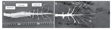
Figure 2 Anatomy of spine and nerve.
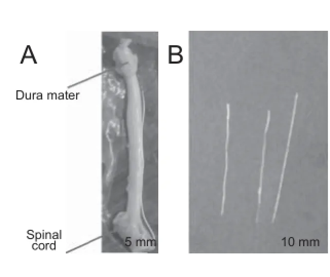
Figure 3 Nerve root and ramus radiculares.
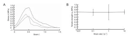
Figure 4 Nerve root tensile test results.
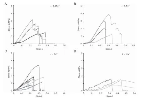
Figure 5 Analysis of stress-strain relationships based on ramus radiculares samples.
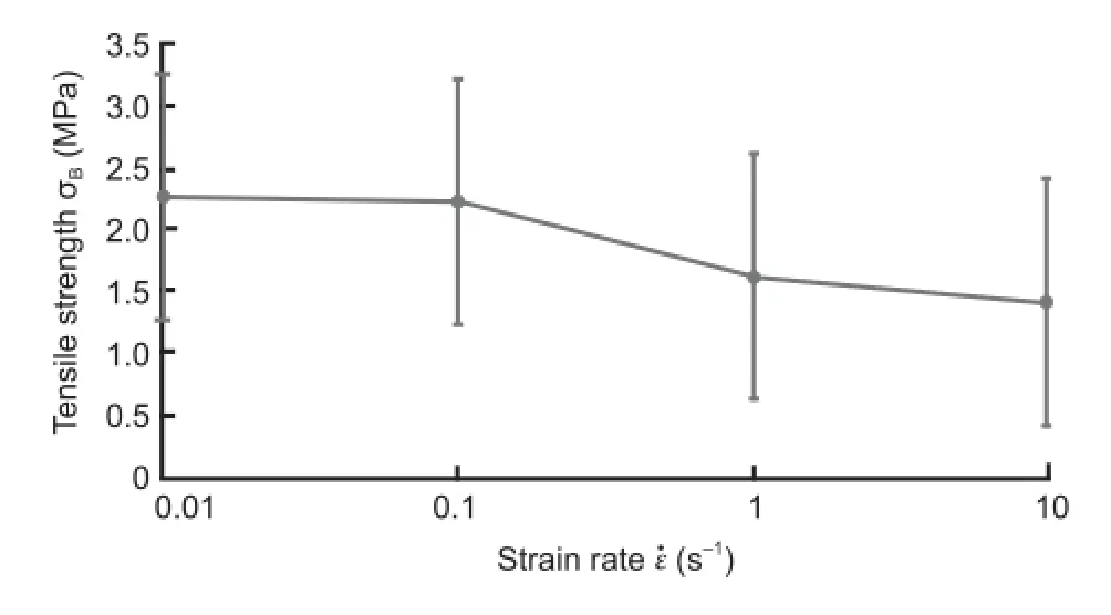
Figure 6 The data for the mean tensile strength (σB) of all ramus radiculares samples in relation to the load strain rate (ε˙).
To investigate damage to the nerve root caused by speed, we further evaluated the results of the tensile test. Figure 4Bshows the data for nerve root tensile strengthin relation to the load strain rateWhenthe relationship betweenandremained relatively constant, despite large variations in these parameters, and the tensile strength was not rate dependent. At each strain rate, the nerve root σB was approximately 0.7 MPa.
Ramus radiculares tensile test results
To examine diff erences between the nerve root and ramus radiculares, we conducted similar tensile tests to investigate the mechanical properties of ramus radiculares. Figure 5A–D shows stress-strain relationships using ramus radiculares samples. Although diff erences indid not infl uence deformation patterns, the response to stress of the ramus radiculares diff ered markedly to that of nerve roots. When= 0.01 s–1the stress decreased in a stepwise manner after reaching a peak, with the decrease becoming more rapid as the strain rate increased.
Rate dependency of ramus radiculares tensile strength
To examine the eff ect of speed on damage to the ramus radiculares, we further evaluated the results of tensile testing. Figure 6 shows results for the mean tensile strength (σB) of all ramus radiculares samples in relation to the load strain rate. The tensile strength of the ramus radiculares tended to decrease as the load strain rate increased.
Discussion
The pathophysiology and classifi cation of lumbar spinal canal stenosis were fi rst reported by Arnoldi (1976). After this initial report, many researchers performed clinical and animal studies examining the symptoms of cauda equina in the context of blood fl ow (Ooi et al., 1990; Takahashi et al., 1993; Ikawa et al., 2005) and dural pressure both in the standing position and at rest (Takahashi et al., 1995a, b). According to these reports, the dural pressure during walking or in the standing position is approximately twice that in the supine position, and the disruption of circulation in patients with cauda equina syndrome is attributable to venous retention rather than to insuffi cient arterial blood supply.
Furthermore, for clarification of the pathophysiology of compressive radiculopathy, experiments on acute compression (Delamarter et al., 1990; Olmarker et al., 1991; Kobayashi et al., 1993) and chronic compression (Kobayashi et al., 2004a, b) have been carried out to evaluate nerve degeneration and pathological changes. In addition, evaluation of nerve root traction and compression has also been conducted frequently. Singh et al. (2015) reported that the morphological changes in nerve roots vary depending on the type of external force. Moreover, Jancalek et al. (2007) conducted histological evaluations of the internal structures of nerves exposed to external force. Reports have also described studies of molecules involved in pain, such as tumor necrosis factor (TNF)-α during nerve root compression (Obata et al., 2004; Sekiguchi et al., 2009). These experiments were designed to create a similar environment of entrapment neuropathy by applying a constant pressure to the nerve.
While previous reports have evaluated blood fl ow or pressure, histological features or degeneration, and the pathology/chemistry, no studies have examined the mechanical properties of nerve roots or rami radiculares. When discussing the occurrence of paralysis arising from compression or trauma, mechanical properties also need to be taken into consideration. Although previous studies investigated the pathological and chemical changes of nerve roots following compression, they did not evaluate damage to the nerve caused by force and speed of injury. While this can be described with respect to future changes in the nerve, it is not possible to estimate the degree of nerve damage or to predict the eff ects of speed and traction. Our analysis is capable of shedding light on this. For example, after trauma to the thoracolumbar vertebral junction, symptoms are diverse because this area is governed by the upper and lower motor neurons of the spinal cord parenchyma, cauda equina, and nerve roots, thus making diagnosis diffi cult (Elsberg, 1996). The symptoms in these tissues may be associated with the strength characteristics of the cauda equina, in addition to the anatomical complexity and the manner of compression (Wall et al., 1990a, b). For these reasons, we attempted tensile tests to clarify the mechanical properties of these tissues.
A similar evaluation of the strength characteristics of nerves was previously reported by Ichihara et al. (2001) who analyzed the material characteristics of the spinal cord (gray and white matter). Based on data describing these mechanical properties, simulation by the fi nite element method has been conducted for analysis of the features of spinal cord disease, yielding results that were very similar to clinical knowledge (Kato et al., 2008, 2009, 2010; Nishida et al., 2011, 2012, 2013, 2014). In the present study, the nerve root tensile strength showed no rate dependency and was about 0.7 MPa; this value is 16 times the gray matter strength and 47 times the white matter strength reported by Ichihara et al. (2001). These results indicated that the nerve root has higher tensile strength than the spinal cord. This feature may be associated with the observation that severe injury of the nerve root in the medullary cone is unlikely to occur. Regarding the rami radiculares, the present study suggested the possibility that the nerve may be injured suddenly when exposed to certain stress. As the pulling rate increased, the likelihood for injury tended to increase as well. Additionally, the stepwise decrease in stress in the nerve root, unlike that in the ramus radiculares, seemed to be attributable to the composite structure of the nerve root, which is comprised of multiple rami, connective tissue, and fat tissue.
Since it is diffi cult to acquire human nerve root or ramus radiculares for study, we used tissues from the pig as an alternative. Clinical symptoms of cauda equina, radiculopathy and the medullary cone can be evaluated in relation to results obtained with the fi nite element method. If the fi nite element method analyses based on the mechanical properties of pig tissues correlate with the clinical symptoms of the patient, this may help to understand the underlying pathology and therefore help to choose the best therapeutic strategy.
This study had a few limitations. First, we did not performevaluation of blood fl ow. Additionally, the pig specimens may have undergone histological degeneration during the 12-hour period from collection to examination. However, the fi nding that the nerve roots and rami radiculares had higher strength than the spinal cord may be important for analyzing the medullary cone and cauda equina and for understanding the clinical symptoms of diseases involving this areas.
Taken together, we performed tensile load tests in nerve roots and rami radiculares. The tensile strength of nerve roots was not rate dependent, while the tensile strength of rami radiculares tended to decrease as the strain rate increased. Our data showed that nerve roots and rami radiculares exhibited higher strength than the gray and white matter of the spinal cord. These fi ndings provide important insight into the clinical symptoms of cauda equina syndrome and of diseases associated with the nerve roots and medullary cone.
Acknowledgments: Special thanks to Mr. Syota Tahara, Mr. Yoshitaka Kanda, Mr. Yuuta Haruna, Mr. Kunitaka Fukuda, Mr. Hidetaka Morita and Mr. Seiya Miyazaki who were cooperator and enforcer of the experiments.
Author contributions: NN and JO wrote the paper. JO and KI performed experiments. TK, XC, and TT guided the research. All authors approved the fi nal version of this paper. Confl icts of interest: None declared.
Plagiarism check: This paper was screened twice using Cross-Check to verify originality before publication.
Peer review: This paper was double-blinded, stringently reviewed by international expert reviewers.
Arnoldi CC, Brodsky AE, Cauchoix J, Crock HV, Dommisse GF, Edgar MA, Gargano FP, Jacobson RE, Kirkaldy-Willis WH, Kurihara A, Langenskiöld A, Macnab I, McIvor GW, Newman PH, Paine KW, Russin LA, Sheldon J, Tile M, Urist MR, Wilson WE, Wiltse LL (1976) Lumbar spinal stenosis and nerve root entrapment syndromes. Defi -nition and classifi cation. Clin Orthop Relat Res (115):4-5.
Delamarter RB, Bohlman HH, Dodge LD, Biro C (1990) Experimental lumbar spinal stenosis. Analysis of the cortical evoked potentials, microvasculature, and histopathology. J Bone Joint Surg Am 72:110-120.
Elsberg C (1996) Diagnosis and treatment of surgical disease of the spinal cord and its membranes. pp63-65.WB saunders, Philadelphia.
Ichihara K, Taguchi T, Yoshinori S, Ituo S, Shunichi K, Shinya K (2001) Gray matter of the bovine cervical spinal cord is mechanically more rigid and fragile than the white matter. J Neurotrauma 18:361-367.
Ikawa M, Atsuta Y, Tsunekawa H (2005) Ectopic fi ring due to artifi cial venous stasis in rat lumbar spinal canal stenosis model: a possible pathogenesis of neurogenic intermittent claudication. Spine 30:2393-2397.
Jancalek R, Dubovy P (2007) An experimental animal model of spinal root compression syndrome: an analysis of morphological changes of myelinated axons during compression radiculopathy and after decompression. Exp Brain Res 179:111-119.
Kato Y, Kanchiku T, Imajo Y, Ichihara K, Kawano S, Hamanaka D, Yaji K, Taguchi T (2009) Flexion model simulating spinal cord injury without radiographic abnormality in patients with ossifi cation of the longitudinal ligament: the infl uence of fl exion speed on the cervical spine. J Spinal Cord Med 32:555-559.
Kato Y, Kanchiku T, Imajo Y, Kimura K, Ichihara K, Kawano S, Hamanaka D, Yaji K, Taguchi T (2010) Biomechanical study of the eff ect of the degree of static compression of the spinal cord in ossifi cation of the posterior longitudinal ligament. J Neurosurgery Spine 12: 301-305.
Kato Y, Kataoka H, Ichihara K, Imajo Y, Kojima T, Kawano S, Hamanaka D, Yaji K, Taguchi T (2008) Biomechanical study of cervical fl exion myelopathy using a three-dimensional fi nite element method. J Neurosurgery Spine 8:436-441.
Kobayashi S, Yoshizawa H, Yamada S (2004a) Pathology of lumbar nerve root compression. Part 1: Intraradicular infl ammatory changes induced by mechanical compression. J Orthop Res 22:170-179.
Kobayashi S, Yoshizawa H, Yamada S (2004b) Pathology of lumbar nerve root compression. Part 2: morphological and immunohistochemical changes of dorsal root ganglion. J Orthop Res 22:180-188.
Kobayashi S, Yoshizawa H, Hachiya Y, Ukai T, Morita T (1993) Vasogenic edema induced by compression injury to the spinal nerve root. Distribution of intravenously injected protein tracers and gadolinium-enhanced magnetic resonance imaging. Spine 18:1410-1424.
Nishida N, Kato Y, Imajo Y, Kawano S, Taguchi T (2011) Biomechanical study of the spinal cord in thoracic ossifi cation of the posterior longitudinal ligament. J Spinal Cord Med 34:518-522.
Nishida N, Kato Y, Imajo Y, Kawano S, Taguchi T (2012) Biomechanical analysis of cervical spondylotic myelopathy: The infl uence of dynamic factors and morphometry of the spinal cord. J Spinal Cord Med 35:256-261.
Nishida N, Kanchiku T, Kato Y, Imajo Y, Kawano S, Taguchi T (2013) Biomechanical analysis of the spinal cord in Brown-Séquard syndrome. Exp Ther Med 6:1184-1188.
Nishida N, Kanchiku T, Kato Y, Imajo Y, Yoshida Y, Kawano S, Taguchi T (2014) Biomechanical analysis of cervical myelopathy due to ossifi cation of the posterior longitudinal ligament: eff ects of posterior decompression and kyphosis following decompression. Exp Ther Med 7:1095-1099.
Obata K, Yamanaka H, Kobayashi K, Dai Y, Mizushima T, Katsura H, Fukuoka T, Tokunaga A, Noguchi K (2004) Role of mitogen-activated protein kinase activation in injured and intact primary aff erent neurons for mechanical and heat hypersensitivity after spinal nerve ligation. J Neurosci 24:10211-10222.
Olmarker K (1991) Spinal nerve root compression. Nutrition and function of the porcine cauda equina compressed in vivo. Acta Orthop Scand Suppl 242:1-27.
Ooi Y, Mita F, Satoh Y (1990) Myeloscopic study on lumbar spinal canal stenosis with special reference to intermittent claudication. Spine 15:544-549.
Sekiguchi M, Sekiguchi Y, Konno S, Kobayashi H, Homma Y, Kikuchi S (2009) Comparison of neuropathic pain and neuronal apoptosis following nerve root or spinal nerve compression. Eur Spine J 18:1978-1985.
Singh J, Hussain F, Decuzzi P (2015) Role of diff erential adhesion in cell cluster evolution: from vasculogenesis to cancer metastasis. Comput Methods Biomech Biomed Engin 18:282-292.
Takahashi K, Kagechika K, Takino T, Matsui T, Miyazaki T, Shima I (1995a) Changes in epidural pressure during walking in patients with lumbar spinal stenosis. Spine 20: 2746-2749.
Takahashi K, Miyazaki T, Takino T, Matsui T, Tomita K (1995b) Epidural pressure measurements. Relationship between epidural pressure and posture in patients with lumbar spinal stenosis. Spine 20:650-653.
Takahashi K, Olmarker K, Holm S, Porter RW, Rydevik B (1993) Double-level cauda equina compression: an experimental study with continuous monitoring of intraneural blood fl ow in the porcine cauda equina. J Orthop Res 11:104-109.
Tsuge K, Yoshikazu I, Matsuishi Y (1977) Repair of fl exor tendons by intratendinous tendon suture. J Hand Surg Am 2:436-440.
Wall EJ, Cohen MS, Massie JB, Rydevik B, Garfin SR (1990a) Cauda equina anatomy. I: Intrathecal nerve root organization. Spine 15:1244-1247.
Wall EJ, Cohen MS, Abitbol JJ, Garfi n SR (1990b) Organization of intrathecal nerve roots at the level of the conus medullaris. J Bone Joint Surg Am 72:1495-1499.
Copyedited by Tang Z, Yang S, Li CH, Song LP, Zhao M
*Correspondence to: Norihiro Nishida, M.D., nishida3@yamaguchi-u.ac.jp.
orcid: 0000-0001-7754-6579 (Norihiro Nishida)
10.4103/1673-5374.170319 http://www.nrronline.org/
Accepted: 2015-08-10

- 中国神经再生研究(英文版)的其它文章
- Intracellular sorting pathways of the amyloid precursor protein provide novel neuroprotective strategies
- The role of the Rho/ROCK signaling pathway in inhibiting axonal regeneration in the central nervous system
- VEGF in the nervous system: an important target for research in neurodevelopmental and regenerative medicine
- Studying neurological disorders using induced pluripotent stem cells and optogenetics
- Ef cacy of glucagon-like peptide-1 mimetics for neural regeneration
- Compliant semiconductor scaf olds: building blocks for advanced neural interfaces