
Left armpit subcutaneous metastasis of gastric cancer:A case report
2019-04-22 09:01FengJunHePengZhangMoJinWangYiChenWenZhuang
World Journal of Clinical Cases 2019年23期
Feng-Jun He, Peng Zhang, Mo-Jin Wang, Yi Chen, Wen Zhuang
Feng-Jun He, Peng Zhang, Mo-Jin Wang, Yi Chen, Wen Zhuang, Department of Gastrointestinal Surgery, West China Hospital Sichuan University, Chengdu 610041, Sichuan Province, China
Abstract
Key words: Stomach neoplasms; Neoplasm metastasis; Subcutaneous; Case report; Cancer therapy; Skin neoplasms
INTRIDUCTION
Gastric cancer is prevalent worldwide, with an average of approximately 990000 new cases per year from 182 countries and 30 world regions[1].The highest incidence is observed in Eastern Asia[2,3].According to the Eindhoven Cancer Registry statistics,between 1995 and 2012, about 40% of gastric cancer patients had one metastasis at least[4].The most common metastasis sites of gastric carcinoma are the liver (48%),peritoneum (32%), lung (15%), and bone (12%); however, relevant data indicated that the incidence of subcutaneous metastasis of gastric cancer is about 0.8%[5,6].Today,there is no data referring to the left armpit metastasis of gastric carcinoma.Here we report the case of a patient with stage III gastric carcinoma who underwent curative intent resection (R0) and D2 lymph node dissection and received eight cycles of chemotherapy post-surgery.However, left armpit subcutaneous metastasis occurred in the fifth year after surgery.We report the case to promote the exploration and monitoring of unusual rare metastatic sites of advanced gastric cancer, and provide clinical evidence for the diagnosis and treatment of metastasis of gastric cancer.
CASE PRESENTATION
Chief complaints
A 69-year-old man was re-admitted to West China Hospital of Sichuan University due to an asymptomatic lump in his left armpit for 3 mo (Figure 1).
History of present illness
The patient had a history of gastric neoplasms.Five years ago, he had undergone curative gastrectomy, followed by eight cycles of oral chemotherapy.
History of past illness
The patient had a free previous medical history.
Physical examination
Physical examination after admission showed that the patient’s body temperature was 36 °C, heart rate was 106 bpm, respiratory rate was 20 breaths per minute, and blood pressure was 140/68 mmHg.A mass of approximately 7.0 cm × 4.5 cm × 5.7 cm mass was observed in the left armpit of the patient.The skin of the mass was reddish, and the temperature was high.There was no skin ulceration or itching.The patient experienced no pain when the mass was pressed.The mass was hard, fixed, and had an unclear boundary.
近年来,各医学院校相继开展机能实验学整合。机能实验学课程的开展,是医学院校实验教学改革的必然要求。该课程除了教师和学生两大主体的参与,也离不开实验技术人员。随着学科建设不断深入,机能实验课程内容变得更加充实和复杂,对参与者提出了更高要求。实验技术人员的工作水平从一定程度上反映了机能实验中心的教学水平、科研水平和管理水平。但是,目前实验技术人员的工作素养与实际工作缺乏有机结合,工作现状亟待改善。
Laboratory examinations
Blood analysis did not reveal raised levels of tumor markers.Prothrombin and partial thromboplastin times were normal and serum C-reactive protein level had increased to 4.5 mg/dL (normal range:< 0.8 mg/dL).
Imaging examinations
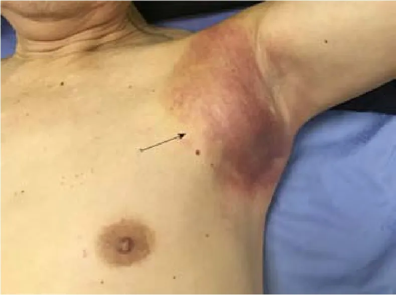
Figure 1 Left armpit subcutaneous metastasis arising from primary gastric cancer.
Initial color Doppler ultrasound imaging of the left axillary lump showed a heterogeneous echo pattern sized approximately 4.2 cm × 2.4 cm × 3.8 cm, with an unclear boundary and irregular shape.There was linear blood flow signal observed in the mass.Several abnormally enlarged lymph nodes were observed around the mass.After three months, color Doppler examination revealed that the mass grew to a size of 6.9 cm × 4.4 cm × 5.7 cm (Figure 2).A chest computed tomography (CT) scan revealed a 3.9 cm × 5.7 cm soft tissue lump in the left armpit.
Further diagnostic work-up
Fine needle aspiration biopsy of the mass guided by color Doppler ultrasound found cancer cells in the mass.Immunohistochemical examination showed that the mass was CDX2-positive, PCK-positive, CK20-positive, CK7-negative, and TTF-negative;this confirmed gastric cancer metastasis (Figure 3).
Positron emission tomography/CT (PET/CT) identification of the distant metastasis
PET/CT examination showed a soft tissue mass sized approximately 6.2 cm × 5.5 cm in the left axilla.The internal density was uneven, and 18F-fludeoxyglucose uptake was abnormally high.The maximum SUV was 11.13 (Figure 4).
MULTIDISCIPLINARY EXPERT CONSULTATION
Wen Zhuang, MD, PhD, Professor and Chief Physician, West China Hospital of Sichuan University
The patient should undergo surgical resection of the left axillary tumor and adjuvant radiotherapy, chemotherapy, or targeted therapy.
Jie Chen, MD, PhD, Professor, Department of Breast Surgery, West China Hospital of Sichuan University
The patient should undergo surgical treatment of the left mass in his left armpit and total excision of the lesion.If necessary, we will assist in the operation.
Zhi-Xing Chen MD, PhD, Associate Professor, Department of Plastic Surgery, West China Hospital of Sichuan University
Surgical resection of the left axillary subcutaneous tumor may require skin grafting.
FINAL DIAGNOSIS
Left axillary subcutaneous metastasis of gastric cancer.
TREATMENT
Due to the adhesion between the large subcutaneous mass of the left axilla and surrounding tissues and severe local inflammation, skin grafting might be required after operation.We asked plastic surgery experts to assist in the operation of tumor resection.However, considering the risk of resection, the patient did not agree to undergo surgical treatment.

Figure 2 Color Doppler ultrasound images.
OUTCOME AND FOLLOW-UP
After discharge, the patient was lost to follow-up.However, through telephonic communication, we know that he is on long-term medication and is alive.
DISCUSSION
Epidemiological studies on metastasis of gastric cancer are rare.Currently, the TNM system is used to stage malignant tumors, and cancer registries often only use “M0”and “M1” to indicate the absence or presence of distant metastasis.Therefore, there is a lack of information regarding specific distant metastasis sites[5].
The five-year cumulative risk of relapse (restricted to patients who undergo R0 resections and excluding in-hospital deaths) for patients with pathological stage T3 tumors is 83% for D1 dissection and 72% for D2 dissection[7].Although considered a“localized tumor”, gastric cancer may show locoregional metastasis and this can be the most important signal of negative prognosis[8-10].
Metastasis is mostly driven by the acquisition of genetic and/or epigenetic alterations within tumor cells and the formation of the tumor microenvironment[11].Metastasis of malignant tumors can occur at an early stage of primary tumorigenesis[12].If metastasis of cancer cells occurs before clinical detection, surgical resection may not prevent recurrence, invasion, and further metastasis.In our case,the patient was followed regularly and monitored through dynamic imaging, and no sign of recurrence was observed.However, in the fifth year, he was diagnosed with distant subcutaneous metastasis.Because of mild clinical manifestations and metastasis into a rare site, the lump was not considered severe.Consequently, we missed the best surgical window.
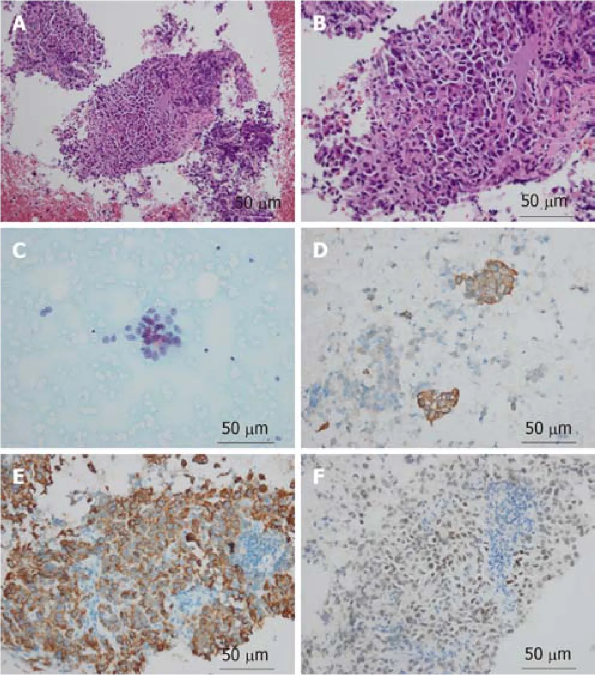
Figure 3 Pathological images of the axillary mass.
Currently, ultrasound and color Doppler are the preferred non-invasive imaging modalities of choice allowing to diagnose superficial masses, which can not only differentiate the nature of masses, but also provide detailed information about vascular anatomy[13-15].Color Doppler, in particular, is highly specific in the identification of benign and malignant nodules of the skin and subcutaneous tissue[16,17].In addition, high-resolution ultrasound may contribute to the differential diagnosis of skin and subcutaneous lesions[17].
Treatment of even well-confined tumors can become difficult due to repeated changes in molecular phenotypes[18], immune evasion[19], and drug resistance[20].Through a long-term observation, it has been shown that some types of malignant tumors metastasize only to specific target organs[20].Cutaneous or subcutaneous tissues may not provide a better growth microenvironment than the liver or peritoneum.However, several cases of subcutaneous metastasis of gastric cancer,including scalp metastasis and mandibular metastasis and so like, have been reported in succession[21,22].Here we report a rare case that provides clinical evidence for studying the specific metastatic sites of gastric cancer.
CONCLUSION
We should pay enough attention to any local mass developing in patients with a history of gastric cancer.We believe that in the future more sensitive, specific, and inexpensive techniques, such as nanoparticles[23]and liquid biopsy, would contribute to the detection of metastasis[24].
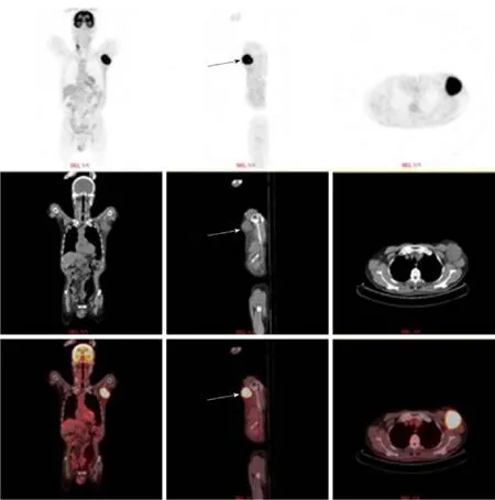
Figure 4 Positron emission tomography computed tomography examination showed that a 6.2 cm × 5.5 cm soft tissue mass was visible in the left axilla(arrow).
猜你喜欢
运动精品(2022年3期)2022-08-12
小学教学研究(2022年21期)2022-07-28
昆明医科大学学报(2022年2期)2022-03-29
河北画报(2021年2期)2021-05-25
课程教育研究(2021年24期)2021-04-14
昆明医科大学学报(2021年1期)2021-02-07
武术研究(2020年3期)2020-04-21
福建基础教育研究(2019年3期)2019-05-28
cookie world(2010年5期)2010-06-10
决策与信息·下旬刊(2009年4期)2009-10-10
 World Journal of Clinical Cases2019年23期
- World Journal of Clinical Cases的其它文章
- Overview of organic anion transporters and organic anion transporter polypeptides and their roles in the liver
- Value of early diagnosis of sepsis complicated with acute kidney injury by renal contrast-enhanced ultrasound
- Value of elastography point quantification in improving the diagnostic accuracy of early diabetic kidney disease
- Resection of recurrent third branchial cleft fistulas assisted by flexible pharyngotomy
- Therapeutic efficacy of acupuncture combined with neuromuscular joint facilitation in treatment of hemiplegic shoulder pain
- Comparison of intra-articular injection of parecoxib vs oral administration of celecoxib for the clinical efficacy in the treatment of early knee osteoarthritis