
Thoracoscopic resection of a huge esophageal dedifferentiated liposarcoma:A case report
2020-05-13 07:34YiWangYeMengYingLiaoZhiMinMouXiaoXinShiYuanCaiXie
World Journal of Clinical Cases 2020年9期
Yi-Wang Ye, Meng-Ying Liao, Zhi-Min Mou, Xiao-Xin Shi, Yuan-Cai Xie
Yi-Wang Ye, Zhi-Min Mou, Yuan-Cai Xie, Department of Thoracic Surgery, Peking University Shenzhen Hospital, Shenzhen 518036, Guangdong Province, China
Meng-Ying Liao, Xiao-Xin Shi, Department of Pathology, Peking University Shenzhen Hospital,Shenzhen 518036, Guangdong Province, China
Abstract
Key words: Thoracoscopic surgery;Esophageal liposarcoma;Dedifferentiated liposarcoma;Huge esophageal tumor;Case report
INTRODUCTION
Esophageal liposarcoma is a rare malignant tumor, with a reported incidence of 0.1%-1.5% of all esophageal neoplasms.Most cases are well-differentiated liposarcoma[1].Dedifferentiated liposarcoma (DDL) has a lower incidence in patients with esophageal liposarcoma.We present a patient with a large esophageal liposarcoma treated by thoracoscopic resection.
CASE PRESENTATION
Chief complaints
A 38-year-old woman with dysphagia for 6 mo and dyspnea for 1 wk.
History of past illness
The patient had no previous medical history.
Imaging examinations
Computed tomography (CT) showed a large mass in the posterior mediastinum,which may have been caused by a large cyst or effusion inside the esophagus.The trachea and left and right main bronchi were subjected to external pressure stenosis(Figure 1).Esophagogastroscopy revealed a large mass pushing against the esophagus.Esophagogastric ultrasonography showed a homogeneous, low echogenic mass outside the esophagus, which ranged from 20 to 40 cm below the incisor, and had a cross-sectional area of 21.5 mm × 15.9 mm (Figure 2).Upper gastrointestinal angiography also showed external pressure stenosis of the esophagus (Figure 3).
The patient’s dyspnea became more serious due to the obstruction caused by tracheal compression.She developed respiratory failure and it was unclear whether this was caused by a mass inside or outside the esophagus.Exploratory thoracoscopy was performed to relieve the tracheal compression obstruction.
A large, smooth-surface mass occupying the posterior mediastinum was observed(Figure 4).The upper segment of the esophagus was split longitudinally, and most of the mass was extracted from the esophageal lumen to the thoracic cavity.The pedicle was excised by linear cutting closers under mirrors.Little residual mass was noted by gastroscopy (Figure 5).We considered this to be a benign tumor of the esophagus and the pedicle of the tumor was near the throat;thus, it was difficult to confirm complete resection by biopsy under endoscopy.In addition, the patient developed severe respiratory insufficiency during surgery.Due to the above reasons we were unable to confirm complete resection by biopsy pathology.The mucous and muscular layers were interruptedly sutured.
Pathological examination
Pathological examination showed some discrete spindle cells, and a little adipose tissue (less than 5%).Immunohistochemical analysis revealed that some discrete spindle cells were positive for Desmin and CD34, and negative for S-100, SMA, DOG-1 and STAT-6.Fatty tissue was positive for S-100 protein.CD117 highlighted the intermixed mast cells and plasma cells.MDM2gene amplification was confirmed by fluorescence in situ hybridization analysis (Figure 6).
高校图书馆资源库是浩瀚无穷的,包括工科、农科、医学类、管理类等大类资源,建设内容不仅仅拘泥于高校图书馆现有的馆藏资源,还包括一些核心期刊的文献资源,以便为智库研究人员提供必要的理论支撑。智库成果库收录了国内外众多珍贵的文献数据,包括短讯、摘要纪实,期刊要闻等,具备一定的理论前瞻性。学术网站数据库也收录了不同机构的资料分析,为智库人员提供最新的学术观点、时事评论等内容。
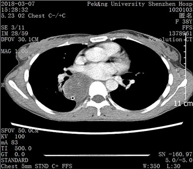
Figure 1 Computed tomography showed an expanded esophagus and low density shadow.
FINAL DIAGNOSIS
The pathologist suspected that the mass was a DDL of the esophagus.Cleveland Medical Center Pathology Consultation considered the mass to be DDL.
TREATMENT
We recommended that the patient undergo extended resection of the tumor pedicle with cervical incision or adjuvant treatment.
OUTCOME AND FOLLOW-UP
The patient did not have any dysphagia or dyspnea 2 wk postoperatively and refused any further treatment, but underwent CT and esophagoscopy every three months.The patient‘s last CT and esophagoscopy did not find any sign of recurrence at 20 mo post-operation (Figure 7).
DISCUSSION
A liposarcoma originates in the mesenchymal layer, and is one of the most common sarcomas in adults, but is rarely found in the esophagus[2].Liposarcomas can be divided into four subtypes:Well-differentiated liposarcoma, pleomorphic liposarcoma, myxoid/round cell liposarcoma, and DDL;the latter is a rare type with a prevalence of approximately 6% of all liposarcomas[1,2,3,4].We found only 60 reported cases of esophageal liposarcomas and just one case of esophageal DDL.Here, we present a case with minimally invasive treatment of a large esophageal DDL by thoracoscopy.
An esophageal liposarcoma grows slowly and presents with symptoms caused by compression or invasion of intrathoracic structures, most commonly dyspnea, weight loss, throat discomfort and dysphagia.Esophagoscopy reveals a submucosal lesion or extrinsic compression encompassing the esophageal lumen.CT shows a soft tissue lesion in the posterior mediastinum[1,5].Differential diagnosis of the disease from benign esophageal tumors, such as a fibrovascular polyp, is often difficult by imaging or endoscopy[6].We were unable to make a definitive diagnosis by frozen section analysis during the operation.In the present case, we diagnosed a large mass in the esophagus before surgery, and frozen section diagnosis was a spindle cell tumor.Esophagoscopic biopsy to distinguish liposarcoma from polyps before surgery is necessary.However, in this case we did not perform preoperative biopsy as the patient underwent emergency, life-saving surgery for obstructive respiratory dysfunction.
Surgery is the best choice of treatment for DDL[7], including aggressive open transcervical, transthoracic, and transabdominal total or subtotal esophagectomy[4,8-10].
There are four case reports of the treatment of esophageal liposarcoma with endoscopic submucosal dissection[4,7,11,12].However, it is not possible to resect large esophageal tumors with endoscopic submucosal dissection.Other approaches have been attempted, but no thoracoscopic approaches have been reported.In the present case, our surgical approach was thoracoscopic esophagotomy and the patient did not have any postoperative discomfort.

Figure 2 Esophagogastric ultrasonography showed a homogeneous, low echogenic mass outside the esophagus.
It is not possible to give an authoritative assessment regarding the prognosis of esophageal liposarcoma after surgery as the number of cases published with longterm follow-up is insufficient for rigorous analysis.Rivaet al[13]reported local recurrence in 13% of patients with esophageal liposarcoma and the average time between first treatment and recurrence was 127.67 mo[13].Our patient is in good condition and did not have any recurrence 20 mo after surgery.
In our patient, non-curative resection of the esophageal DDL may have been performed, as we intended taking a margin biopsy by endoscopy for frozen pathology to confirm complete resection, but the tumor pedicle was near the throat, and it was difficult to confirm complete resection by biopsy under endoscopy.In addition, the patient developed severe respiratory insufficiency during surgery.Thus, we were unable to confirm complete resection by biopsy pathology during surgery.
We could not completely resect the possible residual tumor under endoscopy as the tumor pedicle was near the throat.It is unclear whether the patient should have undergone extended resection of the tumor pedicle by cervical incision as she has a high quality of life to date and we would not be able to guarantee no recurrence of the tumor even with extended resection.Therefore, we respect the choice of the patient and she is being followed up closely.
CONCLUSION
Esophageal DDL is an extremely rare tumor.We present a patient who underwent thoracoscopic resection of a large esophageal liposarcoma who had severe compressive obstructive respiratory insufficiency.Thoracoscopy can be used to treat large esophageal masses.

Figure 3 Upper gastrointestinal angiography showed external pressure stenosis of the esophagus.

Figure 4 lntraoperative photograph showing a large, smooth-surface mass (orange arrow) in the posterior mediastinum and the esophageal muscle layer was intact.
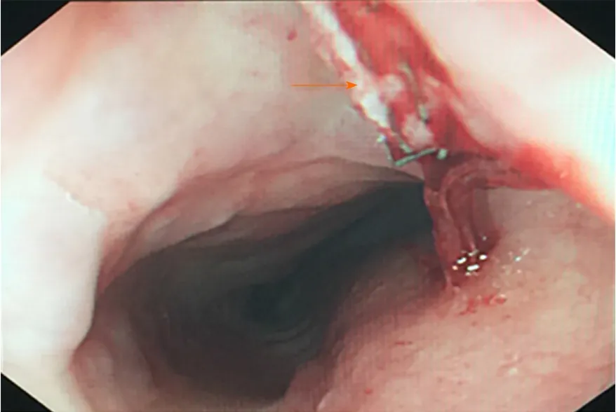
Figure 5 Little residual mass was seen by gastroscopy.

Figure 6 lmmunohistochemical analysis revealed some discrete spindle cells which were positive for Desmin and CD34, and negative for S-100, SMA,DOG-1 and STAT-6.
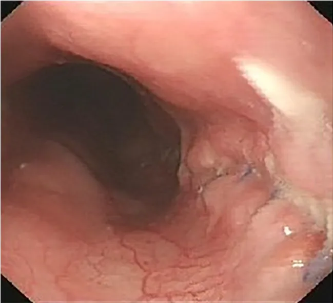
Figure 7 Esophagoscopy did not find any mass in the esophageal lumen 20 mo after surgery.
猜你喜欢
魅力中国(2021年41期)2021-11-28
——以莆田学院继续教育学院为例
莆田学院学报(2021年4期)2021-11-28
大学(2021年2期)2021-06-11
武汉冶金管理干部学院学报(2019年2期)2019-12-24
无人机(2018年1期)2018-07-05
无人机(2017年10期)2017-07-06
汽车与新动力(2013年3期)2013-03-11
汽车与新动力(2012年2期)2012-03-25
 World Journal of Clinical Cases2020年9期
- World Journal of Clinical Cases的其它文章
- Nutrition management in acute pancreatitis:Clinical practice consideration
- Bone disease in chronic pancreatitis
- Role of microRNAs in the predisposition to gastrointestinal malignancies
- Recurrent anal fistulas:When, why, and how to manage?
- Removal of biofilm is essential for long-term ventilation tube retention
- Neutrophil gelatinase-associated lipocalin does not predict acute kidney injury in heart failure