BDNF对缺糖缺氧小鼠mPFC脑区神经元突触形成的影响
2020-10-20 04:48喻晓路傅慧
医学信息 2020年17期
喻晓路 傅慧
摘要:目的 探讨脑源性神经生长因子(BDNF)在缺糖缺氧复灌条件下对mPFC脑区突触前膜蛋白Synapsin-1的保护作用。方法 将18只小鼠随机分为正常组、缺糖缺氧复灌组、缺糖缺氧复灌给药组,每组6只。正常组mPFC脑区低糖DMEM培养基培养,缺糖缺氧复灌组(OGD/R)mPFC腦区EBSS培养基培养15 min后复灌1 h,缺糖缺氧复灌给药组(OGD/R+BDNF)mPFC脑区EBSS培养基中加入BDNF培养15 min后复灌1 h,采用免疫印迹分析和RT-qPCR检测分析突触前膜蛋白Synapsin-1的表达量和mRNA表达量。结果 缺糖缺氧复灌组Synapsin-1蛋白和mRNA表达量低于正常组,差异有统计学意义(P<0.05);缺糖缺氧复灌给药组Synapsin-1蛋白表达量高于缺糖缺氧复灌组,差异有统计学意义(P<0.05);缺糖缺氧复灌给药组Synapsin-1蛋白和mRNA表达量与正常组比较,差异无统计学意义(P>0.05)。结论 缺糖缺氧对mPFC脑区神经元突触有损伤作用,BDNF对神经元突触起到保护作用。
关键词:脑源性神经生长因子;mPFC;缺糖缺氧;突触前膜蛋白
中图分类号:R614 文献标识码:A DOI:10.3969/j.issn.1006-1959.2020.17.018
文章编号:1006-1959(2020)17-0063-04
Abstract:Objective To investigate the protective effect of brain-derived nerve growth factor (BDNF) on the presynaptic membrane protein Synapsin-1 in mPFC brain under the condition of hypoglycemia and hypoxia reperfusion.Methods 18 mice were randomly divided into normal group, hypoglycemia and hypoxia reperfusion group, hypoglycemia and hypoxia reperfusion administration group, 6 mice in each group. Normal group mPFC brain area was cultured in low-sugar DMEM medium, hypoglycemia hypoxia reperfusion group (OGD/R) mPFC brain area EBSS medium was cultured for 15 min and then reperfused for 1 h, hypoglycemia hypoxia reperfusion administration group (OGD/R+BDNF) mPFC brain area EBSS medium was cultured with BDNF for 15 min and reperfused for 1 h.Western blot analysis and RT-qPCR were used to analyze the expression and mRNA expression of the presynaptic membrane protein Synapsin-1.Results The expression of Synapsin-1 protein and mRNA in the hypoglycemia and hypoxia reperfusion group was lower than that of the normal group,the difference was statistically significant (P<0.05);Synapsin-1 protein expression in the hypoglycemia and hypoxia reperfusion administration group was higher than that of the hypoglycemia hypoxia reperfusion group, the difference was statistically significant (P<0.05); Synapsin-1 protein and Compared with the normal group, the mRNA expression level was not statistically different (P>0.05).Conclusion Glucose and hypoxia could damage neuronal synapses in the mPFC brain region, and BDNF could protect neuronal synapses.
Key words:Brain-derived nerve growth factor;mPFC;Hypoglycemia and hypoxia;Presynaptic membrane protein
1.3统计学处理 采用统计学软件SPSS 13.0进行处理,计量资料采用(x±s)表示,采用单因素方差分析,one-way ANOVE进行组间比较,P<0.05 为差异有统计学意义。
2结果
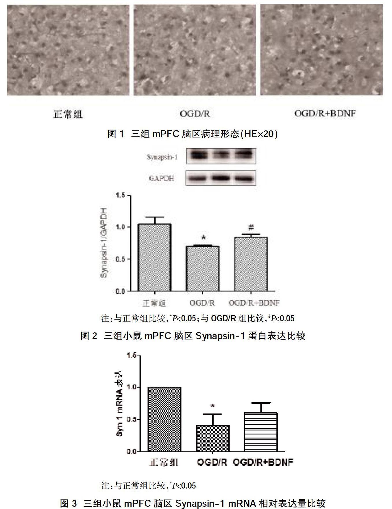
2.1三组小鼠mPFC脑区病理形态 镜检结果显示,三组脑片中细胞核形态清晰、完整,提示模型制作成功,短时间的缺塘缺氧没有造成大面积的组织坏死和细胞凋亡,完全符合后续实验要求,见图1。
2.2三组小鼠mPFC脑区Synapsin-1蛋白表达比较 WB检测结果显示,正常组Synapsin-1蛋白的相对表达量为(1.0573436±0.171485464),缺糖缺氧复灌组Synapsin-1蛋白的相对表达量为(0.702825767±0.03653467),缺糖缺氧复灌给药组Synapsin-1蛋白的相对表达量为(0.845764±0.073608716);缺糖缺氧复灌组Synapsin-1蛋白的表达量低于正常组、缺糖缺氧复灌给药组,差异有统计学意义(P<0.05);缺糖缺氧复灌给药组与正常组Synapsin-1蛋白表达量比较,差异无统计学意义(P>0.05),见图2。
2.3三组小鼠mPFC脑区Synapsin-1 mRNA相对表达量比较 正常组Synapsin-1 mRNA的相对表达量为(1.00000±1.00000),缺糖缺氧复灌组Synapsin-1 mRNA的相对表达量为(0.4126427±0.299804),缺糖缺氧复灌给药组Synapsin-1 mRNA的相对表达量为(0.604788±0.271342)。正常组Synapsin-1 mRNA的相对表达量高于缺糖缺氧复灌组,差异有统计学意义(P<0.05);缺糖缺氧复灌给药组Synapsin-1 mRNA相对表达量与正常组比较,差异无统计学意义(P>0.05),见图3。
3讨论
突触前膜蛋白Synapsin-1是一种进化中高度保守的蛋白质,定位于突触囊泡膜上,是一种与突触结构和功能密切相关的特征性膜蛋白,其数量和分布密度可以间接反映突触的密度[11,12]。脑源性神经生长因子在中枢神经系统中广泛表达,不仅能促进神经元的发育和分化、细胞存活及突触可塑性,同时具有抗凋亡、抗氧化、抑制自噬等神经保护作用,目前已有的研究表明BDNF与多种退行性疾病有关,可以通过多种途径保护受损的脑组织[13,14]。脑部缺糖缺氧是脑缺血病患的主要症状,可以导致患者残疾或者死亡[15,16]。众多研究表明,脑组织缺糖缺氧会诱发一系列的病理变化,甚至短暂性的缺糖缺氧也会对神经元造成一定的损伤,以致患者行为改变。目前,研究认为BDNF在神经系统中起保护作用,但对于其在mPFC脑区缺糖缺氧的治疗效果研究较少[17,18]。本次实验通过模拟短暂性脑缺血疾病,旨在观察外源性BDNF的治疗效果。
本研究结果显示,15 min的缺糖缺氧脑片没有对神经元的形态和结构产生破坏性的影响,说明短暂性脑缺血不会造成大面积的脑组织坏死。在对突触前膜蛋白表达量和mRNA的检测结果显示,缺糖缺氧复灌组与正常组相比,Synapsin-1蛋白和mRNA的表达量降低;缺糖缺氧复灌给药组与缺糖缺氧复灌组相比,Synapsin-1的蛋白表达量升高;与正常组相比,Synapsin-1的蛋白和mRNA表达量基本一致。有报道显示,Synapsin-1与脑缺血引起的神经元损伤和脑梗塞有关,并通过Bcl-XL来影响其病理改变[19]。缺糖缺氧对脑组织的损伤主要表现在突触形成上,在与运动、学习记忆等功能密切相关的mPFC脑区,可反映突触形成的突触前膜蛋白Synapsin-1的表达量存在明显降低,说明缺糖缺氧条件下突触形成降低,从而影响患者行为学的改变。通过本次研究可以发下,BDNF可在短暂性缺糖缺氧mPFC脑区中上调突触前膜蛋白和mRNA的表达,促进神经损伤后突触形成的恢复,其原因可能是BDNF作为一种重要的脑源性神经生长因子对于脑内神经元具有保护和营养作用,其不仅参与神经元突触的形成,还可影响突触形成后的功能[20]。
综上所述,缺糖缺氧对mPFC脑区神经元突触有損伤作用,BDNF对神经元突触起到保护作用。
参考文献:
[1]任艺,崔伟华,王珊珊,等.不同浓度吗啡对新生小鼠皮层神经元活力和突触可塑性的影响[J].中华麻醉学杂志,2018,38(10):1190-1193.
[2]Patzke C,Brockmann MM,Dai J,et al.Neuromodulator Signaling Bidirectionally Controls Vesicle Numbers in Human Synapses[J].Cell,2019,179(2):498-513.e22.
[3]Song M,Martinowich K,Lee FS.BDNF at the synapse:why location matters[J].Molecular Psychiatry,2017,22(10):1370-1375.
[4]BjRkholm C,Monteggia LM.BDNF-a key transducer of antidepressant effects[J].Neuropharmacology,2016(102):72-79.
[5]吕秀芳,胡宝英,巩秀,等.内侧前额叶皮层神经元可塑性改变在吗啡奖赏记忆形成中的作用[J].中华行为医学与脑科学杂志,2014,23(12):1061-1064.
[6]Cho JH,Deisseroth K,Bolshakov V.Synaptic Encoding of Fear Extinction in mPFC-amygdala Circuits[J].Neuron,2013,80(6):1491.
[7]Liu X,Tian F,Wang S,et al.Astrocyte Autophagy Flux Protects Neurons Against Oxygen-Glucose Deprivation and Ischemic/Reperfusion Injury[J].Rejuvenation Res,2017(2017):1999.
[8]Ryou MG,Mallet RT.An In Vitro Oxygen-Glucose Deprivation Model for Studying Ischemia-Reperfusion Injury of Neuronal Cells[J].Methods Mol Biol,2018(1717):229-235.
[9]Fu-Sheng Chou,Pei-Shan Wang.Neuronal Precursor Migration in Ex Vivo Brain Slice Culture[J].Cell Migration,2018(1749):135-143.
[10]Christian,Humpel.Organotypic Brain Slice Cultures[J].Current Protocols in Immunology,2018.
[11]Maesako M,Zoltowska KM,Berezovska O.Synapsin 1 promotes Aβ generation via BACE1 modulation[J].PLoS One,2019,14(12):e0226368.
[12]Navya A,Bianca F,Hari P,et al.Neonatal anesthesia impairs synapsin 1 and synaptotagmin 1,two key regulators of synaptic vesicle docking and fusion[J].Neuroreport,2019(2019):544-549.
[13]Zaletel I,Filipovi D,PuKa N.Hippocampal BDNF in physiological conditions and social isolation[J].Reviews in the Neuroences,2017,28(6):675-692.
[14]Walsh JJ,Tschakovsky ME.Exercise and circulating BDNF:Mechanisms of release and implications for the design of exercise interventions[J].Applied Physiology,Nutrition,and Metabolism,2018,43(11):1095-1104.
[15]Tasca CI,Dal-Cim T,Cimarosti H.In Vitro Oxygen-Glucose Deprivation to Study Ischemic Cell Death[J].Methods in Molecular Biology,2015(1254):197-210.
[16]Povysheva N,Nigam A,Brisbin AK,et al.Oxygen-Glucose Deprivation Differentially Affects Neocortical Pyramidal Neurons and Parvalbumin-Positive Interneurons[J].Neuroscience,2019(412):72-82.
[17]Cheriyan J,Sheets PL.Altered Excitability and Local Connectivity of mPFC-PAG Neurons in a Mouse Model of Neuropathic Pain[J].J Neurosci,2018,38(20):4829-4839.
[18]Lieberman MD,Straccia MA,Meyer ML,et al.Social,Self,(Situational),and Affective Processes in Medial Prefrontal Cortex(MPFC):Causal,Multivariate,and Reverse Inference Evidence[J].Neuroscience&Biobehavioral Reviews,2019.
[19]Kilic E,Hermann DM,Kügler S,et al.Adenovirus-mediated Bcl-X(L)expression using a neuron-specific synapsin-1 promoter protects against disseminated neuronal injury and brain infarction following focal cerebral ischemia in mice[J].neurobiology of disease,2002,11(2):275-284.
[20]Leal G,Bramham CR,Duarte CB.BDNF and Hippocampal Synaptic Plasticity[J].Vitamins and Hormones,2017(104):153-195.
收稿日期:2020-06-04;修回日期:2020-06-14
編辑/成森