
Effect of electroacupuncture pretreatment on the protein expression of c-fos in fastigial nucleus and lateral hypothalamus area in rats with acute myocardial ischemia-reperfusion injury
2021-02-05 09:36CaiRonglin蔡荣林ShaoXuefang邵雪芳YuQing余情ZhangYating张娅婷WeiXiaotong魏小桐HuLing胡玲
Cai Rong-lin (蔡荣林), Shao Xue-fang (邵雪芳), Yu Qing (余情), Zhang Ya-ting (张娅婷), Wei Xiao-tong (魏小桐), Hu Ling (胡玲)
1 College of Acupuncture and Moxibustion, Anhui University of Chinese Medicine, Hefei 230012, China
2 Research Institute of Acupuncture and Moxibustion, Anhui University of Chinese Medicine, Hefei 230038, China
3 Graduate School, Anhui University of Chinese Medicine, Hefei 230012, China
4 Anhui Key Laboratory of Acupuncture and Moxibustion, Hefei 230038, China
Abstract
Keywords: Acupuncture Therapy; Electroacupuncture; Heart Meridian; Myocardial Ischemia; Myocardial Reperfusion Injury; Cerebellum; Hypothalamus; Rats
Many studies have shown that electroacupuncture (EA) pretreatment has protective effects on myocardial ischemia-reperfusion injury (MIRI)[1-2], and we have found its mechanism closely related to the regulation of hypothalamus, hippocampus and other central areas in the previous studies[3-4]. Dopamine, noradrenaline and other neurotransmitters in hypothalamus participate in the process of EA at Heart Meridian improving acute myocardial ischemia[5-6]. Previous studies on the mechanism of hypothalamus involved in the improvement of MIRI by acupuncture mainly focused on the paraventricular nucleus. Whether other nuclei are involved in this protective effect and the possible mechanism still require in-depth study.
In this study, we aimed to further explore the role of FN and LHA in EA improving MIRI on the basis of our previous studies, and preliminarily analyze the possible mechanism of cerebellar hypothalamic neural circuit in the improvement of MIRI by EA.
1 Materials and Methods
1.1 Experimental animal
Specified pathogen free male Sprague-Dawley rats, weighing 220-250 g, were provided by Anhui Experimental Animal Center [SCXK (Wan) 2018-007]. In the same quiet environment, they were raised in separate cages and adaptively fed for one week. The rats were randomly divided into six groups: 14 rats in the sham operation group, model group, EA-Heart Meridian group and EA-Lung Meridian group respectively, and 7 rats each in LHA+EA-Heart Meridian group and FN+EA-Heart Meridian group. Animal experiments strictly adhered to the Guiding Opinions on the Treatment of Experimental Animals issued in 2006 by the Ministry of Science and Technology of the People's Republic of China and the ethics regulations of animal experiment.
1.2 Main reagents and instruments
Pentobarbital sodium (Batch No.: 20160720, Sinopharm Chemical Reagent Co., Ltd., China); Kainic acid (Batch No.: MKCD0368, Sigma, America); DAB chromogenic kit (Batch No.: ZLI-9019, Zhongshan Golden Bridge Bio-technology Co., Ltd., China); anti c-fos antibody (Batch No.: ab209794, Abcam, UK).
DP70 optical microscope (Olympus Corporation, Japan); MP100 PowerLab physiological recording system (ADInstruments Co., Ltd., Australia); SDZ-V EA instrument (Suzhou Medical Appliance Factory, China); RM2025 paraffin embedding station (Leica Biosystems, Germany); 69100 brain stereotactic apparatus (RWD Life Science Co., Ltd., China); CM1900 frozen section machine (Leica Biosystems, Germany).
1.3 Model establishment and evaluation
The acute MIRI model was prepared by ligation of the left anterior descending coronary artery according to the reference[1]. The rats were first anesthetized by intraperitoneal injection of 0.3% pentobarbital sodium [10 mL/(kg·bw)] after the last EA session. The rats were fixed in a supine position. Standard lead Ⅱ electrocardiogram (ECG) was recorded by Powerlab physiological recorder. After tracheotomy, the rats were connected to the animal ventilator [respiratory frequency 70 times/min, respiratory ratio 1:2 and tidal volume 20 mL/(kg·bw)]. An incision was made on the skin between the third and fourth ribs on the left side of the chest, and the local muscles were bluntly dissected. The pericardium was gently opened to expose the left atrial appendage using a cotton swab and to locate the left anterior descending coronary artery. At about 1-2 mm outside the origin of the left anterior descending coronary artery, a 5-0 medical suture with needle was used for ligation (a polyethylene tube was placed between the thread and heart muscles). After ligation, bulging and cyanosis of the lower left coronary artery, cyanosis of the left ventricular anterior wall or ST-segment elevation with T-wave high amplitude in standard lead Ⅱ ECG were taken as the indications of myocardial ischemia. After 30-minute ligation, the silicone tube was loosened, and the left anterior descending artery was allowed reperfusion for 120 min. After loosening of the ligation, the blood supply was recovered, and thus the color of the lower left coronary artery changed from cyanosis to light dark or dark red and the T wave descended by more than 0.2 mV, which indicated successful reperfusion. At the end of the modeling, the muscles and skin were sutured layer by layer. In the sham operation group, no ligation was performed after thoracotomy, and the corresponding position was only pierced with a needle once. The rats that died before they reached the end of the observation or failed in modeling were excluded. At last, there were 14 rats in the sham operation group, 12 rats in the model group, 13 rats in the EA-Heart Meridian group, 12 rats in the EA-Lung Meridian group, 6 rats each in the LHA+EA-Heart Meridian group and the FN+EA-Heart Meridian group.
1.4 Intervention methods
The acupoints were located according to theExperimental Acupuncture Science[11]combined with the method of animal comparative anatomy. Disposable acupuncture needles (0.25 mm in diameter and 13 mm in length) were inserted to a depth of 2-3 mm, and then connected to SDZ-V EA instrument to conduct EA stimulation (frequency 2 Hz, intensity 1 mA), 20 min each time, once a day for 7 d before modeling. EA was not used in the sham operation group or the model group.
Shenmen (HT 7) and Tongli (HT 5) were selected in the EA-Heart Meridian group.
Taiyuan (LU 9) and lieque (LU 7) were selected in the EA-Lung Meridian group for EA stimulation.
Kainic acid (KA) was injected into bilateral FN of rats in the FN+EA-Heart Meridian group and bilateral LHA of rats in the LHA+EA-Heart Meridian group. EA intervention was carried out three days after KA injection. The acupoints and EA parameters were same as those in the EA-Heart Meridian group.
1.5 Nucleus lesion
The rats were anesthetized by intraperitoneal injection of 0.3% pentobarbital sodium (10 mL/kg·bw) and fixed on the stereotaxic instrument. According to the coordinates of FN and LHA (FN: anterior fontanelle posterior (AP) 11.6 mm, right or left (RL) 1.0 mm, height (H) 5.6 mm; LHA: AP 2.8 mm, RL 1.5 mm, H 8.3-8.5 mm), the location of nucleus was located by the stereotaxic instrument and then the holes were drilled with a cranial drill. Slowly inserted a microinjector to inject KA (1 g/L) into the predetermined position, 0.4 μL for each side of the relevant nucleus, and retained the needle for 5 min after injection, and then slowly withdrew the needle and sutured the wound. At the end of the experiment, the brain tissues at the injection site were fixed in paraformaldehyde solution, frozen and sectioned, and the injection sites were confirmed by referring the atlas.
1.6 ECG
The rats were connected to the Powerlab physiological recorder to record the standard lead ⅡECG before modeling, 30 min after ligation and 120 min after reperfusion. The deviation value of ST segment within 15 min was analyzed each time.
Curtis and Walker arrhythmia scoring method was used to assess the cardiac arrhythmia score in each group after reperfusion[12]. Scoring standard: 0 point for normal, 1 point for atrial arrhythmia or incidental ventricular extrasystole, 2 points for frequent ventricular extrasystole, 3 points for ventricular tachycardia (1-2 strings), and 4 points for ventricular tachycardia (more than 3 strings) or ventricular fibrillation.
1.7 Brain tissue sampling and detection
At the end of the experiment, the rats were executed with excessive pentobarbital sodium and the corresponding brain tissues were taken out and fixed in 4% paraformaldehyde solution. The brain tissues in the LHA and FN areas were located and collected by a size of 0.5 cm × 0.5 cm × 0.2 cm. After dehydration, transparence and wax immersion, the tissues were embedded in paraffin and then made into continuous coronal sections at about 4 μm. The brain slices containing the target area were selected for immunohistochemical staining. The positive expression of c-fos protein was observed by optical microscope. In the negative control, the first antibody was replaced by normal serum and diaminobenzidine (DAB) was used as the chromogenic agent. The expression of c-fos was brownish yellow. In Leica Qwin image analysis system, the integral optical density (IOD) of immunoreactive positive substance was calculated from five high-power fields in FN and LHA which were not overlapped, and the mean value was used for statistical analysis.
1.8 Statistical methods
SPSS 17.0 software was used for statistical analysis. The measurement data were presented as mean ± standard deviation (±s), and analyzed by one-way analysis of variance and Tukey's multiple comparisons test was selected for comparison among groups.P<0.05 indicated that the difference was statistically significant.
2 Results
2.1 Comparison of ST-segment deviation
There were no significant between-group differences in ST-segment deviation before modeling (P>0.05). Except the sham operation group, the ST-segment deviation values at 30 min after ligation and 120 min after perfusion were significantly higher than the value before modeling in the other groups (allP<0.05). At 120 min after reperfusion, the ST-segment deviation value in the EA-Heart Meridian group, LHA+EA-Heart Meridian group and FN+EA-Heart Meridian group decreased significantly compared with the model group (allP<0.05); the ST-segment deviation value was significantly higher in the LHA+EA-Heart Meridian group and FN+EA-Heart Meridian group than in the EA-Heart Meridian group (bothP<0.05), (Figure 1).
2.2 Comparison of the cardiac arrhythmic score
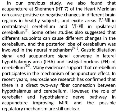
Compared with the sham operation group, the cardiac arrhythmia score was significantly higher in the model group (P<0.05). Compared with the model group, the arrhythmia score was significantly lower in the EA groups (allP<0.05). Compared with the EA-Heart Meridian group, it was significantly higher in the EA-Lung Meridian group, LHA+EA-Heart Meridian group and FN+EA-Heart Meridian group (allP<0.05), (Figure 2).

Figure 1. Comparison of ST-segment deviation
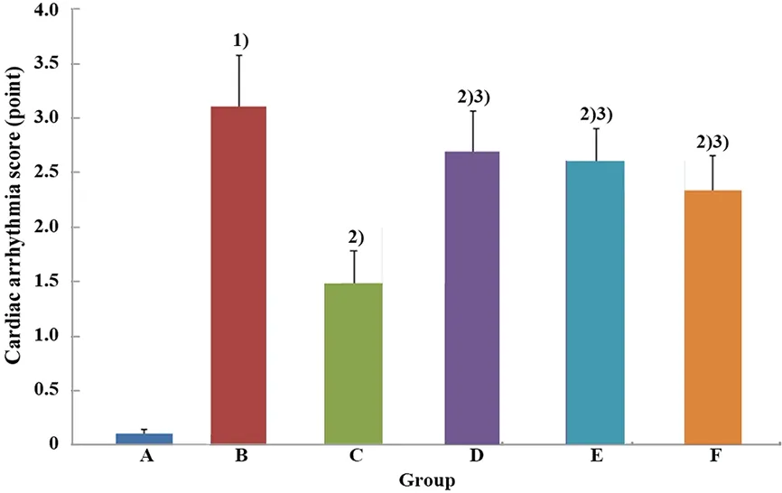
Figure 2. Comparison of the cardiac arrhythmia score
2.3 Comparison of the c-fos protein expression in FN
Compared with the sham operation group, the positive protein expression of c-fos in FN increased significantly in the model group (P<0.05). Compared with the model group, the protein expression of c-fos in FN was significantly lower in the EA-Heart Meridian group (P<0.05). The protein expression of c-fos in FN was significantly higher in the EA-Lung Meridian group and the LHA+EA-Heart Meridian group than in the EA-Heart Meridian group (bothP<0.05), (Figure 3 and Figure 4).
2.4 Comparison of the c-fos protein expression in LHA
Compared with the sham operation group, the protein expression of c-fos in LHA was significantly higher in the model group (P<0.05). Compared with the model group, the protein expression of c-fos in LHA was significantly lower in the three groups intervened by EA (allP<0.05). Compared with the EA-Heart Meridian group, the protein expression of c-fos in LHA was significantly higher in the EA-Lung Meridian group and FN+EA-Heart Meridian group (bothP<0.05), (Figure 5 and Figure 6).

Figure 3. The protein expression of c-fos in FN in each group (immunohistochemical staining, ×200)
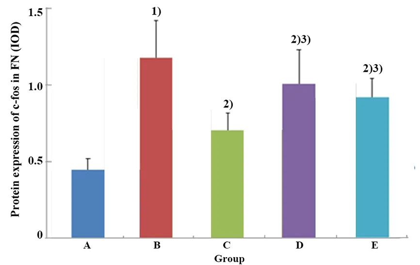
Figure 4. Comparison of the protein expression of c-fos in FN
Figure 5. The protein expression of c-fos in LHA in each group (immunohistochemical staining, ×200)

Figure 6. Comparison of the protein expression of c-fos in LHA
3 Discussion
Studies have shown that acupuncture can protect the cardiac function[13-16]and MIRI also can be mitigated by acupuncture. Many studies have discussed the effect and mechanism of acupuncture in intervening MIRI from gene expression, signal pathway and inflammatory factors[17-21]. Our previous studies demonstrated that EA pretreatment could effectively ameliorate MIRI, and its mechanism was closely related to the regulation of hypothalamus, hippocampus and other central areas[3-4]. It was suggested that central nervous regulation should be one of the important mechanisms in EA regulating visceral function.
Neural circuitry is a universal structure in the brain, and plays a very important role in brain information transmission and processing. Neurons with different properties and functions in the brain form neural circuits and networks at different levels through various forms of complex connections. Many studies have found that the pathogenesis and development of some diseases are closely associated with the establishment and regulation of neural circuits. It is of great significance to study the pathogenesis, diagnosis and treatment mechanisms of diseases at the level of neural circuits[22-23]. Early neuroanatomical studies showed that there were fibrous connections between cerebellum and some visceral nuclei in brainstem. In recent years, research has showed that cerebellum also participates in the regulation of non-somatic activities, such as cardiovascular, respiratory, urinary, immune, learning and cognitive functions[24]. Some of the visceral regulation of cerebellum may be related to the high central nervous system[25]. There is a direct two-way fibrous connection between hypothalamus and cerebellum, that is the hypothalamic-cerebellar projection and the cerebellar-hypothalamic projection, which constitute the hypothalamic-cerebellar neural circuit[26]. The two-way and direct fibrous projections between cerebellum and hypothalamus provide a structural basis for cerebellum to participate in the regulation of non-somatic activities.
Many studies have approved that cerebellum participates in the mechanism of EA effects. Cerebello- thalamo-cortical circuit is involved in the regulation mechanism of acupuncture[27]. A study of resting-state functional magnetic resonance imaging (fMRI) showed that acupuncture decreased blood pressure and this was related to hypothalamus functional connectivity with cerebellum and insula[28]. The activation of c-fos in the brain was first described almost a decade ago and it is one of the most studied immediate-early genes in the brain. C-fos protein is not only the activation marker of neurons after stimulation, but also mediates a series of subsequent reactions[29-30]. Acupuncture or EA can affect the neuronal activity and protein expression of c-fos in specific brain areas, which may be an important way for acupuncture to regulate visceral function and treat diseases[31-32].
This experimental study found that the ST-segment deviation increased significantly in the MIRI rats, and the protein expression of c-fos in FN and LHA also increased significantly, indicating that the myocardial blood supply in the MIRI rats was insufficient, and the neuronal activity in FN and LHA increased significantly. The ST-segment deviation and c-fos protein expression in FN and LHA in the MIRI rats decreased significantly after the pretreatment with EA at the Heart Meridian, suggesting that EA may improve myocardial function by regulating the activity of neurons in FN and LHA. In addition, the protein expression of c-fos in FN and ST-segment deviation were significantly higher in the EA-Lung Meridian group and LHA+EA-Heart Meridian group than in the EA-Heart Meridian group, indicating that when LHA was damaged, the regulative effects of EA on the cardiac function and activity of neurons in FN in the MIRI rats were weakened. The LHA plays an important role in the effect of EA at the Heart Meridian in improving MIRI. At the same time, compared with the EA-Heart Meridian group, the protein expression of c-fos in LHA and ST-segment deviation were significantly higher in the EA-Lung Meridian group and FN+EA-Heart Meridian group, which indicated that when FN of the cerebellum was damaged, the regulation of cardiac function and activity of neurons in LHA in the MIRI rats by the pretreatment with EA at the Heart Meridian was also weakened. It is suggested that FN also plays a role in improving MIRI effect. It should be noted that EA at the Lung Meridian can also mitigate MIRI and reduce the expression of c-fos protein, indicating that acupoint stimulation may have a certain broad-spectrum effect.
In summary, FN and LHA are involved in the mechanism of EA in improving myocardial injury. EA plays an important role in myocardial protection by regulating the activities of neurons in LHA and FN. In addition, it has been confirmed that cerebellum participates in the regulation of non-somatic visceral activity through cerebellar-hypothalamic projection, high-frequency electrical stimulation to FN may inhibit the electrical activity of neurons in LHA through cerebellar-hypothalamic direct fibrous projection[33]. The regulative effect of acupuncture on gastric motility may be related to its regulative action on neurons in FN and LHA[10]. According to the results of this study, we believe that the FN and LHA neural circuitry should be involved in the effect of EA in improving the MIRI, and cerebellum may be involved in the process of EA improving the cardiac function through the cerebellar- hypothalamic projection. With the rapid development of neuroscience and technology, we should actively use the novel neuroscience research method when conducting in-depth exploration of the mechanism of acupuncture in improving visceral function, which is of great significance to help understand the acupuncture mechanisms.
Conflict of Interest
There is no potential conflict of interest in this article.
Acknowledgments
This work was supported by National Natural Science Foundation of China (国家自然科学基金项目, No. 81774414, No. 82074536); Key Projects of Natural Science Research in Universities of Anhui Province (安徽省高校自然科学研究重点项目, No. KJ2019A0455); Supported Project for the Cultivation of Outstanding and Top Talents in Universities of Anhui Province (安徽省高校优秀拔尖人才培育资助项目, No. gxgwfx2019025); National Training Program for Innovative Backbone Talents of Traditional Chinese Medicine (全国中医药创新骨干人才培训项目).
Statement of Human and Animal Rights
The treatment of animals conformed to the ethical criteria in this experiment.
Received: 11 February 2020/Accepted: 21 April 2020
猜你喜欢
少儿画王(3-6岁)(2022年6期)2022-07-19
英语文摘(2022年4期)2022-06-05
国际放射医学核医学杂志(2021年6期)2021-11-30
国际放射医学核医学杂志(2021年1期)2021-11-30
家教世界(2021年7期)2021-03-23
家教世界(2021年5期)2021-03-11
家教世界(2021年2期)2021-03-03
西安航空学院学报(2018年5期)2018-10-15
商周刊(2018年13期)2018-07-11
商周刊(2018年10期)2018-06-06
 Journal of Acupuncture and Tuina Science2021年1期
- Journal of Acupuncture and Tuina Science的其它文章
- Effect of electroacupuncture on calcium-activated chloride channel currents in interstitial cells of Cajal in rats with diabetic gastroparesis
- Effect of herb-partitioned moxibustion in improving tight junctions of intestinal epithelium in Crohn disease mediated by TNF-α-NF-κB-MLCK pathway
- Ginger-partitioned moxibustion plus pediatric massage for treating infantile diarrhea due to spleen deficiency: a randomized controlled clinical trial
- Efficacy observation of long-retaining scalp acupuncture plus interactive training for upper-extremity dysfunction after cerebral stroke
- Observation on therapeutic efficacy of thunder-fire moxibustion for hypomenorrhea after induced abortion
- Clinical observation on the time-effect relationship of moxibustion for primary dysmenorrhea due to stagnation and congelation of cold-damp