
Oxidation degree dependent adsorption of ssDNA onto graphene-based surface∗
2021-10-28 07:13HuishuMa马慧姝JigeChen陈济舸HaipingFang方海平andXiaolingLei雷晓玲
Chinese Physics B 2021年10期
Huishu Ma(马慧姝) Jige Chen(陈济舸) Haiping Fang(方海平) and Xiaoling Lei(雷晓玲)
1Division of Interfacial Water and Key Laboratory of Interfacial Physics and Technology,Shanghai Institute of Applied Physics,Chinese Academy of Sciences,Shanghai 201800,China
2University of Chinese Academy of Sciences,Beijing 100049,China
3Department of Physics,East China University of Science and Technology,Shanghai 200237,China
4Shanghai Synchrotron Radiation Facility,Zhangjiang Laboratory,Shanghai Advanced Research Institute,Chinese Academy of Sciences,Shanghai 201210,China
Keywords: single-strand DNA(ssDNA),molecular dynamics simulation,oxidation degrees,graphene-based surfaces
1. Introduction
DNA, the carrier of genetic information, participates in most of the bio-functional processes such as the replication, transcription and plays a pivotal role in living organisms.[1,2]Recently, with the rapid advance in biotechnology and nanotechnology,DNA-functionalized nanomaterials, such as DNA/GO,[3]DNA/Au,[4]and DNA/MFe2O4,[5]exhibit extensively development as new applications of DNA in biosensors,[3,6,7]biomedicine,[8,9]nanotechnology,[10]and materials science.[11–13]Among these applications, the combination of DNA and graphene,the first two-dimensional(2D)atomic crystal available to us, attracts a great deal of interest. Graphene exhibits many extraordinary properties,such as high surface area,[14]fast electron mobility,[15]good optical transparency,[16,17]high Young’s modulus,[18]and excellent thermal conductivity,[19]which can be exploited for numerous applications in energy, environment,[20]and biomedicine.[21]To enhance its dispersion in aqueous solution for numerous applications,graphene is usually oxidized to be graphene oxide(GO),which mainly bears carboxylic groups on its edges,hydroxyl and epoxy groups on its basal plane.These oxidative functional groups increase the biocompatibility of GO through the formation of both covalent and non-covalent bonds with biomolecules.[22]
Recently,the combination of DNA and GO has inspired a lot of innovative work,[23,24]and practical applications,[25,26]especially in the biosensors. The application of DNA/GO nanomaterials is based on that single-stranded DNA (ss-DNA)has a preferential binding on GO compared to doublestranded DNA (dsDNA), because of non-covalent interactions with the surface.[27–30]The main interactions between ssDNA molecules and GO have been recognized as hydrophobic,[31,32]hydrogen bonding,[21,33]andπ–πstacking interactions[29,33]by both theoretical and experimental methods. The dynamic interactions between DNA molecule and GO surface are complex and depend on the sequences and length of DNA[34,35]as well as the surface defects and surface properties of GO.[36]
As we known, the oxidized groups are not evenly distributed on the GO surface.[37,38]Within the GO plane, there is the coexistence of both large unoxidized and oxidized regions proved by experimental and theoretical works. Even in oxidized regions,there are some small areas of sp2-hybridized domains, similar to “islands”,[39–43]because of the steric effects. This uneven oxidized groups distribution on GO makes its surface polarity complex. Generally, surface polarity is known as an important factor for the dynamic adsorption process and the configurations of biomolecules.[44–46]Yanet al.investigated the effects of the oxidation degree (OD) on MB adsorption behavior systematically, indicating that the dye uptakes of GO exponentially increase with the increase of OD.[46]The relationship among the degree of oxidation,defects of GO and the adsorption performance was investigated by Tanet al.The results showed that the adsorption of Cu2+on GO was strongly dependent on the oxidation degree and independent of the defects under various pH levels and ionic strength.[47]Tuet al.found that the complex surface polarity of GO was crucial to the antibacterial activity of the material based on the experiments and the molecular dynamic simulation.[48]For DNA-surface interactions,experimental study demonstrated that the formation of interfacial hydrogen bonds between DNA and GO was crucial for the attachment of ssDNA and the detachment of hybridized dsDNA during the sensing process.[27]It was obvious that the density of polar groups on the graphene surface or oxidation rate have a direct implication on physisorption process and these interactions can either enhance or reduce the biocompatibility and stability of ssDNA on graphene-based surfaces. Most of the previous studies about the DNA adsorption on GO did not consider the effect of oxidation degree of GO.Kimet al.performed a detailed investigation of the effect of surface oxidation rate of graphene surfaces on the physisorption process of poly(T)20based on all-atomic MD simulations.However,they only considered the GO sheets that the hydroxyl and epoxy groups were grafted on both sides of the surface in a random manner.[49]
Here, based on Shi–Tu model, we performed all-atom molecular dynamics (MD) simulations to investigate the physisorption of ssDNA onto graphene-based surfaces with oxidation degrees from 0 to 25%. From the results, we found ssDNA molecule was absorbed in most stable state on the GO surface with the oxidation degree around 15% due to the maximum value of interaction. By analyzing the distinguished interaction between graphene-based surfaces and ss-DNA molecules,we found that the electrostatic interaction increased with the oxidation degree of graphene-based surface.And the Van Der Walls (vdW) interaction first increases and then decreased with the oxidation degree, holding the greatest value for the GO of 15% oxidation degree. The result was also confirmed by the number ofπ–πstacking between them. Further analysis showed that,the less oxidation degree on graphene-based surface,the greater probability for ssDNA to form the approximately parallel configuration,regardless of the coexistence of both stretched and curved configurations.Our work revealed the oxidation degree dependent absorption of ssDNA molecules onto graphene-based surface and provided theoretical guidance for the application of DNA/GO nanomaterials.
2. Methods
2.1. Structure of ssDNA and graphene-based surfaces
In this study,the ssDNA molecule with the base sequence 5’-ATGCATGCATGC-3’, (ATGC)3, was used. The initial(ATGC)3structure was built using nucleic acid builder(NAB)in AMBER 14 package. The model of graphene-based materials was constructed based on the high correlation between oxidation loci,as reported by Yanget al.[50]Using this model,various oxidation degrees(from 0 to 25%,in 5%increments),dO=nO/nC, wherenOandnCwere the number of oxygen atoms and carbon atoms, respectively, explored in this study were achieved. Representative snapshots of the surfaces were shown in Fig.S1.
The ssDNA molecule was placed on the graphenebased surface with the helix axis parallel to the basal plane of graphene-based which had dimensions of 10.084×10.224 nm2. The centroid distance between the graphenebased surface and ssDNA molecule was 3 nm. To investigate the effects of oxidation degree on adsorption, eight samples of each oxidation degree were constructed. The whole system was filled with water molecules,and sodium ions were added to neutralize the negative charges of the ssDNA molecule.
2.2. MD simulation
The simulation box was 10.084×7.00×10.224 nm3inx,yandzdirection, and the periodic boundary was applied to allx,yandzthree directions. All simulations were performed in the NVT ensemble at 300 K by GROMACS 4.5.4.[51]We chose the Amber03 force filed[52]to simulate the ssDNA molecule, which had been widely used in DNA simulations.[53–56]The detailed force field parameters of graphene-based surfaces were consistent with our previous publication.[34,35,57]As for the simulation model of graphene-based materials, the carbon atoms were presented as the uncharged Lennard–Jones particles with the vdW parameters ofσCC= 3.58 °A andεCC=0.0663 kcal·mol−1.[58–60]The balance length of C–C bond was set to be 0.142 nm by the harmonic potentials with the spring constants of 322.55 kcal·mol−1·°A−2. The vibration of the C–C–C angle was also simulated by the harmonic potentials with the balanced angle of 120°and spring constants of 53.35 kcal·mol−1·rad−2. And the planar dihedral angles between C–C–C–C was kept by the spring constants of 3.15 kcal·mol−1. The simulation parameters of–OH,–O–,and –COOH groups on the graphene-based surface were set according to the Amber03 force filed. The TIP3P model was used.[61]The long-range electrostatic interaction was treated with the particle-mesh Ewald method[62]with a real space cutoff of 1.2 nm. The cutoff distance of the vdW interaction was set up to be 1.2 nm as well.
Initially, the system equilbration was performed in the NPT ensemble and was simulated for 5 ns,where the ssDNA molecule was constrained by position restraints. Then, the constraints were removed and the production simulations were run in the NVT ensemble for 400 ns. The time step in all simulations were set to be 2 fs and the data were collected every 2 ps.
3. Results and discussion
The control MD simulation systems were designed to investigate the ssDNA physisorption dynamics on the pristine graphene (PG) and GO with various oxidation degrees from 5%to 25%(GO-5%,GO-10%,GO-15%,GO-20%,and GO-25%). GO surfaces were constructed based on the Shi–Tu structure model,on which the distribution of oxidized groups was generated according to the rate-constant ratios from the computation by combining the density functional theory and conventional transition-state theory. As shown in Fig. S1(b),the GO nanosheets could be divided into low oxidation degree(PG and GO-5%),moderate oxidation degree(GO-10%,GO-15%)and high oxidation degree(GO-20%,GO-25%).For low oxidation degree GO nanosheets,the unoxidized region dominated the GO surface. For moderate oxidation degree GO nanosheets,both large unoxidized and oxidized regions coexisted on the surface.For high oxidation degree GO nanosheets,the oxidized region dominated the GO surface. However,even though the oxygen groups distributed over all the surface of GO-25%, small areas of sp2-hybridized domains, similar to“islands”,were formed over all the surface. In all systems,the ssDNA molecules were initially placed above the graphenebased surface with the helix axis parallel to the basal plane and the centroid distance of 3 nm inyaxis(Fig.S1(a)).
3.1. Interaction between ssDNA and graphene-based surface
Figure 1(a)showed the typical snapshots of adsorbed ss-DNA molecules on various oxidation degrees GO surfaces from 0 to 25% at 400 ns and all snapshots were shown in Fig. S2. It was clearly that ssDNA molecules could be adsorbed on graphene-based surface as various kinds of configurations.And even on the same graphene-based surface,the absorbed ssDNA molecules could display as stretched or curved configurations. We calculated the averaged total interaction energy between ssDNA and graphene-based surface, including electrostatic and vdW interactions, both of which dominated the adsorption process. As shown in Fig.1(b),we found that the total interaction energy between ssDNA molecules and GO-15% held the value of−1161.87 kJ/mol, which was not only larger than that of other oxidation degree GO surfaces but also of PG.The result suggested that ssDNA molecule was absorbed in the most stable state on the GO surface with the oxidation degree around 15%,rather than PG or other GO surfaces.
In order to analyze the detailed interaction between adsorbed ssDNA and graphene-based surface, we analysed the changing of the electrostatic and vdW interactions individually with the oxidation degrees. From the result,it was noted that the electrostatic interaction increased with the oxidation degree of surface from 0 kJ/mol to−258.43 kJ/mol due to the increased number of oxidized groups on GO surfaces. For the vdW interaction energy, the PG held the maximum value of−999.99 kJ/mol, larger than that of any GO surfaces. Because of the steric effect formed by the oxidized groups on GO surface, the moving area was prevented when the aromatic rings of the nucleobases adjusted its configurations to formingπ–πstacking structure with the GO surface. Interestingly, we found that the vdW interaction between ssDNA and GO surface did not increase or decrease monotonically with the oxidation degree of GO. GO-15% held the value of−992.15 kJ/mol, which was the greatest vdW interaction among all GO surfaces except the PG surface. Due to the energetic properties can be further decomposed into the contribution of the oxygen-containing and non-oxygen-containing groups on the surface, we also calculated the averaged total interaction energy between ssDNA and graphene-based surface,including oxygen-containing and non-oxygen-containing groups regions interactions. As shown in Fig. S7, for the non-oxygen-containing groups regions interaction energy,the PG held the maximum value of−960.82 kJ/mol, larger than that of any GO surfaces. Interestingly,we also found that the non-oxygen-containing groups regions interaction between ss-DNA and GO surface did not increase or decrease monotonically with the oxidation degree of GO. GO-15% held the value of−686.61 kJ/mol,which was the greatest non-oxygencontaining groups regions interaction among all GO surfaces except the PG surface. And the oxygen-containing groups regions interaction increased with the oxidation degree of surface from 0 kJ/mol to−679.10 kJ/mol due to the increased number of oxidized groups on GO surfaces.
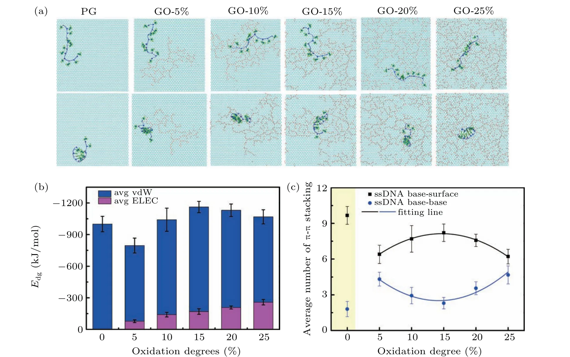
Fig.1. (a)Typical simulation snapshots of stretched and curved structures of ssDNA adsorbed on the PG,GO surface around 5%, 10%, 15%, 20%and 25%oxidation degree. The oxygen and hydrogen atoms on GO were shown as red and white spheres, respectively. The carbon atoms were represented in the cyan line. The phosphate backbone and nucleobases of ssDNA were shown by the blue lines and green bonds,respectively. (b)Average interaction energy between ssDNA and graphene-based surface with variable oxidation degrees. The vdW and electrostatic contributions to non-bonded energy were colored as blue and magenta,respectively. (c)Average number of the intra π–π stacking structures of ssDNA molecules(base stacking,blue circles)and the inter π–π stacking structures between ssDNA molecules and graphene-based surface(base-surface stacking,black squares).
To clearly characterize the vdW interaction, we calculated the number ofπ–πstacking between ssDNA molecule and graphene-based surface,as shown in Fig.1(c). We found that PG nanosheet processes the maximum number ofπ–πstacking with ssDNA molecule. It was noted that the relation between the number ofπ–πstacking and oxidation degree was not monotonous but similar to that between the vdW interaction and oxidation degree. Moreover, the quantitative functional relationship between them could be concluded asy1=−0.018x2+0.538x+4.175. Furthermore, the number ofπ–πstacking inside the ssDNA molecules held the opposite trend with that between ssDNA and GO of the relation function ofy2=0.021x2−0.592x+6.729. As for the PG,the number ofπ–πstacking with ssDNA molecules was greater than any GO surfaces, which was consistent with previous studies[63,64]and also the result of vdW interaction. From the changing trend of the electrostatic and vdW interaction, the GO-15%surface processed the most stable adsorption with the ssDNA molecules.
3.2. Dynamic processes of ssDNA adsorption on graphenebased surface
In order to figure out the underlying function mechanism of the oxidized groups on the dynamic adsorption process of ssDNA molecules on graphene-based surface, we calculated the interaction energy(Edg)together with the number ofπ–πstacking and the hydrogen bond. The absorbed ssDNA had been distinguished as stretched or curved configurations. The results of PG and GO-5%were shown in Fig.2 and the other results were shown in Figs.S4 and S5. From these figures,we noted thatEdgdecreased with the simulation time, indicating the dynamic adsorption process of ssDNA molecule onto the graphene-based surface. The stepwise decrease inEdgwas accompanied with the forming ofπ–πstacking configurations between ssDNA molecules and graphene-based surface, suggesting that the absorbed configuration became more stable step by step.Electrostatic interaction mainly contributed to the physisorption process by forming of hydrogen bonds, which was greatly dependent on the oxidation degree of graphenebased surface. With the increasing of the oxidation degree,the electrostatic interaction between ssDNA and GO surface increased,which was companied with the increasing number of hydrogen bonds.
As for the PG,ssDNA molecule could be stably absorbed on its surface in less than 65 ns as stretched configuration and keep stable to 400 ns with some fluctuation. As curved configuration,the stable adsorption time was much longer of 156 ns.By comparing these two absorption configurations, we figured out that ssDNA molecule was more likely to be absorbed

Fig.2. Analysis of the entire dynamic process of ssDNA adsorption on PG with the adsorption configuration of stretched(a)or curved(c). Analysis of the entire dynamic process of ssDNA adsorption on GO-5%with the adsorption configuration of stretched (b) or curved (d). Black curve was Edg, red curve was the number of π–π stacking between the ssDNA and the graphenebased surface,blue curve was the number of hydrogen bonds between ssDNA and GO surface.
on PG surface as the stretched configuration than the curved due to largerEdgof about−1187.68 kJ/mol and greater number ofπ–πstacking between PG and ssDNA of 11. There was no hydrogen bond forming between PG and ssDNA molecules because of the lack of the oxidized groups on PG surface.
As for the absorption process of ssDNA molecules onto GO-5%surface,the stable adsorption time were about 269 ns and 378 ns for the stretched and curved configurations,respectively,which were much longer compared to that onto PG surface. As shown in Figs.2(b)and 2(d),it was noted that there was a great difference of hydrogen bonds between ssDNA molecules and GO-5% surface as the stretched and curved configurations. For the stretched configuration, the number of hydrogen bonds kept around 0 with some fluctuation along whole 400 ns. This meant that ssDNA molecules mostly were adsorbed in the unoxidized region of the GO-5%surface with the stretched configuration.
Due to the atomic level smooth of the unoxidized region,ssDNA molecules could move in the unoxidized region during the dynamic adsorption process,and then occasionally contact the boundary of the oxidized region. This was the reason that the hydrogen bonds fluctuated with 0. While for the curved configuration,the number of hydrogen bonds increased first to 3, kept relatively stable, and then decreased to about 1. The ssDNA molecules were adsorbed totally or partly on the oxidized regions.During the dynamic adsorption process,ssDNA molecules were adsorbed on the GO surface with the cooperation of hydrogen binding andπ–πstacking. As for GO-10%,GO-15%,GO-20%and GO-25%,the calculation results ofEdg,number ofπ–πstacking and hydrogen bonds between ssDNA and GO surfaces were shown in supporting information(Figs.S4 and S5).
3.3. Analysis of ssDNA after stable adsorption
3.3.1. Structural characteristics of ssDNA adsorbed on graphene-based surface
Based on the distinguished adsorption configurations of ssDNA molecule,we then carefully analyzed their adsorption orientation by calculating the distribution of angleαbetween the interπ–πstacking structures of nucleobases and graphenebased surfaces within last 20 ns. The result was shown in Fig. 3(a). There was an obvious difference between the GO-25% surface and the other graphene-based surface. For GO-25% surface, it was a broad distribution from 0°to 90°, and two peaks appeared around 15°and 45°. For PG and GO surface with oxidation degree from around 5%to 20%,αintensively distributed within 0°–10°and a single peak appeared around 10°. For PG/GO-5% nanosheet, 89.6%/80.4% ofαwas distributed within 0°–10°,indicating that most of the adsorbed nucleobases formed approximately parallel structure on the PG/GO-5%surface. Then, the percentage ofαwithin 0°–10°for the GO-10% and GO-20% was about 67.0%, but 71.2%for GO-15%. As for the GO-25%,theαaccounted for 38.5% at 0°–10°and 34.6% at 10°–20°. For moderate and high oxidation degrees, more adsorbed nucleobases formed approximately parallel structure on the GO-15%surface.
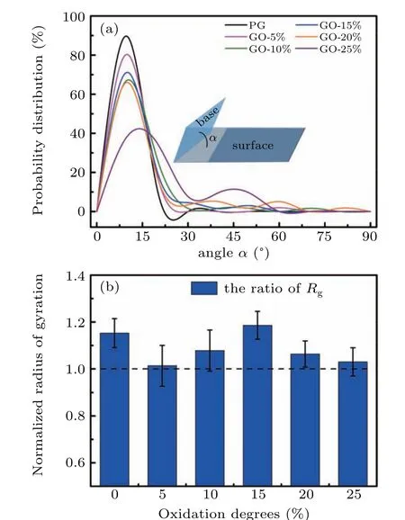
Fig.3. (a)Distribution of angle α between the inter π-π stacking structures of nucleobases and graphene-based surfaces in last 20 ns. (b)Normalized radius of gyration for ssDNA molecules which adsorption on graphene-based surface with variable oxidation degrees.
To characterize the stretched and curved configuration of absorbed ssDNA molecules on graphene-based surface, we analyzed the radius of gyration(Rg)for the ssDNA molecules in every simulation sample and normalized according to their initial configurations. As shown in Fig. S3(b),Rgoscillated from about 1.3 nm to a stable value with a maximum of 2.0 nm and a minimum of 0.8 nm. It was shown that there were both stretched and curved configurations compared with the initial ssDNA configuration on all graphene-based surfaces. The averagedRgfor different oxidized degree GO nanosheets were shown in Fig. 3(b). The averageRgof PG approximately equaled to that of GO-15%. TheRgof GO-10%and GO-20%had the similar value, so did the GO-5% and GO-25%. This unexpected symmetry was resulted from the similar number of the curved configurations. There were only one sample of ss-DNA molecule showing a curved configuration,which means ssDNA tends to be stretched on PG or GO-15%. For GO-5%and GO-25%, ssDNA showed curved configuration in three samples. For GO-10% and GO-20%, ssDNA showed curved configuration in two samples. The trend of the averagedRgdid not follow that of the oxidized degree, but similar to that of theπ–πstacking and the vdW interaction, except for that on the PG.Furthermore,we also found the normalizedRgwas maximum when ssDNA adsorption on GO-15% as shown in Fig.3(b), indicating ssDNA molecules adsorbed on GO-15%had the most relax configuration.
3.3.2. In-plane movement of the absorbed ssDNA molecules on graphene-based surface
Since the absorption of ssDNA on graphene-based surface was a dynamically stable state with some fluctuation,we then analyzed the movement of ssDNA on surface by calculating its displacement within last 20 ns. The displacement of ssDNA centroid for each simulation sample and the average result were shown in Figs.4(a)and 4(b),respectively. We noted that ssDNA molecule was more likely to move on the surface of low oxidation degree,especially on PG.As the oxidation degree of the surface increased, the adsorption of ss-DNA was in more stable state,making it harder for ssDNA to move.

Fig.4. (a)Displacement of ssDNA molecule on each graphene-based surface within last 20 ns. (b)Averaged displacement of the ssDNA centroid.
4. Conclusion
In this work, based on Shi–Tu model, we performed the all-atom MD simulations to investigate the ssDNA physisorption dynamics on the graphene-based surfaces with the oxidation degree ranging from 0 to 25%. By analyzing the stable configurations of the absorbed ssDNA molecules, we found that it could be absorbed in the most stable state on GO-15%according due to the total interactions between the ssDNA molecules and GO that included both vdW and electrostatic interactions. We also found that vdW interaction between them did not vary monotonously with the oxidation degree and the GO-15% held the greatest value. This result was consistent with the number ofπ–πstacking between ssDNA and graphene-based surface. Our analysis also showed that, the less oxidation degree on graphene-based surface relates to the greater probability for ssDNA forming the approximately parallel structure,regardless of the coexistence of both stretched and curved configurations. Our work revealed the detailed absorbed configurations of ssDNA molecules onto the graphenebased surface and provided a theoretical guidance for the applications of DNA/GO nanomaterials.
Acknowledgments
We gratefully acknowledge Prof. Guosheng Shi, Dr.Rongzheng Wan and Dr. Yongshun Song for the helpful discussions and their crucial reading of this manuscript. This work is also supported by the supercomputer Center of the Chinese Academy of Sciences, and the Shanghai Supercomputer Center of China.

- Chinese Physics B的其它文章
- Physical properties of relativistic electron beam during long-range propagation in space plasma environment∗
- High winding number of topological phase in non-unitary periodic quantum walk∗
- Widely tunable single-photon source with high spectral-purity from telecom wavelength to mid-infrared wavelength based on MgO:PPLN∗
- Control of firing activities in thermosensitive neuron by activating excitatory autapse∗
- Adaptive synchronization of chaotic systems with less measurement and actuation∗
- Dynamics analysis of a 5-dimensional hyperchaotic system with conservative flows under perturbation∗