
Effects of electroacupuncture on angiogenesis and cortical VEGF and BDNF expression in rats with focal cerebral ischemia
2022-04-18 06:51LIMengxing李梦醒WANGYu王玉GAOYunyun高云云YAOXiaowen姚晓雯LANWei兰崴TANGWei唐巍
LI Mengxing (李梦醒), WANG Yu (王玉), GAO Yunyun (高云云), YAO Xiaowen (姚晓雯), LAN Wei (兰崴),TANG Wei (唐巍)
1 College of Acupuncture and Moxibustion, Anhui University of Chinese Medicine, Hefei 230012, China
2 Graduate School, Anhui University of Chinese Medicine, Hefei 230012, China
Abstract
Keywords: Acupuncture Therapy; Electroacupuncture; Brain Ischemia; Ischemic Stroke; Cerebrovascular Circulation;Vascular Endothelial Growth Factors; Brain-derived Neurotrophic Factor; Rats
Cerebral ischemic stroke (CIS) is a localized necrosis of brain tissues caused by cerebral blood supply disorders,resulting in progressive aggravation of brain damage and various degrees of neurological dysfunction[1].Studies have shown that angiogenesis is an endogenous protective mechanism for brain tissues to regain oxygen and nutrients after ischemia[2]. Angiogenesis promotes a highly coupled neural repair process, which is beneficial to the recovery of neural function[3].Acupuncture is an effective method to prevent and treat cerebral ischemia[4]. Previous studies have found that the mechanism of acupuncture in CIS treatment is closely related to angiogenesis[5], but there are few studies on the acupuncture treatment method for stroke and the expression changes of angiogenesisrelated factors in different periods. “Concurrent treatment of the brain and heart” is a new idea for the treatment of cardiovascular and cerebrovascular diseases based on the theory of “treating different diseases with the same approach” in traditional Chinese medicine. Therefore, this study focused on the expression of vascular endothelial growth factor (VEGF)and brain-derived neurotrophic factor (BDNF) in the cortex of rats with focal cerebral ischemia and explored the effect of “concurrent treatment of the brain and heart” EA on promoting angiogenesis in CIS. It can provide some reference for EA treatment of cerebral ischemia.
1 Material and Methods
1.1 Experimental animals and groups
A total of 108 specific-pathogen-free male Sprague-Dawley rats (body mass: 280-320 g), were included in this experiment, with the experimental animal production license number of [SCXK (Lu) 20190003],ambient humidity of 45%-55%, room temperature of 20-25℃. Rats were free access to food and water, and fasted for 12 h before surgery. According to the random number table method, 27 rats were divided into the sham-operation group, and the rest of rats were subjected to the right middle cerebral artery occlusion(MCAO) model preparation. The successful models were randomly divided into a model group, a traditional acupoint group, and a concurrent treatment of the brain and heart group (n=27). This experiment was approved by the Animal Experiment Management Committee of Anhui University of Traditional Chinese Medicine, and the experimental process complied with theGuiding Principles for the Care of Experimental Animalsformulated by the Ministry of Science and Technology of the People’s Republic of China.
1.2 Main reagents and instruments
Mouse anti-rat VEGF monoclonal antibody (Cat. No.sc-7269, SANTA CRUZ, USA); mouse anti-rat BDNF monoclonal antibody (Cat. No. ab205067, Abcam, UK);PV-6000 universal secondary antibody kit (Cat. No.D1007, Beijing Zsbio, China); FITC-goat anti-rabbit fluorescent secondary antibody (Cat. No. D1007, Beijing Zsbio, China); rabbit anti-CD34 polyclonal antibody (Cat.No. bs-2038R, Beijing Boaosen Biotechnology Co., Ltd.,China); quantitative reverse transcription-polymerase chain reaction (qRT-PCR) kit (Cat. No. ZB-2305, TaKaRa Bio Inc, China); total RNA extraction reagent (Cat. No.15596026, Life Technologies, USA).
PeriFlux 5000 laser Doppler tissue flow instrument[Perimed (China), China]; H-800 transmission electron microscope (Leica, Germany); PIKOREAL 96-well real-time fluorescent quantitation PCR instrument(Thermo Fisher Scientific China Co., Ltd., China); Eclipse 80i confocal fluorescence microscope, optical microscope (Nikon Corporation, Japan); JD-801 gel imaging analysis system, DP-801 morphological microscopic image analysis system (Jiangsu Jetta Technology Development Co., Ltd., China); Image Pro Plus image processing system (Media Cybernetic, USA);SDZ-V multifunctional EA therapeutic apparatus(Suzhou Medical Products Factory Co., Ltd., China);PIKOREAL 96 PCR meter (Thermo Fisher Scientific China,China).
1.3 Model preparation
The MCAO model was prepared with reference to the relevant literature[6]. After isoflurane inhalation anesthesia, the rat skin was incised in the middle of the neck slightly to the right, and then the common carotid,the external carotid, and the internal carotid arteries were separated and exposed layer by layer. Slowly pushed the thread from the bifurcation of the external carotid and internal carotid artery into the internal carotid artery, and stopped at about 18-20 mm, fixed and sutured.
The steps of the sham-operation group were the same as above, while only the carotid artery was separated, and routinely fixed and sutured without nylon thread insertion.
The degree of rat’s neurological functional impairment was assessed with an 18-point modified neurological severity score (mNSS) 4 h after surgery[7].The lowest score of mNSS was 0 point and the highest score was 18 points. A higher score indicated a more serious neurological functional impairment. In this experiment, the mNSS of 2-18 points indicated a successful model.
1.4 Therapeutic methods
EA therapy was performed 4 h after the operation.
Shuigou (CV26), the left Quchi (LI11), Hegu (LI4), and Zusanli (ST36) in the traditional acupoint group, while Fengfu (GV16), Baihui (GV20), and the left Xinshu (BL15)and Neiguan (PC6) in the concurrent treatment of the brain and heart group were positioned referring to the relevant literature[8].
The rat was fixed on the rat board. Needles of 0.25 mm in diameter and 15 mm in length were connected to the EA therapeutic apparatus for acupuncture after insertion. The sparse-dense wave with a frequency of 4 Hz/20 Hz and a stimulating current intensity of about 0.5 mA was used. It was appropriate to see slight shaking at the acupuncture site. The treatment lasted 20 min/time, once a day for 14 consecutive days.
Rats in the sham-operation group and the model group were grasped and fixed under the same conditions without treatment.
1.5 Experimental indicators and detection methods
1.5.1 Neurological severity score
Before and after treatment, the 18-point mNSS was used to evaluate the rat’s neurological functional impairment and recovery.
1.5.2 Detection of regional cerebral blood flow (rCBF)in the ischemic side by laser Doppler
The rCBF was detected 5 min after wire insertion and after EA on the 3rd, 7th, and 14th days, respectively.Nine rats were randomly selected from each group.After isoflurane inhalation anesthesia, the head was fixed to the brain stereotaxic instrument, a cranial window was drilled along the coronal suture and sagittal suture, and the Doppler fiber optic probe was fixed to the cranial window. The ischemic regional cerebral blood flow was dynamically monitored, and the average blood flow was calculated using software,and recorded.
1.5.3 Detection of micro-vessel density (MVD) by immunohistochemistry
After the treatment, 3 rats were randomly selected from each group, and the rats were fixed in the same anesthesia procedure as above. After perfusion with 0.9% physiological saline, the brains were separated and fixed in paraformaldehyde solution for 48 h. Followed by dehydration, embedding, sectioning (about 4 μm in thickness), spreading, and baking in sequence. Added CD34 primary and secondary antibodies to the sections,incubated, color-developed, and mounted. The sections were observed under a light microscope, and the endothelial cells stained with brownish-yellow were used as the positive staining standard, and the average count of 5 randomly selected fields was used as the MVD value.
1.5.4 Detection of VEGF and BDNF positive expression by immunofluorescence
After treatment, 3 rats were randomly selected from each group, and paraffin sections were made with the same steps as above. The slices were soaked and rewarmed for 20 min, immersed in PBS for 3 times,3 min/time. After blotting the PBS with absorbent paper,goat serum was added dropwise, the slides were then blocked at room temperature for 30 min. The primary antibody, secondary antibody, and DAPI were added dropwise according to the steps and incubated in the dark. Nuclei were stained, dehydrated, mounted, and observed under a confocal fluorescence microscope.Visible green fluorescent immunoreactive positive products labeled with fluorescein isothiocyanate (FITC)in the neuron cytoplasm of the cortex were VEGF and BDNF proteins. The scanned images were analyzed by Image J software. The average optical density (AOD) of immunopositive reactions was calculated.
1.5.5 Detection of VEGF and BDNF mRNA expression by qRT-PCR
After treatment, 3 rats were randomly selected from each group and received anesthesia with the same steps as above. The brains were separated on ice after decapitation, with RIPA lysate added dropwise, and ground on ice for 20 min followed by centrifugation for protein extraction. Total RNA from cortical tissues was extracted with TRIzol according to the instruction, and the cDNA was reverse transcribed and stored at -80°C for later use. The β-actin is an internal reference gene,referring to the Genbank nucleotide sequence data. The probes for mRNA detection are shown in Table 1. A gel imaging analysis system was used to scan the electrophoresis film. The absorbance value of the target gene/the absorbance value of the internal reference gene was used as the relative expression level of the target gene.
Table 1. Primers for mRNA detection
1.6 Statistical analysis
Data processing was performed using SPSS version 22.0 statistical software. Measurement data conforming to normal distribution were presented as mean ±standard deviation (±s), one-way analysis of variance was used for comparisons among multiple groups, and the least significant difference test was used for pairwise comparisons between two groups. Repeated measures analysis of variance was used to compare data at multiple time points for the same indicator. The test level wasα=0.05, andP<0.05 indicated that the difference was statistically significant.
2 Results
2.1 Comparison of mNSS
There were significant differences in the mNSS among different groups (P<0.01); there were significant differences in the mNSS changes in each group between different time points (P<0.01), and there was a significant interaction between grouping and time(P<0.01). The time factor was controlled for comparison between groups. Compared with the sham-operation group at the same time point, the mNSS of the model group significantly decreased (P<0.01); on the 3rd, 7th,and 14th days of treatment, the mNSS of the traditional acupoint group and the concurrent treatment of the brain and heart group was lower than that of the model group (P<0.01); on the 14th day, the mNSS of the concurrent treatment of the brain and heart group was lower than that of the traditional acupoint group(P<0.05).
The grouping factor was controlled for comparing different time points within the group. Except for the sham-operation group, the mNSS in the other three groups significantly decreased with the extension of time within the group (P<0.01), and the scores were the lowest on the 14th day (P<0.01).
Please see Table 2 for details.
2.2 Comparison of rCBF in the ischemic side of the brain
There were significant differences in the rCBF between groups (P<0.01); there were significant differences in the rCBF of each group between different time points (P<0.01), and there was a significant interaction between grouping and time (P<0.01).
Compared with the sham-operation group at the same time point, the rCBF in the model group significantly decreased (P<0.01); the rCBF of the two EA groups was higher than that of the model group on the 7th and 14th days of treatment (P<0.01); the rCBF of the concurrent treatment of the brain and heart group was higher than that of the traditional acupoint group on the 14th day (P<0.05).
The grouping factor was controlled, and the comparisons between different time points within the group were carried out. Except for the sham-operation group, the rCBF significantly increased on the 3rd, 7th,and 14th days in the other three groups over time(P<0.01). Compared with the 3rd and 7th days in the two EA groups, the rCBF reached the highest on the 14th day (P<0.05 orP<0.01). These results suggest that a significant improvement in the cerebral blood flow on the 3rd day and accumulate with time.
Please see Table 3 for details.
2.3 Comparison of cortical MVD of the ischemic side
There were significant differences in the MVD among different groups (P<0.01); the change of MVD at each time point was statistically significant in each group(P<0.01), and there was a significant interaction between grouping and time (P<0.01).
The time factor was controlled for comparison between groups. Compared with the sham-operation group at the same time point, the MVD in the model group significantly decreased (P<0.01). The MVD in the two EA groups was higher than that in the model group on the 7th and 14th days of treatment (P<0.05 orP<0.05). The MVD of the concurrent treatment of the brain and heart group was higher than that of the traditional acupoint group (P<0.05 orP<0.01).
Controlling the grouping factor and comparing between different time points within the group, there was no statistical difference at each time point in the MVD in the sham-operation group (P>0.05); the MVD on the 7th and 14th days was higher than that on the 3rd day in the other three groups (P<0.05 orP<0.01).
Please see Figure 1-Figure 3 and Table 4 for details.
Table 2. Comparison of the mNSS among groups (±s point)
Table 2. Comparison of the mNSS among groups (±s point)
Note: mNSS=Modified neurological severity score; compared with the sham-operation group at the same time point, 1) P<0.01;compared with the model group at the same time point, 2) P<0.01; compared with the traditional acupoint group at the same time point, 3) P<0.05; compared with the same group at 4 h, 4) P<0.01; compared with the same group on the 3rd day, 5) P<0.01;compared with the same group on the 7th day, 6) P<0.01
Group n 4 h 3 d 7 d 14 d Sham-operation 9 0 0 0 0 Model 9 9.33±0.711) 8.78±0.441) 7.67±1.121)4)5) 5.11±0.781)4)5)6)Traditional acupoint 9 9.11±0.60 7.89±0.932)4) 5.44±0.532)4)5) 4.00±0.872)4)5)6)Concurrent treatment of the brain and heart 9 9.00±0.71 7.56±0.732)4) 5.00±0.872)4)5) 3.33±0.712)3)4)5)6)Grouping F=1 036.03, P<0.01 Time F=221.29, P<0.01 Interaction between grouping and time F=28.38, P<0.01
Table 3. Comparing regional cerebral blood flow (rCBF) in the ischemic side of the rat brain among groups (±s PU)

Table 3. Comparing regional cerebral blood flow (rCBF) in the ischemic side of the rat brain among groups (±s PU)
Note: rCBF=Regional cerebral blood flow; compared with the sham-operation group at the same time point, 1) P<0.01; compared with the model group at the same time point, 2) P<0.01; compared with the traditional acupoint group at the same time point, 3) P<0.05;compared with the same group at 5 min, 4) P<0.01; compared with the same group on the 3rd day, 5) P<0.01; compared with the same group on the 7th day, 6) P<0.01
Group n 5 min 3 d 7 d 14 d Sham-operation 9 132.10±1.35 133.22±1.78 133.15±1.59 133.95±2.58 Model 9 8.93±0.751) 90.00±1.541)4) 90.23±1.721)4) 91.08±1.551)4)Traditional acupoint 9 8.82±1.61 91.70±2.624) 104.07±2.532)4)5) 110.34±3.682)4)5)6)Concurrent treatment of the brain and heart 9 8.80±1.03 91.67±2.114) 105.20±1.942)4)5) 113.12±1.662)3)4)5)6)Grouping F=3 856.92, P<0.01 Time F=15 220.20, P<0.01 Interaction between grouping and time F=1 696.53, P<0.01
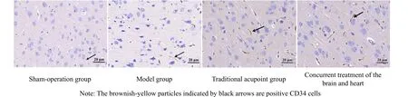
Figure 1. Micro-vessel density (MVD) counts in the ischemic side of each group on the 3rd day (immunohistochemistry, ×400)
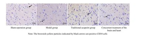
Figure 2. Micro-vessel density (MVD) counts in the ischemic side of each group on the 7th day (immunohistochemistry, ×400)

Figure 3. Micro-vessel density (MVD) counts in the ischemic side of each group on the 14th day (immunohistochemistry, ×400)
Table 4. Comparison of the MVD counts in the cerebral ischemic side of rats in each group (±s number)
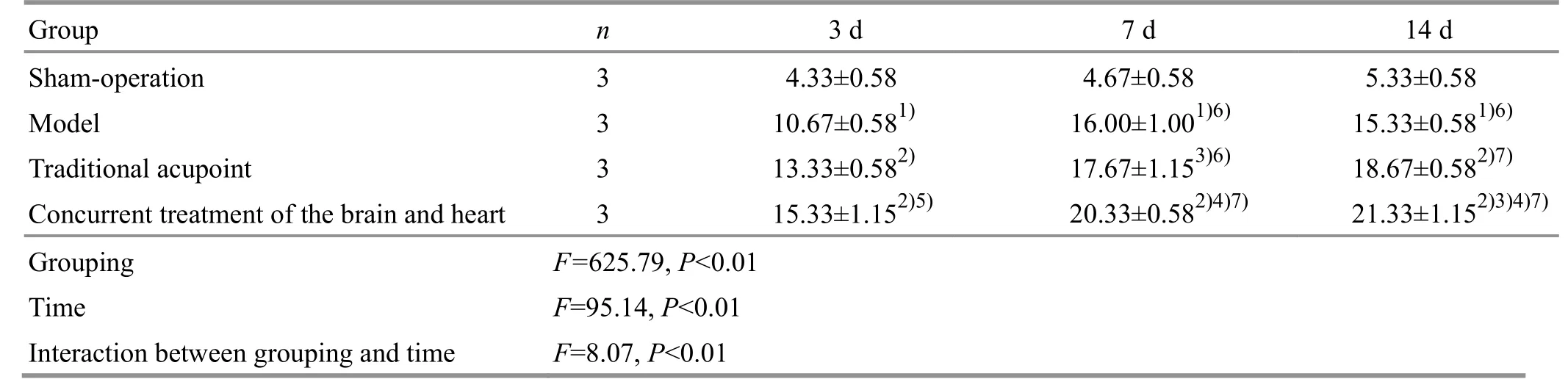
Table 4. Comparison of the MVD counts in the cerebral ischemic side of rats in each group (±s number)
Note: MVD=Micro-vessel density; compared with the sham-operation group at the same time point, 1) P<0.01; compared with the model group at the same time point, 2) P<0.01, 3) P<0.05; compared with the traditional acupoint group at the same time point, 4)P<0.01, 5) P<0.05; compared with the same group on the 3rd day, 6) P<0.01, 7) P<0.05
Group n 3 d 7 d 14 d Sham-operation 3 4.33±0.58 4.67±0.58 5.33±0.58 Model 3 10.67±0.581) 16.00±1.001)6) 15.33±0.581)6)Traditional acupoint 3 13.33±0.582) 17.67±1.153)6) 18.67±0.582)7)Concurrent treatment of the brain and heart 3 15.33±1.152)5)20.33±0.582)4)7) 21.33±1.152)3)4)7)Grouping F=625.79, P<0.01 Time F=95.14, P<0.01 Interaction between grouping and time F=8.07, P<0.01
2.4 Comparison of the positive expression of VEGF and BDNF in the ischemic side of cerebral cortex
There were significant differences in the positive expression of VEGF and BDNF among different groups(P<0.01); the positive expression of VEGF and BDNF changed significantly over time in each group (P<0.01).There was a significant interaction between grouping and time (P<0.01).
The time factor was controlled for comparing the positive expression of VEGF and BDNF between groups.Compared with the sham-operation group at the same time point, the positive expression of VEGF and BDNF significantly increased in the model group (P<0.01).Compared with the model group, the positive expression significantly increased in the two EA groups(P<0.01). The VEGF expression in the concurrent treatment of the brain and heart group was higher than that in the traditional acupoint group on the 7th and 14th days (P<0.05), and the BDNF expression in the concurrent treatment of the brain and heart group was significantly higher than that in the traditional acupoint group on the 14th day (P<0.01).
The grouping factor was controlled, and the comparison between different time points within the group was carried out. There was no significant difference in the BDNF expression between the shamoperation group and the model group at each time point (P>0.05). The positive VEGF expression increased on the 7th and 14th days in the model group (P<0.05).On the 7th day in the traditional acupoint group, the positive expression of VEGF and BDNF reached the highest (P<0.05). The positive VEGF expression was the highest on the 7th day in the concurrent treatment of the brain and heart group, and the positive BDNF expression on the 7th and 14th days was higher than that on the 3rd day (P<0.05).
Please see Figure 4-Figure 9 and Table 5-Table 6 for details.
2.5 Comparison of the VEGF and BDNF mRNA expression in the ischemic side of cerebral cortex
There were significant differences in the VEGF and BDNF mRNA expression among different groups(P<0.01). The changes in the VEGF and BDNF mRNA expression were statistically significant in each group over time (P<0.01). There was a significant interaction between grouping and timeP<0.01).
The time factor was controlled for comparing the mRNA expression of VEGF and BDNF between groups.Compared with the sham-operation group at the same time point, the mRNA expression of VEGF and BDNF significantly increased in the model group (P<0.01).Compared with the model group, the expression significantly increased in both EA groups (P<0.01). The expression levels were significantly higher in the concurrent treatment of the brain and heart group than in the traditional acupoint group on the 7th day of treatment (P<0.01).
Controlling the grouping factor and comparing between different time points within the group, there was no significant difference in the VEGF and BDNF mRNA expression between the sham-operation group and the model group at each time point (P>0.05); the mRNA expression levels of VEGF and BDNF were the highest in both the traditional acupoint group and the concurrent treatment of the brain and heart group on the 7th day (P<0.05 orP<0.01). The above results suggest that EA regulates the expression of key angiogenesis factors VEGF and BDNF; different acupoint combinations can produce different regulatory effects,and the concurrent treatment of the brain and heart group has a better result.
Please see Figure 4-Figure 9 and Table 7-Table 8 for details.

Figure 4. Positive VEGF expression in the rat ischemic cortex of each group on the 3rd day (immunofluorescence, ×200)
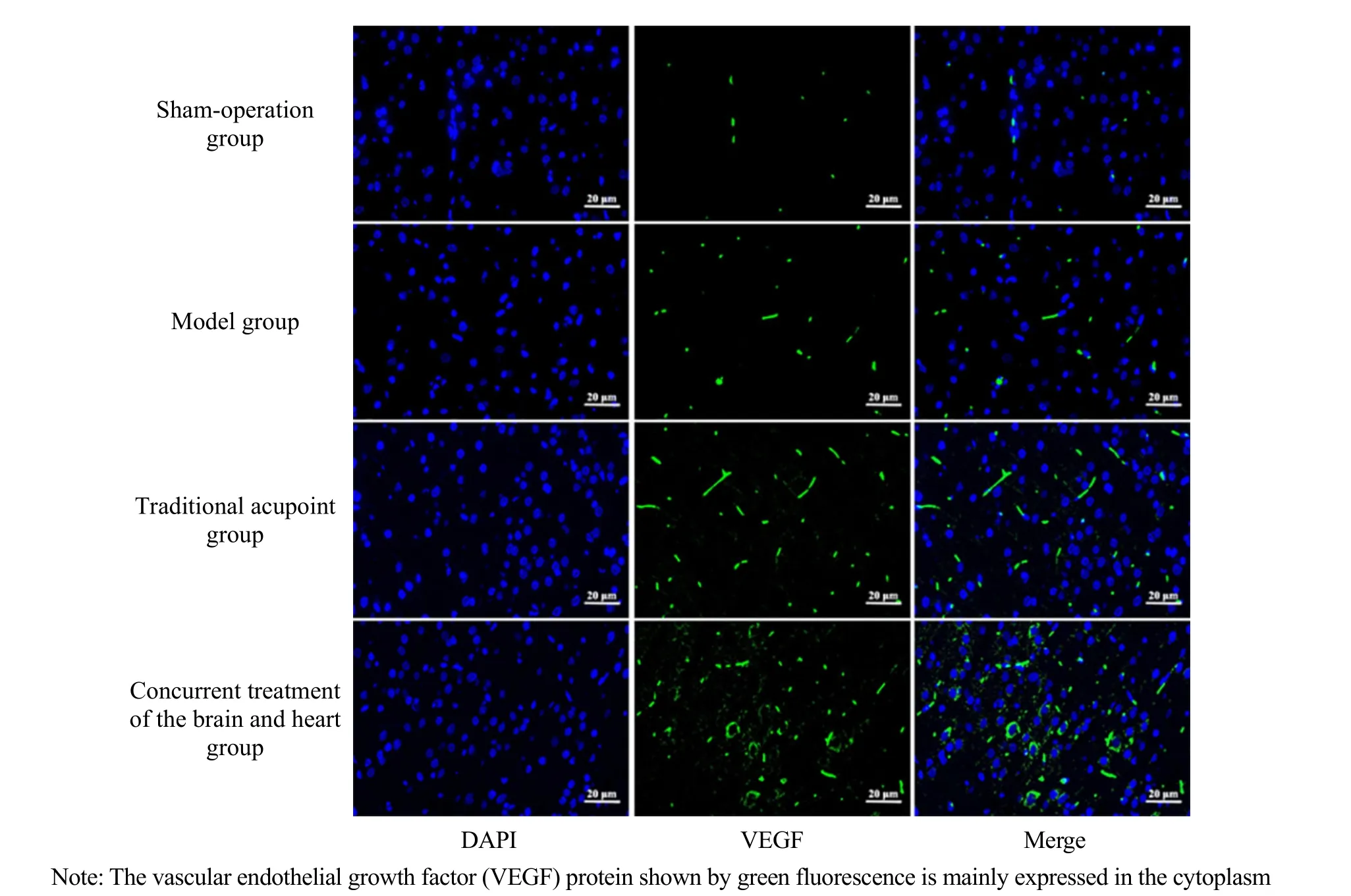
Figure 5. VEGF positive expression in the ischemic cortex of each group on the 7th day (immunofluorescence, ×200)
Figure 6. Positive VEGF expression in the ischemic cortex of each group on the 14th day (immunofluorescence, ×200)
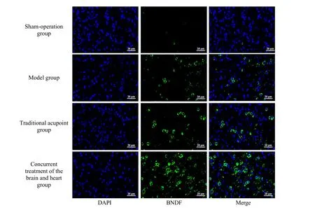
Figure 7. Positive BNDF expression in the ischemic cortex of each group on the 3rd day (immunofluorescence, ×200)
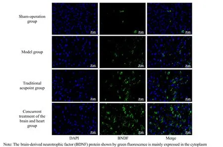
Figure 8. Positive BNDF expression in the ischemic cortex of each group on the 7th day (immunofluorescence, ×200)

Figure 9. Positive BNDF expression in the ischemic cortex of each group on the 14th day (immunofluorescence, ×200)
Table 5. Comparison of the positive VEGF expression in the cerebral ischemic cortex (±s)
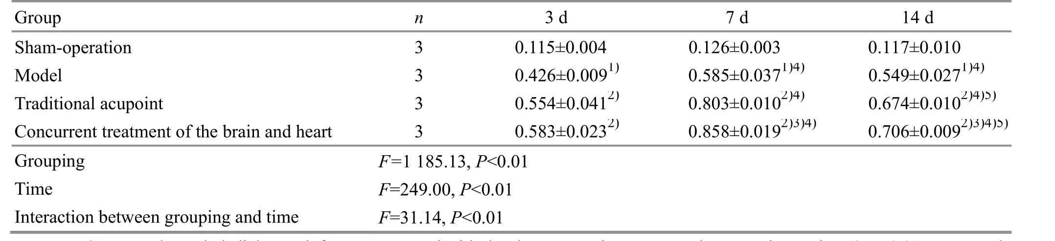
Table 5. Comparison of the positive VEGF expression in the cerebral ischemic cortex (±s)
Note: VEGF=Vascular endothelial growth factor; compared with the sham-operation group at the same time point, 1) P<0.01; compared with the model group at the same time point, 2) P<0.01; compared with the traditional acupoint group at the same time point, 3)P<0.01; compared with the same group on the 3rd day, 4) P<0.05; compared with the same group on the 7th day, 5) P<0.05
Grouping F=1 185.13, P<0.01 Time F=249.00, P<0.01 Interaction between grouping and time F=31.14, P<0.01
Table 6. Comparison of the positive BDNF expression in the cerebral ischemic cortex (±s)

Table 6. Comparison of the positive BDNF expression in the cerebral ischemic cortex (±s)
Note: BDNF=Brain-derived neurotrophic factor; compared with the sham-operation group at the same time point1) P<0.01; compared with the model group at the same time point, 2) P<0.01; compared with the traditional acupoint group at the same time point, 3)P<0.01; compared with the same group on the 3rd day, 4) P<0.05; compared with the same group on the 7th day, 5) P<0.05
Group n 3 d 7 d 14 d Sham-operation 3 0.114±0.003 0.115±0.005 0.111±0.011 Model 3 0.389±0.0181) 0.470±0.0531) 0.438±0.0631)Traditional acupoint 3 0.547±0.0272) 0.794±0.0182)4) 0.635±0.0142)5)Concurrent treatment of the brain and heart 3 0.581±0.0422) 0.829±0.0342)4) 0.797±0.0102)3)4)Grouping F=641.49, P<0.01 Time F=71.09, P<0.01 Interaction between grouping and time F=16.12, P<0.01
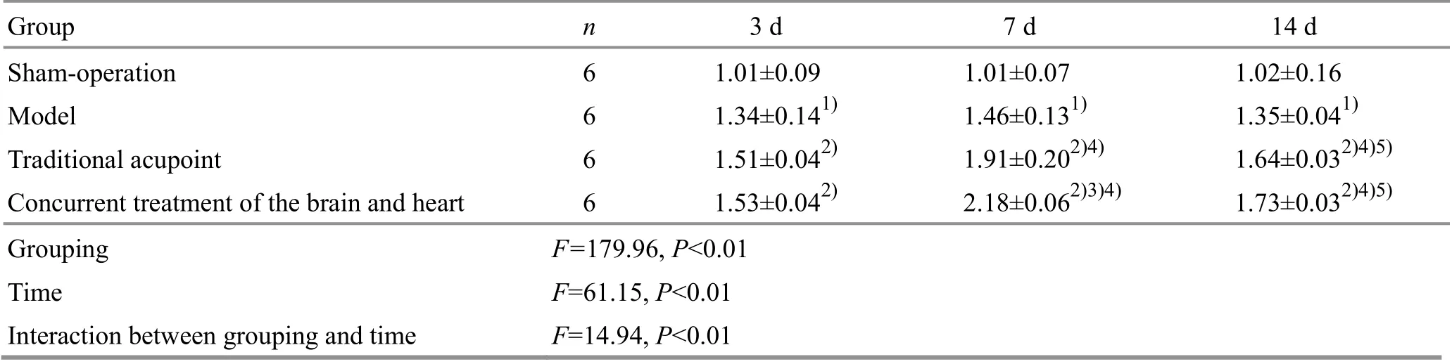
Table 7. Comparison of the VEGF mRNA expression in the cerebral ischemic cortex (x ±s, bp)
Table 8. Comparison of the BDNF mRNA expression in the cerebral ischemic cortex (±s bp)
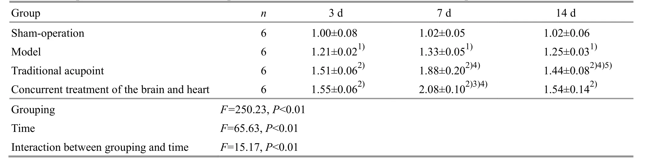
Table 8. Comparison of the BDNF mRNA expression in the cerebral ischemic cortex (±s bp)
Note: BDNF=Brain-derived neurotrophic factor; compared with the sham-operation group at the same time point, 1) P<0.01;compared with the model group at the same time point, 2) P<0.01; compared with the traditional acupoint group at the same time point, 3) P<0.01; compared with the same group on the 3rd day, 4) P<0.05; compared with the same group on the 7th day, 5) P<0.05
Group n 3 d 7 d 14 d Sham-operation 6 1.00±0.08 1.02±0.05 1.02±0.06 Model 6 1.21±0.021) 1.33±0.051) 1.25±0.031)Traditional acupoint 6 1.51±0.062) 1.88±0.202)4) 1.44±0.082)4)5)Concurrent treatment of the brain and heart 6 1.55±0.062) 2.08±0.102)3)4) 1.54±0.142)Grouping F=250.23, P<0.01 Time F=65.63, P<0.01 Interaction between grouping and time F=15.17, P<0.01
3 Discussion
CIS belongs to the category of “stroke disease” in traditional Chinese medicine, and its pathogenesis includes wind, fire, phlegm, blood stasis, Qi, deficiency,disorders of Zang-Fu organs, Yin-Yang, and flow of Qi-blood, which directly attack the brain and block the cerebral vessel, leading to unconsciousness. Phlegm and blood stasis bind together, blocking the collaterals and subsequently leading to this disease. Researchers have proven that acupuncture-moxibustion in CIS treatment is effective to improve clinical symptoms and reduce disability rate[9]. At present, most scholars believe that repairing or rebuilding the homeostasis of the central nervous system as soon as possible is the key to cerebral ischemia treatment[10]. The neovascularization in the ischemic side and the establishment of collateral circulation contribute to the repair of damaged but not dead neurons in the ischemic penumbra of the brain,thereby playing a positive role in improving neurological function[11]. Therefore, vascular endothelial cell activation, proliferation, and microvascular formation are new ways to promote the recovery of neurological function after stroke.
Angiogenesis is regulated by many complementary and complex signaling pathways. Among them, VEGF is a highly specific vascular endothelial growth-promoting cytokine that participates in the angiogenesis process of ischemic and hypoxic tissues or organs[12-13]. BDNF is a neurotrophic factor, which is greatly stimulated in the ischemia-hypoxia microenvironment. It can not only protect nerve cells from damage and enhance plasticity,but also regulate the downstream relevant signaling pathways to induce angiogenesis[14-15].
Although the mechanism of promoting angiogenesis is activated immediately after cerebral ischemia, the body's self-repair ability is limited, and therapeutic angiogenesis can make up for the lack of its own mechanism response. In this experiment, it was found that the expression levels of VEGF and BDNF in the postoperative model group increased, indicating that the expression of VEGF and BDNF increased under the stimulation of ischemia and hypoxia[16]. Since the EA intervention started 4 h after the operation, the two factors’ expression continued to increase, and peaked on the 7th day, suggesting that VEGF and BDNF may play a role in the early and middle stages of cerebral ischemia. EA intervention can up-regulate their expression and advance the expression phase.
VEGF and BDNF mediate the interaction of blood vessels and nerves, which can be reflected by increased cortical positive CD34 cells and local rCBF. After CIS occurs, CD34 can homing to the cerebral infarction site along with endothelial progenitor cells, differentiate and proliferate into mature endothelial cells to participate in angiogenesis[17]. It is a sensitive factor for detecting angiogenesis[18]. The results of this study showed that the MVD of the model group and the two EA groups were increased with the treatment time and reached the highest on the 14th day in the concurrent treatment of the brain and heart group. The microcirculation of blood in brain tissue was measured by laser Doppler blood flow detector, showing that the rCBF in the model group decreased significantly 5 min after insertion, indicating that the model was successful.On the 3rd day of treatment, there were obvious changes in the rCBF, and during the 14th day of treatment, the rCBF in the two EA groups continued to increase, and the rCBF in the concurrent treatment of the brain and heart group obtained the highest on the 14th day. At the same time, the mNSS of the two EA groups decreased significantly 3 d after the operation.This suggests that the earlier the intervention of acupuncture treatment, the better the effect in promoting angiogenesis after cerebral ischemia, as it can increase the rCBF, reduce the degree of neurological functional impairment, and expand the treatment time window[19]. Moreover, the therapeutic effect within the effective time was continuously accumulated, which is consistent with previous reports[20].
In this study, guided by the basic principle of“concurrent treatment of the brain and heart”, four acupoints, Fengfu (GV16), Baihui (GV20), Xinshu (BL15)and Neiguan (PC6), were used. This group of acupoints can improve endothelial function and regulate the central activity of hypothalamus[21]. The traditional acupoint group used Shuigou (CV26), Quchi (LI11), Hegu(LI4) and Zusanli (ST36) according to the modern clinical acupuncture treatment for stroke. This study found that compared with the traditional acupoint group, the concurrent treatment of the brain and heart group showed a better improvement in promoting angiogenesis and cortical VEGF and BDNF expression in CIS rats.
In conclusion, after cerebral ischemia, the body initiates self-healing angiogenesis in the ischemiahypoxic microenvironment. EA therapy can up-regulate the expression levels of VEGF and BDNF in the cortex,release the angiogenesis effect after cerebral ischemia,and play a role in remodeling the neurovascular unit after brain injury. Further, the acupoint combination guided by the theory of “concurrent treatment of the brain and heart” is superior to the traditional acupoint group in promoting angiogenesis.
Conflict of Interest
The authors declare that there is no potential conflict of interest in this article.
Acknowledgments
This work was supported by Projects of National Natural Science Foundation of China (国家自然科学基金项目,No. 81373711, No. 81874500); Youth Project of Anhui Provincial Natural Science Foundation of China (安徽省自然科学研究青年基金项目, No. 1808085QH268); Key Project of Natural Science Research Program in Colleges and Universities in Anhui Province (安徽省高校自然科学研究项目重点项目, No. KJ2020A0377).
Statement of Human and Animal Rights
The treatment of animals conformed to the ethical criteria in this experiment.
Received: 18 February 2021/Accepted: 10 June 2021
猜你喜欢
国际放射医学核医学杂志(2021年6期)2021-11-30
国际放射医学核医学杂志(2021年1期)2021-11-30
商界评论(2021年4期)2021-06-08
商界评论(2021年3期)2021-05-12
商界评论(2021年2期)2021-04-23
商界评论(2021年1期)2021-03-11
校园英语·上旬(2020年2期)2020-05-11
校园英语·上旬(2020年2期)2020-05-11
校园英语·中旬(2019年5期)2019-07-16
校园英语·上旬(2018年11期)2018-11-30
 Journal of Acupuncture and Tuina Science2022年2期
- Journal of Acupuncture and Tuina Science的其它文章
- Effects of ginger-partitioned moxibustion on the expression levels of PGF2α, E2, P, and mRNAs of PGF2αR and E2R in rats with primary dysmenorrhea due to cold-dampness stagnation
- Therapeutic efficacy of acupuncture plus Tuina for spastic cerebral palsy and discussion of its mechanism
- Protocol-optimizing study of combining Tuina and horse-riding squat exercise for knee osteoarthritis
- Clinical observation of pediatric Tuina plus oral Chinese medication for pediatric anorexia due to spleen failing in transportation
- Acupuncture plus naloxone hydrochloride in the treatment of coma after surgery for cerebral hemorrhage: a randomized controlled trial
- Reduction of serum level of interleukin-2 and pruritus severity after acupuncture at Quchi (LI11) in hemodialysis patients: a placebo-controlled randomized clinical trial