
Atypical Adams-Oliver syndrome with typical ocular signs of familial exudative vitreoretinopathy
2022-08-10 01:39EnZhongJinLyuZhenHuangMingWeiZhaoHongYin
INTRODUCTION
Adams-Oliver syndrome (AOS) is a rare inherited condition first described in 1945
. As a multiple malformation syndrome, AOS was characterized by a combination of aplasia cutis congenita (ACC) and variable degree of transverse limb defects
. Several genes associated with AOS including
and
have been reported in the past decade
. In previous studies, the systemic phenotype within families of AOS may range from no obvious clinical manifestations in mutation carriers to severe multiple-system anomalies that can even result in miscarriage or stillbirth
.
1.6 统计学处理 采用SPSS 17.0统计学软件进行统计分析,以P<0.05为差异有统计学意义。计量资料以表示,计数资料以率表示。单因素分析采用Student′s t检验或四格表χ2检验,将P<0.1的因素纳入多因素分析,多因素分析采用二项分类Logistic回归模型,通过多因素分析控制混杂因素,明确胸腰椎结核手术患者早期植骨融合的独立影响因素。
小规模纳税人的科目设置参照《规定》,在“应交税费”科目下设置“应交增值税”、“转让金融商品应交增值税”、“代扣代交税金”明细科目,核算原理同上。
MATERIALS AND METHODS
This study was approved by the Ethical Review Committee of Peking University People’s Hospital(Beijing, China), which was conducted in accordance with the Declaration of Helsinki. Written informed consent for genetic testing and medical photograph collection was obtained from the parents.
“儿童离不开生活,生活离不开健康教育。”晨间户外锻炼作为体育活动的活动形式之一不仅能让幼儿有效地锻炼身体,拥有健康,而且能更多直接地接受阳光、新鲜空气和水分等自然因素的刺激,所以只有让幼儿充分体验晨间户外体育锻炼活动的快乐,形成活泼、向上的性格,才能更好地促进各领域的学习,才能提高幼儿的生活乃至生命的质量。
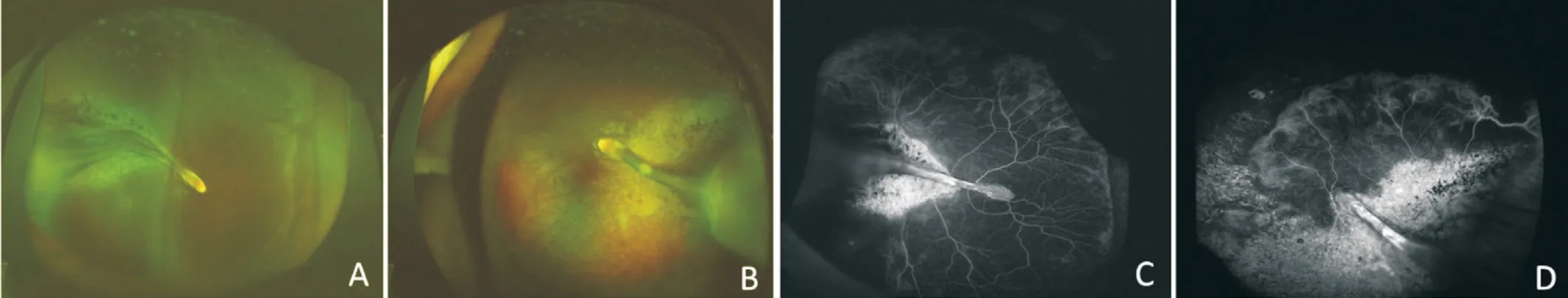
The boy born at full-term with birth weight of 3000 g, and no special prenatal history was recorded. The rough visual acuity of the boy was 20/80 for both eyes with poor cooperation. Noncontact intraocular pressure was 17 mm Hg and 16 mm Hg for right and left eye, respectively. The cycloplegic refraction was measured as follows: -0.25 D for the right eye and -4.00 D for the left eye. No obvious abnormalities can be observed for the examination of the anterior segment of the eyes. For the binocular indirect ophthalmoscope fundus examination, normal boundary and color of the optic discs can be showed, while the avascular area was visible in the periphery retina. Ultrawide-field FA examination was performed for the patient and obvious non-perfusion in the peripheral retina can be defined.Abnormal vessels can be observed at the junction of vascular and avascular retina though no leakage was identified (Figure 1).Fundus color photographs and FA images were also collected for the boy’s elder sister and parents. For the sister, significant peripheral retinal detachment can be observed in both eyes with temporal retinal folds originating from the optic disc, and the temporal retina was gray-white with hyperpigmentation sounded. FA examination showed peripheral retinal nonperfusion area and abnormal vascularization including tortuous veins, arteriovenous shunt, abnormal anastomosis. Retinal atrophy and hyperpigmentation can also be identified in fluorescein angiogram (Figure 2).
该方法根据网络的不同情况将网络进行拆分,再根据每一个部分的特点采用不同的方法进行混合预测.De[13]等人根据节点的属性和网络结构将目标网络分为两层,第一层根据节点获得的信息判断两个节点之间产生连接的可能性,第二层根据网络的结构,结合AA指标,采用协同聚类的方式获取特征值,最后通过支持向量机完成整个预测过程;Chen[14]等人提出网络稀疏化框架,通过四种不同的方法稀疏化目标网络,对稀疏化后的网络逐一训练分类器,并整合在一起进行综合预测.该方法将特定网络以特定方法分解后能有效预测网络链路,但普适性不佳.
Ophthalmologic examination was performed with a standard protocol including best-corrected visual acuity (BCVA), slit-lamp examination, binocular indirect ophthalmoscope fundus examination, ultra-wide-field fundus photography and fluorescein angiography (FA; Optos Daytona, Optos PLC, Dunfermline, UK). All the examination data was collected along with the data of medical history. All the digital images of fundus and FA were blinded reviewed by two experienced ophthalmologists (Yin H and Jin EZ).
Whole exome sequencing was performed to characterize mutations for the proband boy and all his family members. Peripheral blood samples were obtained for the gene sequencing and sent to an external service to be sequenced.
RESULTS
The genetic sequencing was performed for the whole family and the results indicated that the children (both the boy and his sister) had two compound heterozygous mutations (c.1396C>T p.R466X, c.4796G>A p.W1599X) in the gene
associated with AOS type 2, and the family verification results showed the two heterozygous mutations were originated from their father and mother, respectively. Pedigrees of the family and sanger confirmation of the identified
variants in proband with family members are shown in Figure 3.
The AOS associated with ocular findings is rarely reported especially for the patients initially diagnosed in Ophthalmology Department. Almost all reports associated with ocular disorders were cases. In a previous literature, a female AOS case with bilateral congenital cataract was reported
. In another report, a case of AOS mimicked as familial exudative vitreoretinopathy(FEVR) had been reported recently, but genetic testing was not performed in this single case
. In 2012, an AOS case presenting retinal findings consistent with ischemicproliferative retinopathy had also been reported
. Since AOS was found to be associated with several ocular disorders and few cases have already been reported, the relationship between AOS and ocular diseases worth further discussion.In our present report, a family of AOS with ocular signs of FEVR was identified according to the clinical and genetic findings with
mutation. The ophthalmic examinations and gene sequencing were performed for the patient, his elder sister and their parents.
A 5-year-old boy initially diagnosed with FEVR and found to have AOS according to the systemic situation, family history and gene sequencing was described. The individual and family investigation were performed. All family members of the proband boy including the parents and his 10-year-old sister were included in the present study and underwent ocular and genetic examinations for further analysis.
In this case, the parents of the boy showed no obvious ocular symptoms or signs, but the boy showed slightly lower intelligence, poor vision, mild esotropia and large nonperfusion zone at the temporal retina, which suggesting FEVR-like fundus changes. For the family history, it was found that the child’s sister had a history of retinal detachment with retinal fold in both eyes. Fundus examinations showed obvious retinal folds in both eyes, with small palpebral fissure,esotropia, raised outer corner of the eye, right hand penetrating palm, high muscle tension, mild hemiplegia of the left limb,
. There are many similarities between these manifestations and the previously reported systemic and ocular signs of AOS
.
The 5-year-old boy was found to have abnormal eye position and slightly mental retardation from birth according to his parents’ discription. No physical deformitie and mental abnormalities existed among his parents. For further family members’ investigation, his 10-year-old sister previously defined as retinal detachment and clinically diagnosed with FEVR. She was found to have poor vision, esotropia,blepharophimosis, upward of external eye corner, mild mental retardation and left hemiplegia.
DISCUSSION
AOS was not frequently reported in patients with ocular disorders though some isolated cases had been described associated with congenital cataract and retinal findings
. In our case, we described a family of AOS with all the members showing retinal signs similar to FEVR and
mutations.To the best of our knowledge, this is the first report of AOS family identified by gene mutations with mimicking FEVR retinal findings.
As we all know, AOS was first reported in 1945, mainly manifested as scalp defects and varying degrees of limb defects
.The AOS syndrome can be inherited in autosomal dominant manner in most cases, and in autosomal recessive manner for some cases (
and
gene mutations)
. The underlying pathological mechanism of AOS is considered as a congenital vascular disease that may involve multiple systems such as the cardiovascular system, brain, liver, lung,eyes and skin
. In the past, few reports about AOS related to ocular disorders, especially for retinal manifestations
.The insufficient evidence between AOS and retinal disorders like FEVR made the present study more meaningful. But the limitations of our study can not be ignored. First, only one family of AOS mimicked FEVR was included which may reduce the persuasion, more families or cases should be collected and combined analyzed. Second, as a report of family case, no functional validation was performed even though two novel mutations were found. Third, the ocular signs of FEVR of the proband and his sister was typical while the AOS signs of them were atypical, but the genetic sequencing can provide most strong evidence.


For their parents, the FA examinations were also performed.Only avascular zone and abnormal anastomosis at the vascularavascular junction can be identified for them and both of them can reached a BCVA of 20/20. Meanwhile, no physical deformitie and mental abnormalities existed among the parents.
Basing on the ocular and systemic manifestations of the boy and his sister, a whole-exome sequencing analysis of the single-gene disease was performed on the whole family. On one hand, further clarification whether there are FEVR-related genetic changes were performed, and on the other hand, it is also analyzed whether there was genetic mutation associated with AOS. The sequencing revealed that the proband had two heterozygous mutations (c.1396C>T p.R466X, c.4796G>A p.W1599X) in
which is associated with the AOS type 2. Though the variant in
was reported to be associated with AOS type 2 before, these two novel missense mutations identified in this study had not been previously registered in the Ensembl database (http://www.ensembl.org/index.html) or the Human Gene Mutation Database (HGMD, http://www.hgmd.cf.ac.uk/ac/index.php)
. And the compound heterozygous mutations of them were found to be associated with the AOS proband. The further family verification results showed that the two heterozygous mutations came from their parents respectively and were compound heterozygous mutations. The sister also carries the two compound heterozygous mutations.As truncating mutations, the two novel missense mutations show high potential pathogenicity. Our findings expand the mutational spectrum of
and suggest that the two compound heterozygous mutations of
is associated with AOS.
For the previous reports, the retinal fold with or without retinal detachment had been reported in AOS patients, and the retinal folds involving the macular was also described in one case
. A seven-week-old full-term infant within normal birth weight clinically diagnosed with AOS with ocular signs similar to FEVR was reported
. While no remarkable change existed in anterior segment, a radial falciform retinal fold extending from the macula to temporal periphery can be defined in this case. Significant preretinal fibrous proliferation can be noted, but no neovascularization or exudation were observed, and no ophthalmic examination was performed for the parents and three male siblings of the infant
. In our case,the whole family underwent gene sequencing and ophthalmic examinations, and genetic mutations can be identified accurately. Retinal detachment accompanied by retinal fold can also be found in the sister, along with poor vision, mild mental retardation and left hemiplegia. The ocular and systemic disorders of the sister confirmed by the genetic sequencing can further assist the diagnosis of AOS for the whole family.In this case, FEVR-like fundus and fluorescein angiogram can be observed in all family members. Despite the presence of FEVR-like changes existed, they were generally quiet. No obvious peripheral leakage or retinal traction can be observed,and the boy was only asked for closely and regular follow-up without treatment.
As an inherited retinal disorder characterized by abnormal development of retinal vasculature, FEVR was thought to be associated with Wnt signaling pathway
. Several genes including
, and
had been found to be associated with FEVR in previous studies
. On the other hand, these genes were also identified in atypical retinopathy of prematurity (ROP) patients
. The Wnt signaling pathway may play a common pathological role in them, which is a major role for the development of retinal vascular. According to the whole-exome sequencing analysis, no special mutations of these genes can be found in our present family case, which did not support the relationship between AOS and Wnt signaling pathway. Since no association between FEVR and
had been identified before, and the
and
involved in the Notch signaling pathway were found to play a crucial role in developing blood vessel walls, the pathogenesis of AOS may be different with FEVR
. Although a case of AOS with similar changes mimicking FEVR diagnosed basing on clinical systemic characteristics has been reported,the present case is the first confirmed family case basing on genetic sequencing. Our report provides information on genetic mutations and clinical features to assist the ophthalmologists in recognizing AOS patients. Also, this family case can help us further understanding the ocular manifestations of AOS. It can be defined that the ocular phenotype of AOS may mimic that of FEVR. The patients diagnosed with AOS should be further evaluated for the retinal vasculopathy by fundus examinations including fluorescein angiography to make sure whether specific treatment was required. And the FEVR patients should also be carefully evaluated for the potential possibility of suffering from AOS.
ACKNOWLEDGEMENTS
Jin EZ: Project development, data management and analysis, manuscript writing and editing;Huang LZ: Data analysis, manuscript editing; Zhao MW:Manuscript editing; Yin H: Project development, manuscript writing and editing.
2.1.1 子宫出血的特点:月经周期发生紊乱,出血量时少时多,经期的长短不定,有时有大量出血。出血期无下腹痛或其他不适,出血量多或时间长者可伴发贫血症状,大量出血时可导致休克[2]。
传统企业发展过程中,忽略会计成本核算工作的重要性,最终成本支出较高,获得的经济利益较小,难以满足企业发展对资金流的需求。因此,企业应重新审视会计成本核算工作,对其重要性进行了解,以此提升会计成本核算工作地位,培养优秀人才,从而有效地促进会计成本核算工作正常开展,帮助企业进行成本计算,并为企业的资金流动方向做出正确的指引。使企业在激烈的市场竞争中不断提升竞争力,促进我国经济市场体制的改革。
Supported by the National Natural Science Foundation of China (No.81800850).
Jin EZ, None; Huang LZ, None; Zhao MW, None; Yin H, None.
1 Alsulaiman AM, Alsulaiman HM, Almousa A, Alsulaiman SM. Adams oliver syndrome: a mimicker of familial exudative vitreoretinopathy.
2020;19:100715.
2 Dedania VS, Moinuddin O, Lagrou LM, Sathrasala S, Cord Medina FM, del Monte MA, Chang EY, Bohnsack BL, Besirli CG. Ocular manifestations of cutis marmorata telangiectatica congenita.
2019;3(9):791-801.
3 Fayol L, Garcia P, Denis D, Philip N, Simeoni U. Adams-Oliver syndrome associated with cutis marmorata telangiectatica congenita and congenital cataract: a case report.
2006;23(3):197-200.
4 Hassed S, Li SB, Mulvihill J, Aston C, Palmer S. Adams-Oliver syndrome review of the literature: refining the diagnostic phenotype.
2017;173(3):790-800.
5 Hassed SJ, Wiley GB, Wang SF, Lee JY, Li SB, Xu WH, Zhao ZJ,Mulvihill JJ, Robertson J, Warner J, Gaffney PM.
mutations identified in two families affected by Adams-oliver syndrome.
2012;91(2):391-395.
6 Küster W, Lenz W, Kääriäinen H, Majewski F. Congenital scalp defects with distal limb anomalies (Adams-Oliver syndrome):report of ten cases and review of the literature.
1988;31(1):99-115.
7 Cerikan B, Schiebel E. Mechanism of cell-intrinsic adaptation to Adams-Oliver Syndrome gene DOCK6 disruption highlights ubiquitinlike modifier ISG15 as a regulator of RHO GTPases.
2019;10(3):210-217.
8 Schröder KC, Duman D, Tekin M, Schanze D, Sukalo M, Meester J,Wuyts W, Zenker M. Adams-Oliver syndrome caused by mutations of the
gene.
2019;179(11):2246-2251.
9 Huang SQ, Yang L, Zhao LQ, Xu R, Wu YR. Novel In-frame deletion mutation in
in a Chinese sporadic case of Adams-oliver syndrome.
2020;39(5):783-789.
10 Alzahem T, Alsalamah AK, Mura M, Alsulaiman SM. A novel variant in DOCK6 gene associated with Adams-Oliver syndrome type 2.
2020;41(4):377-380.
11 Orstavik KH, Strömme P, Spetalen S, Flage T, Westvik J, Vesterhus P, Skjeldal O. Aplasia cutis congenita associated with limb, eye, and brain anomalies in sibs: a variant of the Adams-Oliver syndrome?
1995;59(1):92-95.
12 Peralta-Calvo J, Pastora N, Casa-Ventura YG, Hernandez-Serrano R, Abelairas J. Peripheral ischemic retinopathy in Adams-oliver syndrome.
2012;130(8):1078-1080.
13 Prothero J, Nicholl R, Wilson J, Wakeling EL. Aplasia cutis congenita,terminal limb defects and falciform retinal folds: confirmation of a distinct syndrome of vascular disruption.
2007;16(1):39-41.
14 Shaheen R, Aglan M, Keppler-Noreuil K,
. Mutations in
confirm the genetic heterogeneity of autosomal-recessive Adamsoliver syndrome.
2013;92(4):598-604.
15 Shaheen R, Faqeih E, Sunker A, Morsy H, Al-Sheddi T, Shamseldin HE, Adly N, Hashem M, Alkuraya FS. Recessive mutations in DOCK6, encoding the guanidine nucleotide exchange factor DOCK6,lead to abnormal actin cytoskeleton organization and Adams-Oliver syndrome.
2011;89(2):328-333.
16 Southgate L, Machado RD, Snape KM,
. Gain-of-function mutations of
, a Cdc42/Rac1 GTPase regulator, cause syndromic cutis aplasia and limb anomalies.
2011;88(5):574-585.
17 Naravane AV, Belin PJ, Bhambhani V, Quiram PA. Adams-Oliver syndrome: a case of bilateral progressive ischemic maculopathy.
2020;24(3):186-188.
18 Meyer BI, Williams PJ, Hanif AM, Lenhart PD, Hubbard GB, 3rd Jain N. Proliferative retinopathy in a 13-year-old with Adams-oliver syndrome.
2020:2020 Dec 7. Online ahead of print.
19 Stittrich AB, Lehman A, Bodian DL,
. Mutations in
cause Adams-oliver syndrome.
2014;95(3):275-284.
20 Wang ZX, Liu CH, Huang S, Chen J. Wnt Signaling in vascular eye diseases.
2019;70:110-133.
21 Tauqeer Z, Yonekawa Y. Familial exudative vitreoretinopathy:pathophysiology, diagnosis, and management.
(
) 2018;7(3):176-182.
22 Wang ZR, Chen CL, Sun LM, Zhang AY, Liu CX, Huang L, Ding XY.Symmetry of folds in FEVR: a genotype-phenotype correlation study.
2019;186:107720.
23 Tang M, Sun LM, Hu A, Yuan ME, Yang Y, Peng XN, Ding XY.Mutation spectrum of the
,
, and
genes in Chinese patients with familial exudative vitreoretinopathy.
2017;58(13):5949.
24 Tian T, Chen CL, Zhang X, Zhang Q, Zhao PQ. Clinical and genetic features of familial exudative vitreoretinopathy with onlyunilateral abnormalities in a Chinese cohort.
2019;137(9):1054-1058.
25 Xiao HT, Tong YN, Zhu YX, Peng M. Familial exudative vitreoretinopathy-related disease-causing genes and norrin/β-catenin signal pathway: structure, function, and mutation spectrums.
2019;2019:5782536.
26 Li JK, Li YA, Zhang X, Chen CL, Rao YQ, Fei P, Zhang Q, Zhao PQ, Li J. Spectrum of variants in 389 Chinese probands with familial exudative vitreoretinopathy.
2018;59(13):5368-5381.
27 Rathi S, Jalali S, Musada GR, Patnaik S, Balakrishnan D, Hussain A, Kaur I. Mutation spectrum of
,
and
genes in Indian patients with retinopathy of prematurity.
2018;102(2):276-281.
28 Li YA, Li JK, Zhang X, Peng J, Li J, Zhao PQ. Identification of gene mutations in atypical retinopathy of prematurity cases.
2020;2020:4212158.
29 Hurtado C, Safarova A, Smith M,
. Disruption of NOTCH signaling by a small molecule inhibitor of the transcription factor RBPJ.
2019;9(1):10811.
30 Giaimo BD, Gagliani E, Kovall R, Borggrefe T. Transcription factor RBPJ as a molecular switch in regulating the Notch response.
2021;1287:9-30.
猜你喜欢
今日财富(2022年15期)2022-05-24
教育界·下旬(2021年7期)2021-08-09
意林·少年版(2021年3期)2021-04-13
财经界·上旬刊(2019年12期)2019-12-20
智富时代(2019年6期)2019-07-24
智富时代(2019年6期)2019-07-24
环球时报(2017-11-23)2017-11-23
作文周刊·小学一年级版(2016年47期)2017-06-06
诗潮(2017年2期)2017-03-16
中国总会计师(2014年10期)2015-03-11
 International Journal of Ophthalmology2022年8期
- International Journal of Ophthalmology的其它文章
- Celastrol inhibits laser-induced choroidal neovascularization by decreasing VEGF induced proliferation and migration
- Vascular endothelial growth factor-165b protects the blood-retinal barrier from damage after acute high intraocular pressure in rats
- Severe unilateral congenital ptosis with poor levator function: tarsoconjunctival mullerectomy plus levator resection vs frontalis sling procedure
- Methotrexate for chronic non-necrotizing anterior scleritis in Chinese patients
- Monitoring of central corneal thickness after phacoemulsification—comparison of statical and rotating Scheimpflug pachymetry, and spectral-domain OCT
- Outcomes of chronic angle-closure glaucoma treated by phacoemulsification and endocyclophotocoagulation with or without endoscopically goniosynechialysis