
A novel mutation of RPGR in a Chinese family with X-linked retinitis pigmentosa
2022-09-14 06:51HuiHuiSunJingCongZhaoSuLingYangJinDouShiYunShuoWeiJianCangWangFengGuLuChen
INTRODUCTION
听说蒂纳兹家族在曼杜里亚还买下另外一个酿酒合作社——Cantine San Giorgio,拥有一个有6层楼,21米高,用于存放木桶的高塔,当地称为Torre Vinaria,重力原理让葡萄酒从不锈钢桶流入下层的容器中,从而免受炎热气候的负面影响,遗憾的是,本次行程太匆忙,最后没能去成Cantine San Giorgio,就得赶往下一个酒庄。
So far, more than 80 genes have been identified which are associated with RP, including 3 genes (
,
,and
) for X-linked retinitis pigmentosa (XLRP)
.Among all the related genes of XLRP, mutations account for over 70% in
and approximately 8% to 15% in
,respectively
. Up to now, 261 mutaions including deletion translocation, point muations (frameshit, nonsence, splice site)and deep intronic mutations has been detected of
(http://www.hgmd). What’s more, the majority of variants occurr in the region of
. Due to
mutations,variable expression and penetrance, even the manifesting of female carriers, it is a challenge to identify the underlying genetic defect and provide appropriate genetic counseling for the individuals with RP
.
R etinitis pigmentosa (RP), is the most prevalent form of inherited retinal degeneration, which represent a spectrum of eye disorders that primarily resulted by retinal photoreceptor cells malfunctioning, and ultimately leading to blindness
. In common, at the initial stages of the disease, male patients exhibit nyctalopia. With progressive photoreceptor degeneration, visual field reduces gradually and eventually reaches blindness in midlife. According to previous reports, in general, there were between 1/3000 and 1/4000 cases of RP. Commonly, the mode of inheritance of this disease can be autosomal dominant (30%-40%), autosomal recessive(50%-60%) and X-linked manner (5%-15%)
.
Herein, we reported a case of Chinese family, consisted of 20 numbers, in which 4 individuals with a novel hemizygous mutation c.2865G>A p.W955X in
gene and 8 carrierswere investigated. Based on genetic analysis, pedigree analysis and comprehensively clinical features, we characterized the phenotypic manifestation associated with the mutation.

SUBJECTS AND METHODS
Meanwhile, it is noting that ORF15, which is the terminal exon of the
containing repetitive glutamic acid and glycinerich sequence, the product that is considered to assist in regulating molecules between the inner and outer segments
.Breuer
described that the special sequence region creates a mutational hotspot, the incidence rate has reached 60% roughly, and majority of these occurs in the region between codons 801 and 1070. The novel mutation in this study was further validating the region of high mutation.
The family, which is a 4 generation pedigree,comprised 4 affected individuals (Figure 1). The proband IV-1 and family members (II-5, III-3, III-5, III-8, III-9, IV-2, IV-3,IV-4, IV-5, IV-6), totally 11, underwent a complete ophthalmic examination. It includes best-corrected visual acuity (BCVA),slit lamp biomicroscopy, intraocular pressure (IOP), cycloplegic refraction by compound tropicamide, detailed fundus photography, full-field electroretinogram (ERG) according to International Society for Clinical Electrophysiology of Vision(ISCEV) standards and optical coherence tomography (OCT),that were performed by ophthalmologists.

Peripheral blood sample of 20 subjects was collected into sample tube which contains ethylenediamine tetraacetic acid (EDTA). Genomic DNA was extracted from peripheral blood leukocytes. Primers were designed from GenBank (OMIM 312610) and adopted the published exon 15 sequence (Table 1)
. The reaction mixture was set up with high fidelity Taq polymerase (Invitrogen,USA). Cycling conditions of the PCR were conducted as follows: (95℃ 30s, 57.5℃ 45s, 72℃ 45s) ×32 cycles, 72℃5min (Bio-Rad, USA). The products were sequenced by company (TSINGKE, China) and visualized by the Finch TV software (Geospiza, USA). The presence of the mutations in
gene was assessed by comparing the patient’s sequence with the reference sequence (Figure 2).
An analysis of the data using SPSS18.0 software was carried out. Two-tailed Student’s
-test was used to determine whether there were significant differences between two groups. The criterion for statistical significance was
<0.05.
As a result of the detailed family history and the clinical presentation of the patients, an X-linked genetic defect was inferred. Hence, direct Sanger sequence was conducted to verify the possible diseasecausing mutations of
and
(data not shown). In the event, no variants were identified, whole exome sequencing(WES) would be conducted. Fortunately, a novel mutation(c.2865G>A p.W955X) of
was identified in the proband, which was not reported in either public databases.What’s more, mutated individuals and carriers can be classified separately according to the same criteria.
In
, there is a novel nonsense mutation W955X(c.2865G>A) that leads to premature termination codons,resulting in truncated proteins.
Among 20 participating individuals, 4 affected patients with hemizygous mutation and 8 carries with heterozygous mutation were confirmed eventually (Table 2). Unfortunately, it’s difficult to obtain the clinical data of II-1 and III-1, because of living so far away. There was, however, a detailed medical history that had shown signs of visual dysfunction of RP.
打造智慧教室,利用移动互联网、云计算、物联网、大数据等前沿信息技术,开展课程线上线下的混合式教学,提供个性化师生服务和智能化管理,将物理环境和虚拟环境有机融合,实现多元化交互教学的学习环境。构建线上资源数据库,综合计量经济学教学内容、测试题目、考评标准等。数据库还需引入人工智能技术,比如当学生线上测试时出现较多错误时,数据库可以自行调低测试难度;对学生在学习中提出的各种疑问,通过问题中的关键词使用机器应答的形式,给出特定范围内可量化的标准答案,若学生仍未能从机器回答中获得满意解答,可连线到教师端人工解答。
The patient (IV-1), a 10-year-old boy, he exhibited typical manifestations of RP. Such as a significant decline in visual acuity (OD: +0.25 DS, -0.75 DC×5°=0.4; OS: -0.50 DS,-1.50 DC×170°=0.4), with initial symptoms being nyctalopia,which presented in the decade of his life. Bilateral fundus changes revealed tilted optic disc, optic disc drusen, myopic maculopathy, macular atrophy, even hyperpigmented deposits in the periphery. There was a drastic reduction in the amplitude of rods and cones on ERGs. During OCT imaging, it was a common to see the degeneration and loss of the outer retinal bands in the peripheral retina, especially in regions near ellipsoid zone (EZ) as well as the inner/outer segments (IS/OS),while relative structure and function was conserved sparing of the central macula (Figure 3C).
围绕四个重大平台 全力做好用地保障(张龙明等) .......................................................................................1-27

According to previous researches, the mutational hotspot of the RPGR
has been reported to cause pathological myopia (PM) in Asians almost exclusively
. Zhang
even suggested that PM appears may be a distinct phenotype that associated with ORF15 nonsense mutations (c.2833G>T p.E945X). There is a possibility that the rehabilitation of cell degeneration may be involved in PM in XLRP patients
. In comparison, patients with RP manifested a refractive error ranging from +1.0 DS to -5.0 DS (media -2.22 D), in which the percent of subjects with moderate myopia is 50%, without high myopia. Meanwhile, myopia was also performed in normal individuals (III-9) and female carriers (IV-4, IV-5, IV-6), in which just only one carrier (IV-4) has moderate myopia and severe astigmatism. In a word, the incidence of PM in affected male and female carriers was lower than ever reports
.Except for myopia, esotropia and exotropia are also obvious association with BCVA. In this study, BCVA deterioration of affected subjects did not correlate with increasing of age, that conclusion was disagree with previous report
. Small sample is to a great extent a limitation of this study.
Individual III-3, available records of the oldest of patients were found at the age of 31. Initial symptoms include poor visual acuity (BCVA OD: 0.4; OS: 0.3), which typically appears in the third decade of life, compared to others. He diagnosed with RP and displayed typical disease characteristics like bone-spicule pigment deposition. Temporal yellow dots in fundus and flat or barely extinguishing ERG were also found. A significant alteration in the organization of the retina, characterized by multiple low reflective cystic spaces and distortion of layers,was also seen in both eye by OCT (Figure 3A). Meanwhile,the results of III-8 ophthalmic examination showed poor visual acuity (OU 0.4) and ametropia (OD: -5.00 DS, -0.75 DC×180°; OS: -4.75 DS, -0.75 DC×180°). The funduscopic,structural and functional changes were also detected, consist with affected individuals (Figure 3B). Exams of both eyes of patients were normal in terms of intraocular pressure and anterior segment (Table 2).
Combined with their family history, genetic testing and detailed ophthalmicexamination were performed to confirm
mutation carrier status. Then we looked into the clinical features of carriers. In summary, individuals with heterozygous mutation had various symptoms, ranging from completely normal condition to slight or mild retinal changes to obvious complaints. Compared to 4 male patients (III-3, III-8, IV-1, IV-2), the visual function of 6 female carries (II-5, III-5, IV-3, IV-4, IV-5, IV-6), was much better. All female carriers (age range from 6 to 35y), had a minimum of 20/50 vision in one eye (BCVA ranged from 0.4 to 1.0). Three female carriers (IV-4, IV-5, IV-6) suffered from myopia, which the power of spherical equivalent (SE) range was -1.0 to -8.75 diopters (D). What’s more, one of the female carriers (IV-4) had high myopia (SE OD -7.5 D, OS -8.75 D).Anisometropia, denoted by >1.5 D of spherical degree or >1.0 D of cylindrical degree, was observed in 2 female carriers(IV-4, IV-6). In addition, half of female carriers (II-5, IV-4)who harbor poor visual acuity suffered esotropia or exotropia,this might also be a factor to influence BCVA.
又譬如,据人民网报道,北京市土地储备中心密云县分中心原主任任明信(已被判刑13年),也是以购房缺钱为由向开发商陈勃羽借款的。陈勃羽在法庭作证时说,他理解任明信就是要让他出钱,自己虽不情愿但又不敢得罪他。即便任明信真还钱,他也不敢要,毕竟业务上要靠他帮忙。

中国农村家庭生育二孩的成本核算及分摊机制研究——基于川渝两地的实地调研....................................................................................................................邱德胜 李诗韵 王志章(8)
DISCUSSION
The development of DNA sequencing technology has made genetic testing more accessible to patients. It is conventional that scholars are passionately interested in next-generation sequencing (NGS) to explore the novel causative gene even unknown mutations
. Surely, it offers great benefits to diagnosis. At the same time, it also has limitations, such as long time consuming, high costing and false error. Herein, we used the direct Sanger sequence and it was reliable and costeffective to focus on the hot point of mutation. History and clinical characteristics had further revealed the true identity of the disease. As a consequence, the result forcefully suggested that the direct sequence is still an effective detection method of special diseases.
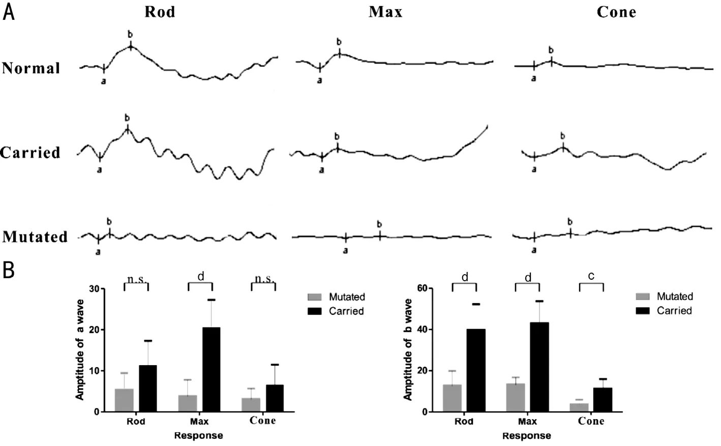
Remarkably, it was noting that a total of 66.7% (8 of 12 eyes) of eyes of female carriers exhibited patchy pigment clumping in the peripheral retina, however, in 4 of the 12 eyes with carriers,the fundus appeared fairly normal (Figure 4). Additionally,there was a dramatic reduction in rod and cone amplitude for affected males, while 83.3% of female carriers were relatively stable, within a relatively normal range (Figure 5). What’s more, 2 female carriers subjects (33.3%) had abnormal OCT findings, such as slight disruption of IS/OS or thinning of the outer retinal layer. Negative traits of OCT were detected from 4 carriers (III-5, IV-3, IV-5, IV-6). The detailed clinical features are summarized in Table 2.

A cone-rod or cone dystrophies due to mutations in the
gene, in which a truncation mutant tends to override endogenous RPGR proteins to affect photoreceptor function.Consistent with previous notion, multiple cystics and distortion of outer layers (external limiting membrane, EZ and IS/OS)were detected in the peripheral retina of affected male subjects,with relative structure of IS/OS line sparing of the macula.In terms of ERG, it showed minimal cone response and extinguished rod response. The results of all those revealed extensive loss of photoreceptors, especially in the peripheral retina rather than the macula, which could account for the remaining visual acuity and flat ERG response.
This study observed the tenets of the Declaration of Helsinki. And it was approved by the Ethics Committee of Children’s Hospital of Hebei Province. Twenty participating individuals agreed study and signed informed consent.
在公路工程建设过程中,施工单位应该坚持依法监督的原则,增强自身的诚信理念与服务意识,注重做好质量安全环节的监督与管理工作。为达到这一目标,施工单位需要注重提高质量安全监管队伍的专业素质与监管水平。因此,施工单位应该注重定期组织培训活动,既丰富监管人员的专业知识,提高其综合素质水平,在执行监管行为的过程中,要严格遵循相关规章制度,全面保障质量安全监管工作的成效。同时,施工单位可以加强文化建设,营造质量安全至上的良好精神氛围,打造公路工程的质量安全监督文化,增强施工人员和质量安全监管人员的责任感,使其自觉地采取行动,落实质量控制工作和安全监督工作,推进公路工程建设获得质的发展。
Some scholars believed that patients with XLRP who have frameshift mutations or nonsense mutations within exons 1 to 14 may develop severe clinical features, it is possibly because of this reason that truncated protein from the RPGR
variant transcript interacts relatively well with other proteins
.However, in the current study, not only mutated males but also some carried females with the W955X (c.2865G>A)mutation had a more significantly severe visual impairment,more varied fundus (including tilted optic disc, bone-spicule pigment deposition, retinal arteriole attenuation and optic disc pallor, even retinal atrophy), faster loss of ERG amplitudes and more thinning of OCT than patients with the mutations in exon 1 to 14 that described in some past reports
. In summary,we found a mutation in the RPGR
region that may have potentially more deleterious clinical features.

Meanwhile, the identical mutation was confirmed in his consanguineous brother (IV-2). He showed poor visual acuity,with mild myopia and myopic astigmatism (OD: -2.50 DS,-2.5 DC×5°=0.3; OS: -2.5 DS, -2.50 DC×175°=0.3). Myopic maculopathy, Bull’s eye, attenuated arteries and bone-spicule hyperpigmentation in retina of fundus were also discovered beginning in the first decade, accompanied by obviously flat ERG and atrophy of outer retinal bands at the (para)fovea of OCT (Figure 3D).
Based on mutagenesis analysis, an exon 15 mutation in
gene has been identified. The gene, which is located on the X chromosome and approximately accounts for 5%-15% of all cases of RP
. Commonly, multiple isoforms of RPGR were detected in functional performance. The RPGR
is one of isoforms, which consists of 15 exons coding 1152-aa protein,enriches in retina and is thought to facilitate the connecting and trafficking of rod and cone photoreceptors
. Previous analysis revealed that the RPGR
variants are associated with significantly dysfunction of retinal defects, ranging from RP to cone-rod or macular degeneration
. In the present study, nearly all the patients suffered from visual disturbance and tended to be worse with increasing age. That was consistent with previous reports
.
Compare with 4 patients, carriers showed a wide range of clinical features. ERG and fundus showed abnormalities in 16.7% and 66.7% of female carriers, respectively, which is lower than previous reports
. Additionally, normal layers of OCT were also more apparent in female carriers. Variability of phenotypic severity in female carriers is attributed to a variety of factors, such as age, environment, skewed X-activation, and genetic modifiers
. As discussed above, RP exhibits a higher degree of phenotypic heterogeneity and variability. Nanda
advocated female carriers who suffer from severe visual impairment and display a retinal phenotype should be considerate to process therapeutic intervention. Specifically,phenotypic follow-up of female carriers must be conducted for a longer period of time
. Hence, it’s significant to discover novel RP mutations, generalize phenotype and assess the process, so that patients can undergo genetic therapy treatment timely
.
In summary, we reported the clinical features of individuals with RP and carriers caused by a novel mutation (c.2865G>A p.W955X) in
, and broadened the spectrum of mutations,which is valuable for provide future genetic counselling and specific gene therapy.
(1)人类活动导致了水土流失。随着人类科学技术的不断进步,城市建设手段越来越先进,建设速度也越来越快,与此同时,不断扩张的城市范围,对自然环境造成了极大的破坏,大量的植被消失导致植物的蒸腾作用减弱,从而造成了水土流失的现象,进而使河流中的泥沙含量大幅度上升,这又导致河床高度上升,河水溢出河道造成了洪水灾害。
We thank all the individuals for participating in this project.
引理为伪BCI-代数X的一个犹豫模糊滤子的充要条件是对任意γ ∈ P([0,1]), 要么 ∅, 要么为X的滤子.
Sun HH and Chen L conceived the idea; Sun HH, Zhao JC, Yang SL, Shi JD, and Wei YS collected clinical message and performed the experiments; Sun HH, Wang JC performed data analyses; Sun HH, Gu F and Chen L wrote the manuscript. All authors approved the manuscript.
Supported by Natural Science Foundation of Hebei Province (No.H2021316006); Hebei Provincial the Ministry of Health Research Fund for Medical Sciences(No.20200638).
None;
None;
None;
None;
None;
None;
None;
None.
1 Hamel C. Retinitis pigmentosa.
2006;1:40.
2 Hartong DT, Berson EL, Dryja TP. Retinitis pigmentosa.
2006;368(9549):1795-1809.
3 Tee JJL, Smith AJ, Hardcastle AJ, Michaelides M. RPGR-associated retinopathy: clinical features, molecular genetics, animal models and therapeutic options.
2016;100(8):1022-1027.
4 Campochiaro PA, Mir TA. The mechanism of cone cell death in retinitis pigmentosa.
2018;62:24-37.
5 Zhang ZM, Dai HH, Wang L, Tao TC, Xu J, Sun XW, Yang LP, Li GL.Novel mutations of RPGR in Chinese families with X-linked retinitis pigmentosa.
2019;19(1):240.
6 Sharon D, Sandberg MA, Rabe VW, Stillberger M, Dryja TP, Berson EL.
and
mutations and clinical correlations in patients with X-linked retinitis pigmentosa.
2003;73(5):1131-1146.
7 Stone EM, Andorf JL, Whitmore SS, DeLuca AP, Giacalone JC, Streb LM, Braun TA, Mullins RF, Scheetz TE, Sheffield VC, Tucker BA.Clinically focused molecular investigation of 1000 consecutive families with inherited retinal disease.
2017;124(9):1314-1331.8 Tsang SH, Sharma T. X-linked retinitis pigmentosa.
2018;1085:31-35.
9 Vervoort R, Lennon A, Bird AC, Tulloch B, Axton R, Miano MG,Meindl A, Meitinger T, Ciccodicola A, Wright AF. Mutational hot spot within a new RPGR exon in X-linked retinitis pigmentosa.
2000;25(4):462-466.
10 Churchill JD, Bowne SJ, Sullivan LS, Lewis RA, Wheaton DK, Birch DG, Branham KE, Heckenlively JR, Daiger SP. Mutations in the X-linked retinitis pigmentosa genes RPGR and RP2 found in 8.5% of families with a provisional diagnosis of autosomal dominant retinitis pigmentosa.
2013;54(2):1411-1416.
11 Appelbaum T, Becker D, Santana E, Aguirre GD. Molecular studies of phenotype variation in canine RPGR-XLPRA1.
2016;22:319-331.
12 Chiang JPW, Lamey TM, Wang NK, Duan J, Zhou W, McLaren TL, Thompson JA, Ruddle J, De Roach JN. Development of highthroughput clinical testing of RPGR ORF15 using a large inherited retinal dystrophy cohort.
2018;59(11):4434-4440.
13 Levy SE, Boone BE. Next-generation sequencing strategies.
2019;9(7):a025791.
14 Zhang QH, Giacalone JC, Searby C, Stone EM, Tucker BA, Sheffield VC. Disruption of RPGR protein interaction network is the common feature of RPGR missense variations that cause XLRP.
2019;116(4):1353-1360.
15 Hong DH, Pawlyk BS, Adamian M, Sandberg MA, Li TS. A single,abbreviated RPGR-ORF15 variant reconstitutes RPGR function
.
2005;46(2):435-441.
16 Koyanagi Y, Akiyama M, Nishiguchi KM,
. Genetic characteristics of retinitis pigmentosa in 1204 Japanese patients.
2019;56(10):662-670.
17 Kurata K, Hosono K, Hayashi T, Mizobuchi K, Katagiri S, Miyamichi D, Nishina S, Sato M, Azuma N, Nakano T, Hotta Y. X-linked retinitis pigmentosa in Japan: clinical and genetic findings in male patients and female carriers.
2019;20(6):E1518.
18 Megaw RD, Soares DC, Wright AF. RPGR: Its role in photoreceptor physiology, human disease, and future therapies.
2015;138:32-41.
19 Breuer DK, Yashar BM, Filippova E,
. A comprehensive mutation analysis of
and
in a North American cohort of families with X-linked retinitis pigmentosa.
2002;70(6):1545-1554.
20 Fahim AT, Bowne SJ, Sullivan LS, Webb KD, Williams JT, Wheaton DK, Birch DG, Daiger SP. Allelic heterogeneity and genetic modifier loci contribute to clinical variation in males with X-linked retinitis pigmentosa due to RPGR mutations.
2011;6(8):e23021.
21 Sandberg MA, Rosner B, Weigel-Difranco C, Dryja TP, Berson EL.Disease course of patients with X-linked retinitis pigmentosa due to RPGR gene mutations.
2007;48(3):1298-1304.
22 Wu ZJ, Hiriyanna S, Qian HH, Mookherjee S, Campos MM, Gao C, Fariss R, Sieving PA, Li TS, Colosi P, Swaroop A. A long-term efficacy study of gene replacement therapy for RPGR-associated retinal degeneration.
2015;24(14):3956-3970.
23 Sheng XL, Li ZL, Zhang XF, Wang J, Ren HW, Sun YB, Meng RH,Rong WN, Zhuang WJ. A novel mutation in retinitis pigmentosa GTPase regulator gene with a distinctive retinitis pigmentosa phenotype in a Chinese family.
2010;16:1620-1628.
24 Jones BW, Pfeiffer RL, Ferrell WD, Watt CB, Marmor M, Marc RE. Retinal remodeling in human retinitis pigmentosa.
2016;150:149-165.
25 Sanchez Tocino H, Diez Montero C, Villanueva Gómez A, Lobo Valentin R, Montero-Moreno JA. Phenotypic high myopia in X-linked retinitis pigmentosa secondary to a novel mutation in the RPGR gene.
2019;40(2):170-176.
26 Talib M, van Schooneveld MJ, Van Cauwenbergh C,
. The spectrum of structural and functional abnormalities in female carriers of pathogenic variants in the RPGR gene.
2018;59(10):4123-4133.
27 Nanda A, Salvetti AP, Clouston P, Downes SM, MacLaren RE.Exploring the variable phenotypes of RPGR carrier females in assessing their potential for retinal gene therapy.
(
)2018;9(12):E643.
28 Cehajic Kapetanovic J, McClements ME, Martinez-Fernandez de la Camara C, MacLaren RE. Molecular strategies for
gene therapy.
(
) 2019;10(9):674.
29 Cehajic-Kapetanovic J, Xue K, Martinez-Fernandez de la Camara C,
. Initial results from a first-in-human gene therapy trial on X-linked retinitis pigmentosa caused by mutations in RPGR.
2020;26(3):354-359.
30 Giacalone JC, Andorf JL, Zhang QH,
. Development of a molecularly stable gene therapy vector for the treatment of
-associated X-linked retinitis pigmentosa.
2019;30(8):967-974.
猜你喜欢
党员生活·下(2020年5期)2020-07-04
财经(2017年2期)2017-03-10
建筑建材装饰(2016年13期)2017-01-04
财经(2016年15期)2016-06-03
财经(2016年3期)2016-03-07
财经(2016年6期)2016-02-24
建筑工程技术与设计(2015年29期)2015-10-21
上海人大月刊(2015年8期)2015-09-10
 International Journal of Ophthalmology2022年9期
- International Journal of Ophthalmology的其它文章
- Clinical analysis of bilateral acute depigmentation of the iris: first reported case in China
- Interferon-gamma release assays in tuberculous uveitis:a comprehensive review
- Ocular stem cells: a narrative review of current clinical trials
- Different compression sutures combined with intracameral air injection for acute corneal hydrops
- Comparison of intravitreal aflibercept and dexamethasone implant in the treatment of macular edema associated with diabetic retinopathy or retinal vein occlusion: a Meta-analysis and systematic review
- Reproducibility of macular perfusion parameters in nonproliferative diabetic retinopathy patients by two different OCTA sweep modes