
Morphological changes in the iridocorneal angle and their relationship with intraocular pressure after infantile cataract surgery
2022-09-14 06:50DanDanWangZhangLiangLiBingZhangZiYiLuWeiChenGuanYunZhao
INTRODUCTION
All surgeries were performed by the same surgeon under general anesthesia.
Gonioscopy is the standard examination technique used to observe the iridocorneal angle structure and provide an overview of the detectable pathological changes in the eyes of children with congenital cataract. Previously
, we have shown that the iridocorneal angle of children with cataracts is open, accompanied by a small amount of pigment and iris process preoperatively, and that the proinflammatory state may be maintained for a long time postoperatively. However,it remains unknown if the aphakic status can lead to structural changes in the iridocorneal angle.
Therefore, the purpose of this study was to investigate the changes in the iridocorneal angle using a gonioscope after primary infantile congenital cataract surgery, and to explore the relationship of morphological changes and intraocular pressure(IOP).
对缺少授粉树,只开花不结果的园,结合开张角度,在大树上直接高接换头。接穗应注意早、中、晚熟品种搭配,或花期一致的品种,选择秦樱一号、先锋、拉宾斯为授粉品种。在3月中旬或9月上旬嫁接为宜,采用双芽切接、方块芽接和“T”字芽接,或将2~4个芽的花枝作接穗进行改良嫁接。接穗成活后,在秋季或次年拉枝,促其开花结果。
事实证明,日常多行善,出门总会遇到贵人。在当天晚上以火锅补充了体力之后,我遇到的贵人就是旅店的老板。这位生在广东梅州的客家人在河北和内蒙接壤的草原上已经享受了多年由越野车带来的乐趣,他车库里两辆伤痕累累的丰田兰德酷路泽似乎也在暗示着他在这里的丰富阅历。出于对全新AMG G 63的好奇,他主动提出成为我们的向导,我们所要付出的代价不过是摄影师在必要时指导他如何拍下一组美丽的雪景而已。
SUBJECTS AND METHODS
All procedures were conducted following the tenets of the Declaration of Helsinki and were approved by the institutional ethics committee (clinical trial number:NCT03778086). Informed consent was obtained from the children’s caregivers.
流坑村的主体,基本保存明后期整治修缮后的仿里坊制的平面格局,依地形地貌,建宗祠、造书院、修街市、筑庙宇、树门楼,基本涵盖我国古代建筑的基本类型。以青砖灰瓦、高马头墙、双坡屋顶为主体建筑形式;楼阁、牌坊多建成飞檐翘角,古典轩昂,构成了富有地方特色的赣派民居。流坑民居的建筑装饰手法,采用木雕、石雕、灰塑、彩画、墨绘和匾额七类,往往雕、塑、绘、书并用,内容丰富、题材多样,使建筑富丽堂皇,富有文化艺术内涵。
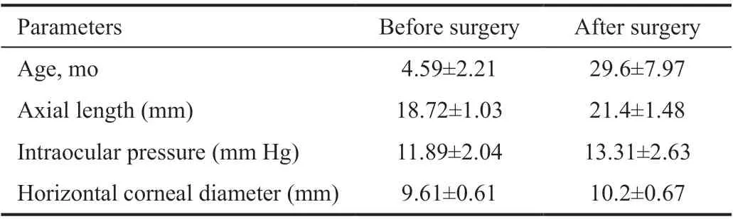
This was a prospective observational study, including 64 eyes of 41 children who underwent congenital cataract surgery (23-gauge lensectomy and anterior vitrectomy) at the Eye Hospital of Wenzhou Medical University, Hangzhou,China from March 2016 to January 2018, with an average age of 4.59±2.21mo. All the children were implanted with IOL in 2y, with an average age of 29.6±7.97mo, and were followed up for 1y after secondary IOL implantation.
Even with the development of modern microsurgical techniques and equipment, complications following pediatric cataract surgery still remain challenging
. The Infant Aphakia Treatment Study (IATS) found that common intraocular complications include visual axis opacification,glaucoma, and intraocular lens (IOL) exchange
. Glaucoma is a major sight-threatening complication of pediatric cataract surgery that can occur from months to decades after surgery
. According to the IATS, the risk of glaucoma increases from 12% one year after the operation to 24% after 5y
. Although several risk factors, such as age at time of cataract surgery, surgical technique used, microphthalmia,persistent fetal vasculature (PFV), and duration of follow-up,have been reported, convincing pathophysiological evidence for glaucoma remains unavailable. Some authors have proposed the collapse of the Schlemm’s canal and decreased outflow of the aqueous humor in post-lensectomy patients with glaucoma
.
二是人口增长因素。有利面是我国人口增长速度得到了有效控制,节水理念得到了一定程度的普及,减轻了资源压力;人口空间流动改善了部分地区的资源过载状况。不利面是人口持续增长,农田灌溉系数仍较低,城市人口舒适性需求持续提升,水资源缺口依然很大。
All patients underwent two indirect gonioscopic examinations(Goldmann 2-mirror lens, VOLK, USA). The first examination was performed before primary lens removal (pre-surgery group), and the second examination (post-surgery group) was performed before secondary IOL implantation.
在女主人公的生活故事中,构成两性关系影响的男人有五位,他们是剑、李木、子墨、老师和“他”(抑或“金岳霖”)。这些男子分作两组,与女主人公形成两个“多角恋”关系。第一个多角恋即构成了“江山娇”叙述线。其恋情关系发生在江山娇与剑、李木和老师之间,时间在江山娇与李木结婚前。最终以江山娇与李木结婚而告结。第二个多角恋构成了“伊一”叙述线。其恋情关系在伊一与子墨、李木和“他”之间进行。这个恋情是无解的,乃至一直到伊一绝尘而去。
All patients underwent a comprehensive ophthalmological examination, including slit-lamp bio-microscopy, dilated fundus examination, A/B-scan ultrasound examination (Quantel Medical, Cournon-d’Auvergne,France), and estimation of IOP (iCare, Vantaa, Finland). Data on the axial length (AL), corneal diameter, age of surgery, and IOP were collected before each operation. IOPs were recorded at 1, 3, 6, and 12mo after the operation.
All gonioscopy examinations were performed before surgery. The iridocorneal angle was graded for each quadrant separately,according to the Shaffer and Spaeth classification system
.The iris insertion was defined as follows: 1) anterior to Schwalbe’s line; 2) between Schwalbe’s line and scleral spur;3) scleral spur visible; 4) deep with ciliary body visible; 5)extremely deep with >1 mm ciliary body band (CBB). The description of iris process insertion was defined as follows:to CBB; to trabeculae; to Schwalbe’s line
. The number of iris process was defined as follows: 0, none; 1, minimal; 2,moderate; 3, marked. Table 1 shows the numerical method for classifying the various features of the iridocorneal angle was based on our previous research month
. Two experienced pediatric ophthalmologist (Wang DD and Zhao YE) graded by reviewing the surgery videos and the iridocorneal angle photographs. Any disagreement was resolved through discussion by the two ophthalmologists.
Primary surgery, which combined 23-gauge lensectomy and anterior vitrectomy using the CENTURION system (Alcon Laboratories, Inc., USA), was performed under general anesthesia by the same surgeon for all patients (Zhao YE). The vacuum was set at 350 mm Hg with a cut rate of 2000 per minute, as previously reported
.No intraoperative complications were noted. At the end of surgery, all eyes received subconjunctival injections of 0.2 mL dexamethasone. Postoperative topical treatment was as follows: levofloxacin eye drops (0.5%, Santen, Japan) four times a day for 2wk; tobramycin dexamethasone eye drops(Alcon, USA) four times a day for 4wk, with dose reduction once a week; and compound tropicamide (Santen, Japan) once a day for 4wk.
Secondary surgery for IOL implantation was considered when the children with unilateral cataract were approximately 1.5 years old and those with bilateral cataract were approximately 2 years old. The capsular bag was reopened with auxiliary instruments, ophthalmic viscosurgical devices or a 23-gauge cutting head, and then the hyperplastic cortex in the capsular bag was removed by automated irrigation/aspiration (Figure 1).The IOL was implanted in the capsular bag for 59 eyes, while the IOL was implanted in the sulcus combined with peripheral iridectomy for 5 eyes.
All data analyses were analyzed using SPSS software, version 26.0 (SPSS Inc., Chicago, Illinois,USA). Shapiro-Wilk test is used to check the normality of continuous variables. If it meets the normality, it is expressed as the mean±standard deviation, and if it does not meet the normality, it is expressed as the median interquartile range(IQR); Categorical variables are expressed as frequency and percentage. The difference between the two groups before and after operation was compared by generalized estimation equation (GEE), and the relationship between IOP and iris angle was analyzed; Categorical variables were compared between preoperative and postoperative groups by generalized linear mixed model (GLMM).
<0.05 was considered statistically significant.


RESULTS
Sixty-four eyes from 41 children (19 boys and 22 girls),including 18 with unilateral cataracts (18 eyes) and 23 with bilateral cataracts (46 eyes), were included in the analysis.Table 2 presents the demographic data of the patients.
路径的经济性受多种因素的影响,而各因素并非一成不变,如运价会随着燃料价格、政策等的变化而变化;受内部或外界的影响,货主集装箱运输需求也会波动.本文选取集装箱运输需求量和运价对路径经济性的影响进行分析.
Another important finding in this study was that the trabecular meshwork pigmentation was obviously increased after surgery, which was consistent with the findings of many cases of postoperative glaucoma. Phelps and Arafat
examined and treated 18 patients who developed open angle glaucoma after an operation for congenital cataract and noted that gonioscopy revealed a heavily pigmented uveal meshwork and an anteriorly inserted iris root. Walton
observed that many patients who had abnormal pigment deposition may also have micro PAS. Asrani and Wilensky
reported that the only abnormal finding in patients with open angle glaucoma after congenital cataract surgery was increased pigmentation of the trabecular meshwork. Therefore, we suspect that one of the causes of secondary glaucoma is closely related to trabecular pigmentation caused by surgery in children with congenital cataract.
We found that all 64 eyes had a wide and open iridocorneal angle before and after surgery, visible CBB was observed in 60 eyes before and 61 eyes after surgery, and there was no significant difference in the iridocorneal angle width in every quadrant after surgery. A multicenter randomized controlled trial showed that glaucoma was mainly of the open-angle kind(19/20 cases, 95%) after congenital cataract, when patients were 1-6 months old at the time of the surgery
. Asrani and Wilensky
evaluated 64 eyes from 48 cases treated for glaucoma after congenital cataract surgery and found that 51 eyes (79.7%) were diagnosed with open angle glaucoma.Kirwan
retrospectively reviewed cases of congenital cataract surgery and found that the angles were open in the cases in which gonioscopy was performed. The results from these previous studies are consistent with those from our prospective study.
The mean preoperative IOP was 11.89±2.04 mm Hg (range 6-16 mm Hg). At 1mo after surgery,IOP showed a statistically significant increase relative to that observed before surgery (13.51±2.20
11.89±2.04 mm Hg,
<0.001), and the highest IOP was 24 mm Hg. The IOP decreased to a level close to the preoperative level at 3mo postoperatively (at 3mo, 12.34±1.90 mm Hg
preoperative,11.89±2.04 mm Hg,
=0.183), and it gradually increased in the following months. The mean IOP at 6 and 12mo was significantly higher than that before the operation (12.62±1.70 and 13.48±2.63 mm Hg, respectively,
<0.05).


The change trend of IOP after two operations is similar. IOP increased gradually at 1mo after IOL implantation (preoperative,13.31±2.63 mm Hg
at 1mo 15.51±4.89 mm Hg,
<0.0001),and returned to the preoperative level at 3mo (at 3mo,13.18±3.90 mm Hg
preoperative, 13.31±2.63 mm Hg,
=0.95),and increased gradually at 6mo and 12mo (14.70±2.75 mm Hg and 15.96±3.26 mm Hg, respectively,
<0.05).
The increase in trabecular meshwork pigmentation and anterior insertion of iris process were positively correlated with the elevated IOP after primary surgery (
²=0.1005,
=0.0265 and
²=0.1052,
=0.0230,respectively; Figure 5).
Table 3 shows the comparison of the changes in the iridocorneal angle between the pre-surgery and post-surgery groups. All 64 eyes had a wide and open iridocorneal angle before and after surgery. A visible CBB was observed in 60 eyes before surgery and in 61 eyes after surgery, with a significant increase in both the superior and inferior quadrant of CBB (
<0.05 for both) at the second examination compared to that at the first examination.Further, at the second examination, the trabecular meshwork pigments (Figure 2) and the number of iris processes (Figure 3)in every quadrant of the iridocorneal angle were significantly increased relative to those at the first examination (
<0.05 for both). However, there was no significant difference in the iridocorneal angle width, iris insertion, or iris process insertion between the two groups. Moreover, there was no significant difference in the iridocorneal angle structure in the two groups between the two examinations.
DISCUSSION
In this study, we performed gonioscopy as a routine examination to observe the changes in the iridocorneal angle in infantile cataracts. To control the influence of surgical procedures on the results obtained, all patients underwent standardized surgery performed by the same surgeon.The main outcomes of this study were that the trabecular meshwork pigments and the number of iris processes increased significantly in every quadrant of the iridocorneal angle after the primary operation, along with an increase in the width of CBB in the superior and inferior quadrants.
The inclusion criteria were as follows: the patient had pediatric cataract with obvious visual opacity (>3-mm central opacity)and the surgical technique performed was lensectomy with limited anterior vitrectomy, without primary IOL implantation.The exclusion criteria were as follows: gestational age at birth <37wk, preoperative glaucoma, high risk of glaucoma(
, a known family history of glaucoma, systemic or topical steroid use before surgery), severe PFV, chronic anterior uveitis, removal of visual axis opacification before secondary IOL implantation, previous ocular surgery, and a history of ocular trauma.
Moreover, we found that the CBB width was wider in the vertical meridian (in the superior and inferior quadrant)postoperatively. Most previous studies have reported that the anterior chamber angle and anterior chamber depth increased after routine cataract surgery in adults
. Petermeier
evaluated 50 pseudophakic eyes in adults and found that the vertical angle-to-angle diameter (AAD) was significantly larger than the horizontal diameter when measured with a very high frequency ultrasound. Baikoff
found that in the 89 adult phakic eyes studied, 74% of the eyes had a vertical AAD at least 100 μm larger than the horizontal diameter as evaluated using the anterior segment ocular coherence tomography system. Our results are consistent with the results of these studies performed in adults.

We hypothesized that the anterior chamber of infants was oval, which was similar to that of adults. Cataract removal may be the reason for releasing all centripetal forces, which led to an increase in the vertical meridian of the AAD relative to the horizontal meridian. Additionally, young infants with fragile eyeballs may experience mild leakage after surgery due to crying, which may lead to anterior iris synechia around incisions, particularly around the two side paracentesis positions, resulting in a narrower horizontal CBB. Therefore, it is very important to suture the incisions and completely avoid incision leakage in the case of pediatric surgery.
In the postsurgery examination, peripheral anterior mini-synechia(PAS) was observed in four eyes by gonioscopy examination.Synechia were also observed in the nasal or temporal clear corneal incisions (8-10 or 2-4 o’clock), and goniosynechialysis was performed during the procedure for the secondary IOL implantation (Figure 4). One eye was observed to have multiple(more than one at different locations) PAS. Thus, peripheral iridectomy combined with goniosynechialysis was performed during the secondary IOL implantation surgery. Small cortical debris in the iridocorneal angle were observed in three eyes.
从表4可知,在3个分布区地枫皮叶片上表皮单位面积的细胞密度数值相差不大,且上表皮细胞的长、宽也均较接近,说明地枫皮叶片上表皮细胞发育较为稳定。而下表皮细胞特征差异较为明显,下表皮细胞密度大小为平果>马山>靖西,下表皮细胞的大小也相应发生变化,即靖西>马山>平果。
In addition, we found that the number of iris processes increased significantly after surgery. Kimura and Levene
reported that more iris processes and more trabecular pigmentation were found in primary open angle glaucoma in patients over the age of 40y. They believed that a congenital angle anomaly was an important factor in the pathogenesis of the elevated IOP. Similar results were also found in our study.We found that the insertion of the iris process became denser after the primary operation, and the marker pigment attached to the iris process forming a “pseudo-membrane.” We speculate that the “pseudo-membrane” may subsequently develop a“pseudoangle”, which is an angle lined by a spot of peripheral anterior synechiae, but seemingly open under the gonioscope.Perry
presented histopathologic findings showing that these anatomical derangements and pseudomembranous were the causes of goniosurgery failure in the newborn glaucoma treatment. Their results supported our postulation that the iridocorneal angle of the paired normal eye of children with unilateral cataract remained unchanged with the extension of follow-up time. This finding explained that the iridocorneal angle change was closely related to the surgery. Similarly,Haargaard
found that none of the children with pediatric cataract who had never undergone surgery had subsequent development of glaucoma. Therefore, we are convinced that the surgery changed the morphology of the iridocorneal angle.Our study indicated that an increase in the number of trabecular meshwork pigments and iris processes was associated with a proinflammatory state. This has been demonstrated in our previous study, which showed a significant increase in proinflammatory cytokine levels in the aqueous humor after congenital cataract surgery
. Some tiny crystalline cortex debris were found in the iridocorneal angle, which came from the hyperplastic cortex. It can not only reduce the outflow facility of the iridocorneal angle but also lead to a microinflammatory state.
Our results showed that IOP gradually increased at 1mo after surgery, both primary and secondary surgery. Some previous studies have reported IOP elevation during the early postoperative period in children and adolescents
.Researchers have suspected that normal trabecular meshwork cells are likely to be infected by the presence of lens epithelial cells, followed by changes in their structural features as well as protein and gene expressions
. However, in the present study, we speculated that it might be due to the corticosteroidinduced mechanisms, which were related to the use of steroid eye drops after surgery, such as dexamethasone, because the IOP decreased gradually after the administration of steroid eye drops was tapered or stopped, and at 3mo post-operatively,the IOP decreased to the preoperative level. Steroid eye drops can reduce the intraocular inflammation, thereby reducing the risks of synechiae formation and angle closure. Therefore, we preferred to use high-efficiency steroid eye drops in the early stage after congenital cataract surgery. With the reduction of inflammation, steroid eye-drop administration should be stopped in time or replaced by that with less potency to prevent the early postoperative IOP increase.
Further, we found that the increase in trabecular meshwork pigmentation and anterior insertion of the iris process were positively correlated with IOP elevation. With the extension of the follow-up duration, the IOP of 6 and 12mo after surgery increased significantly (14.70±2.75 and 15.96±3.26 mm Hg,respectively). Although, there were no glaucoma cases were found in our study, based on our gonioscopy examinations findings, we suggest that more attention should be paid to monitoring the IOP in the future.
我跟乔振宇结婚至今已满四年三个月又两个星期,可每每俩人擦枪走火起争执时,他挂在嘴边那句亘古不变的抱怨仍旧是——我乔振宇堂堂一个大丈夫,当初怎么就猪油蒙心找了你这个油盐不进的“三等女”了呢?
This study has several limitations. First, our study previously excluded eyes with severe PFV, which were reported as a great risk of secondary glaucoma
. Second, this study did not include the long-term follow-up results concerning changes in the IOP after secondary IOL implantation. Thus,a low incidence of glaucoma is expected. However, based on the strict criteria used in this study, this is an ideal method to compare the effects of surgery on the iridocorneal angle and due to the difficulty in infantile gonioscopy, this study lacks age-matched normal children as a comparative group. Finally,the subjective numerical method used in this study is also one of the limitations.
In conclusion, this prospective study revealed the changes in the iridocorneal angle after cataract surgery in infants,including an increase in the trabecular meshwork pigmentation and the number of iris processes. Furthermore, the increase in trabecular meshwork pigmentation and anterior insertion of iris process were positively correlated with the elevated IOP after surgery.
Supported by the National Natural Science Foundation of China (No.81870680); National Science Foundation of Zhejiang Province (No.LQ20H120002).
None;
None;
None;
None;
None;
None.
1 Haargaard B, Ritz C, Oudin A, Wohlfahrt J, Thygesen J, Olsen T,Melbye M. Risk of glaucoma after pediatric cataract surgery.
2008;49(5):1791-1796.
2 Yagev R, Khatib N, Barrett C, Lior Y, Lifshitz T, Tsumi E. Intraocular lens implantation as an isolated risk factor for secondary glaucoma in pediatric patients.
2019;54(5):621-625.
3 Wang JH, Chen JJ, Chen W,
. Incidence of and risk factors for suspected glaucoma and glaucoma after congenital and infantile cataract surgery: a longitudinal study in China.
2020;29(1):46-52.
4 Zhao QH, Zhao YE. Commentary review: challenges of intraocular lens implantation for congenital cataract infants.
2021;14(6):923-930.
5 Plager DA, Lynn MJ, Buckley EG, Wilson ME, Lambert SR; Infant Aphakia Treatment Study Group. Complications in the first 5 years following cataract surgery in infants with and without intraocular lens implantation in the Infant Aphakia Treatment Study.
2014;158(5):892-898.
6 Bothun ED, Wilson ME, Vanderveen DK,
. Outcomes of bilateral cataracts removed in infants 1 to 7 months of age using the toddler aphakia and pseudophakia treatment study registry.
2020;127(4):501-510.
7 Phelps CD, Arafat NI. Open-angle glaucoma following surgery for congenital cataracts.
1977;95(11):1985-1987.
8 Kirwan C, Lanigan B, O’Keefe M. Glaucoma in aphakic and pseudophakic eyes following surgery for congenital cataract in the first year of life.
2010;88(1):53-59.
9 Liu ZZ, Lin HT, Jin GM,
. In-the-bag versus ciliary sulcus secondary intraocular lens implantation for pediatric aphakia: a prospective comparative study.
2022;236:183-192.
10 Zhang ZH, Fu YN, Wang JJ, Ji XP, Li ZL, Zhao YY, Chang PJ, Zhao YE. Glaucoma and risk factors three years after congenital cataract surgery.
2022;22(1):118.
11 Freedman SF, Beck AD, Nizam A,
, Group IATS. Glaucomarelated adverse events at 10 years in the infant aphakia treatment study: a secondary analysis of a randomized clinical trial.
2021;139(2):165-173.
12 Freedman SF, Lynn MJ, Beck AD, Bothun ED, Örge FH, Lambert SR,Group IATS. Glaucoma-related adverse events in the first 5 years after unilateral cataract removal in the infant aphakia treatment study.
2015;133(8):907-914.
13 Daniel MC, Dubis AM, Theodorou M,
. Childhood lensectomy is associated with static and dynamic reduction in schlemm canal size.
2019;126(2):233-241.
14 Wang DD, Li ZL, Zhang F, Zhang YJ, Zhao YY, Chang PJ, Fu YN,Zhao YE. Iridocorneal angle and anterior segment structure of eyes in children with cataract.
2020;63(2):194-202.
15 Zhao YY, Deng XH, Chang PJ, Hu M, Li ZL, Zhang F, Ding XX, Zhao YE. Expression profiles of inflammatory cytokines in the aqueous humor of children after congenital cataract extraction.
2020;9(8):3.
16 Shaffer RN. Primary glaucomas. gonioscopy, ophthalmoscopy and perimetry.
1960;64:112-127.
17 Spaeth GL. The normal development of the human anterior chamber angle: a new system of descriptive grading.
(
) 1971;91:709-739.
18 Kimura R, Levene RZ. Gonioscopic differences between eyes with primary open-angle glaucoma and normal eyes in subjects over the age of forty.
1975;73:74-85.
19 Li ZL, Chang PJ, Wang DD, Zhao YY, Hu M, Ding XX, Yu LQ, Zhao YE. Morphological and biometric features of preexisting posterior capsule defect in congenital cataract.
2018;44(7):871-877.
20 Asrani SG, Wilensky JT. Glaucoma after congenital cataract surgery.
1995;102(6):863-867.
21 Shin HC, Subrayan V, Tajunisah I. Changes in anterior chamber depth and intraocular pressure after phacoemulsification in eyes with occludable angles.
2010;36(8):1289-1295.
22 Lee HB, Zukaite I, Juniat V, Dimitry ME, Lewis A, Nanavaty MA.Changes in symmetry of anterior chamber following routine cataract surgery in non-glaucomatous eyes.
(
) 2019;6:19.
23 Petermeier K, Suesskind D, Altpeter E, Schatz A, Messias A, Gekeler F, Szurman P. Sulcus anatomy and diameter in pseudophakic eyes and correlation with biometric data: evaluation with a 50 MHz ultrasound biomicroscope.
2012;38(6):986-991.
24 Baikoff G, Jitsuo Jodai H, Bourgeon G. Measurement of the internal diameter and depth of the anterior chamber: IOLMaster versus anterior chamber optical coherence tomographer.
2005;31(9):1722-1728.
25 Walton DS. Pediatric aphakic glaucoma: a study of 65 patients.
1995;93:403-413; discussion 413-420.
26 Perry LP, Jakobiec FA, Zakka FR, Walton DS. Newborn primary congenital glaucoma: histopathologic features of the anterior chamber filtration angle.
2012;16(6):565-568.
27 Solebo AL, Rahi JS, Group BCCI. Glaucoma following cataract surgery in the first 2 years of life: frequency, risk factors and outcomes from IoLunder2.
2020;104(7):967-973.
28 Michael I, Shmoish M, Walton DS, Levenberg S. Interactions between trabecular meshwork cells and lens epithelial cells: a possible mechanism in infantile aphakic glaucoma.
2008;49(9):3981-3987.
29 Daniel MC, Mohamed-Noriega J, Petchyim S, Brookes J.Childhood glaucoma: long-term outcomes of glaucoma drainage device implantation within the first 2 years of life.
2019;28(10):878-883.
30 Nihalani BR, VanderVeen DK. Long-term outcomes of secondary intraocular lens implantation in children.
2022;260(5):1733-1739.
猜你喜欢
歌海(2021年2期)2021-06-22
哈哈画报(2021年10期)2021-02-28
保健与生活(2020年14期)2020-07-27
祝您健康·文摘版(2019年4期)2019-06-11
美文(2016年23期)2017-03-22
南都娱乐周刊(2016年48期)2017-01-11
南都娱乐周刊(2016年48期)2017-01-11
饮食科学(2016年3期)2016-07-04
第二课堂(课外活动版)(2015年6期)2015-10-21
建筑遗产(2014年1期)2014-10-21
 International Journal of Ophthalmology2022年9期
- International Journal of Ophthalmology的其它文章
- Clinical analysis of bilateral acute depigmentation of the iris: first reported case in China
- Interferon-gamma release assays in tuberculous uveitis:a comprehensive review
- Ocular stem cells: a narrative review of current clinical trials
- Different compression sutures combined with intracameral air injection for acute corneal hydrops
- Comparison of intravitreal aflibercept and dexamethasone implant in the treatment of macular edema associated with diabetic retinopathy or retinal vein occlusion: a Meta-analysis and systematic review
- Reproducibility of macular perfusion parameters in nonproliferative diabetic retinopathy patients by two different OCTA sweep modes