
缺氧诱导因子-1α对脂多糖所致心肌细胞线粒体功能损伤的影响
2012-09-28 06:12:26杨乐邹晓静
中华急诊医学杂志
2012年8期
关键词:检测
杨乐 邹晓静

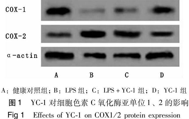

【摘要】目的 探讨心肌细胞缺氧诱导因子-1α(hypoxia-inducible factor-1α, HIF-1α)对脂多糖(lipopolysaccharide, LPS)诱导心肌细胞线粒体功能损伤的影响。方法 培养新生乳鼠心肌细胞,随机(随机数字法)分为4组。① C组:常规心肌细胞培养,不予任何处理;②LPS组:给予1μg/ml LPS,培养2 h;③YC-1+LPS组:预给HIF-1α特异性抑制剂YC-1 4 μmol/L培养8 h,再加入1μg/ml LPS,培养2 h;④YC-1组:仅给予4 μmol/L YC-1培养8h。利用荧光分光光度计测定线粒体膜电位(mitochondrial membrane potential, MMP) ,荧光素酶法测定细胞ATP含量,比色法测定细胞色素C氧化酶(cytochrome c oxidase, COX)活性,Western blot测定细胞色素氧化酶亚单位1,2的蛋白表达水平。结果 与C组相比,LPS显著抑制心肌细胞MMP、COX活性,ATP含量也显著降低(P<0.05),并抑制COX-1蛋白表达(P<0.05),上调COX-2蛋白表达(P<0.05)。与LPS组相比,HIF-1α抑制剂YC-1能进一步降低心肌细胞MMP、COX活性及ATP含量(P<0.01),显著增加COX-1表达并使COX-2表达的下调(P<0.01)。结论 HIF-1α抑制剂YC-1可能通过改变COX-1及COX-2表达,加重LPS所致的心肌细胞线粒体功能的损伤,提示HIF-1α对LPS诱导的心肌细胞线粒体损伤可能具有保护作用。
【关键词】脂多糖;缺氧诱导因子-1α;心肌细胞;线粒体
Effects of HIF-1α on mitochondrial function of cardiomyocytes stimulated by lipopolysaccharideYANG Le, ZOU Xiao-jing* . *Department of Anesthesiology, Union Hospital of Tongji Medical College, Huazhong University of Science and Technology, Wuhan 430022, China
Corresponding author: ZOU Xiao-jing, Email: xjzou68@yahoo.com
【Abstract】Objective To investigate the effects of HIF-1α, on mitochondrial function of cardiomyocytes stimulated by lipopolysaccharide (LPS). Methods Cultured neonatal cardiomyocytes were randomly (random number) distributed to four groups (n=5). Control group: the neonatal cardiomyocytes were cultured in normal condition without any treatmeat; LPS group: cardiomyocytes were exposed to 1μg/ml LPS for 2 h; YC-1+LPS group: cardiomyocytes were exposed to 4 μmol/L YC-1 for 8 h before LPS treatment; YC-1 group: cardiomyocytes were exposed to 4μmol/L YC-1 for 8 h. As indexes of mitochondrial function, MMP, the content of ATP and the activity of cytochrome oxidase were measured, COX1/2 protein expression was determined by Western blotting. Results Compared with control group, LPS decreased MMP, the content of ATP and the activity of COX obviously(P<0.05), depressed the expression of COX-1 protein (P<0.05), induced the expression of COX-2 protein (P<0.05). Compared with the LPS group, MMP, the content of ATP and the activity of COX were markedly attenuated (P<0.01), the expression of COX-1 was significantly increased and the expression of COX-2 was decreased in theYC-1+LPS group (P<0.01).Conclusions YC-1 attributes to the dysfunction of mitochondrial of myocyte by alterating the expression of COX-1 and 2, HIF-1α may attenuate the mitochondrial dysfunction of cadiomyocytes stimulated by LPS.
【Key words】Lipopolysaccharide; Hypoxia inducible factor-1α; Cardiomyocyte; Mitochondrial
脓毒症是感染所致的全身炎症反应综合征,是多器官功能障碍的重要原因[1]。脓毒症患者通常伴有心功能障碍,包括收缩性和舒张性功能障碍,有研究表明脓毒症大鼠心肌出现力学和酶学双重改变[2-3]。目前研究认为脓毒症心肌抑制可能与线粒体呼吸功能障碍及氧化磷酸化功能受损有关[4]。本课题组前期实验发现,脂多糖(LPS)可诱导原代培养乳鼠心肌细胞缺氧诱导因子-1α(hypoxia-inducible factor-1α, HIF-1α)表达明显上调,但HIF-1α与脓毒症心肌抑制的关系尚未可知。HIF-1α可调控与线粒体分子氧利用有关的多个代谢途径,本实验拟建立脓毒症心肌抑制的细胞模型,利用HIF-1α抑制剂YC-1研究HIF-1α对脓毒症心肌细胞线粒体功能的影响。……
登录APP查看全文
猜你喜欢
中国设备工程(2022年12期)2022-07-11 04:33:00
海峡科技与产业(2016年3期)2016-05-17 04:32:12
