
Transplanting neural progenitors to build a neuronal relay across the injured spinal cord
2014-06-01 09:42ChristopherHaas,ItzhakFischer
中国神经再生研究(英文版) 2014年12期
Transplanting neural progenitors to build a neuronal relay across the injured spinal cord
Cellular transplantation for repair of spinal cord injury is a promising therapeutic strategy that includes the use of a variety of neural and non-neural cells isolated or derived from embryonic and adult tissue as well as embryonic stem cells and induced pluripotent stem cells. In particular, transplants of neural progenitor cells (NPCs) have been shown to limit secondary injury and scar formation and create a permissive environment in the injured spinal cord through the provision of neurotrophic molecules and growth supporting matrices that promote growth of injured host axons. Importantly, transplants of NPC are unique in their potential to replace lost neural cells - including neurons, astrocytes, and oligodendrocytes - critical for reconstruction of the normal microenvironment of the spinal cord and restoration of connectivity and function. Different NPC preparations have been used for transplantation experiments in multiple, diverse models of SCI, ranging from the classical work with fetal spinal cord (FSC) that de fi ned the optimal age for donor tissue and its capacity to generate neural cells (Reier et al., 1983; Reier et al., 1986), to studies that demonstrated the formation of a neuronal relay by NPC transplants across the injured spinal cord to reconnect the interrupted sensory system (Bonner et al., 2011). At the developmental stage of embryonic day (E)13.5-14 the FSC is composed primarily of neuronal restricted progenitors (NRP) and glial restricted progenitors (GRP), but also contains a small number of immature neural stem cells, neurons, endothelial cells and fibroblasts (Kalyani et al., 1998; Cai et al., 2002; Lepore and Fischer, 2005; Medalha et al., 2014). While grafts of acutely isolated FSC contain all of these cell subpopulations, the process of isolating and culturing of NRP and GRP from FSC, particularly by adherent, sub-con fl uent culture on a poly-L-lysine/laminin substrate generates pure, de fi ned, and reproducible populations of progenitors (Cai et al., 2002) for cryopreservation or transplantation experiments. NRP and GRP, in contrast to multipotent NSC have a capacity for self-renewal and a restricted differentiation potential, as they are committed to neuronal and glial phenotypes, respectively (Han et al., 2002; Lepore and Fischer, 2005). The injured spinal cord, however, presents a variety of impediments not only to the regeneration of injured host axons in the form of chondroitin sulfated proteoglycans (CSPG) and myelin-associated byproducts, but also signi fi cantly limits the survival and differentiation of graft-derived neurons (Cao et al., 2002; Lepore and Fischer, 2005).
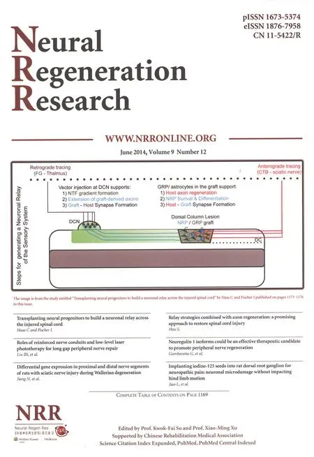
The model that we have proposed focuses on the formation of a functional relay to reconnect the injured spinal cord and requires the formation of two synaptic connections, one between host axons and graft-derived neurons, and the other between graft axons and target sites within the host (Figure 1). The design of such a relay requires speci fi c steps that assure: 1) graft survival and generation of neurons, 2) axon growth into and out of the graft by host axons and graft-derived neurons, respectively and 3) formation of physiologically active synaptic connections and restoration of function. The analysis of relay formation requires the characterization of graft-derived neurons (usually by using cells derived from transgenic animals expressing Alkaline Phosphatase (AP) or GFP), anatomical evidence for connectivity (including tracing showing regeneration of host axons and possibly immune-EM for detection of synaptic structures), physiological evidence of connectivity (expression of c-Fos following stimulation and electrophysiology), and functional analysis of sensory or motor recovery with speci fi c tests depending on the injury model. The generation of a neuronal relay circumvents many of the obstacles inherent in attempts to achieve functional reconnectivity by long-distance regeneration of lesioned host axons, including the limited intrinsic regenerative capacity of many adult neuronal CNS populations and the sensitivity of these populations to inhibitors of axon growth found in the external environment, such as CSPG. Intriguingly, NRP (derived from embryonic spinal cord) express signi fi cantly lower levels of the CSPG receptors Protein Tyrosine Phosphatase σ (PTP σ) and Leukocyte Common Antigen-related Phosphatase (LAR), compared to more mature primary neuronal populations, and concomitantly retain an ability to extend axons in the presence of such inhibitors (Ketschek et al., 2012). In addition, axons generated by NRP-derived neurons are readily guided to target sites utilizing lentiviral-established gradients of neurotrophins, such as BDNF and NT3, highlighting the ability to direct these axons to speci fi c areas of the cord for synapse formation (Bonner et al., 2010; Bonner et al., 2011) without risking unchecked axonal growth and mis-connectivity. Importantly, grafts of GRP recreate the proper structural and physiological environment necessary for synaptic transmission, as supported by GRP-derived astrocytes. Furthermore, grafts composed entirely of GRP are capable of limiting the formation of the glial scar (e.g., preventing the upregulation of host GFAP and CSPG expression) (Hill et al., 2004; Haas, 2013, 2014; Fischer, 2014), modulating the immune environment by limiting invasion of peripheral macrophages (Haas, 2013, 2014; Fischer, 2014), facilitating axonal regeneration into the graft (Haas et al., 2012; Haas and Fischer, 2013), and differentiating into critically necessary astrocytes and oligodendrocytes (Hill et al., 2004; Haas et al., 2012) thus making them an ideal therapeutic platform for use in combinatorial strategies designed to achieve re-connectivity. In contrast, alternative grafting platforms, such as genetically-modi fi ed marrow stromal cells, while providing a bridge for long-distance axonal regeneration, may contribute to conduction failure of regenerating axons due to their inability to generate CNS cell subtypes (e.g., astrocytes and oligodendrocytes) that support synaptic connectivity (Alto et al., 2009), highlighting the importance of recreating the original environment of the spinal cord. Despite the advantages of utilizing NPC as permissive grafts capable of supporting synaptic transmission across the injured spinal cord by way of synaptic relays, grafting of NPC still faces considerable challenges, particularly in the context of severe spinal cord injuries.
The concept of relay formation has been applied not only in reference to neural transplants that reconnect with the original targets, but also for connectivity through propriospinal interneurons generating alternative pathways (Courtine et al., 2008).
This Perspective will be focused on three recent articles that underscore the promise and challenges of relay formation in spinal cord injury. The discussion of one study (Bonner et al., 2011) will demonstrate successful formation of a functional relay in a partial injury of the sensory system, while the other two studies (Lu et al., 2012; Medalha et al., 2014) will present possible solutions when dealing with a severe injury.

Figure 1 Forming a synaptic relay to reconnect the injured sensory system.
In the study that serves as the model of relay formation and analysis (Bonner et al., 2011), a preparation of cultured and banked NRPs and GRPs, derived from E14 spinal cord, was transplanted into a dorsal column lesion that interrupted the connections between sensory host axons (of the fasciculus gracilus) with their dorsal column nucleus (DCN) target (gracile tubercle of the DCN) in the brainstem. Evidence for the formation of a functional relay within the injured spinal cord was demonstrated through a number of steps: the survival and differentiation of graft-derived neurons, guidance of graft-derived axons to specific target sites, synaptic connectivity between graft-derived axons and target neurons, the regeneration of lesioned host axons into the graft, and the establishment of synaptic connectivity between lesioned host axons and graft-derived neurons. The ability of NRP/GRP grafts to generate neurons was based upon a series of studies that demonstrated the challenges of neuronal differentiation in the non-neurogenic adult spinal cord (Cao et al., 2002; Han et al., 2002) and the necessity of including GRP to generate astrocytes that would support neuronal survival and differentiation at the injury site (Lepore and Fischer, 2005). Graft-derived axons were directed by chemotropic cues along a BDNF gradient to the DCN, a strategy that was based on work from Tuszynski and Blesch demonstrating the formation of neurotrophic gradient by lentiviral vectors and their ability to promote regeneration of injured dorsal column axons (Blesch, 2004; Taylor et al., 2006; Alto et al., 2009). Numerous studies have shown that the axons of neurons derived from grafted embryonic tissue can grow long distances along white matter (Davies et al., 1999; Lepore and Fischer, 2005; Lu et al., 2012) due to their intrinsic growth potential (Smith and Skene, 1997) and reduced responsiveness to external inhibitory cues compared to adult neurons (Ketschek et al., 2012). In our study, graft-derived axons were able to form synaptic connections with the neurons of the denervated DCN, creating one of the synaptic connections necessary for relay formation. At the same time, lesioned host sensory axons regenerated into the graft, forming synaptic connections with graft-derived neurons, thus creating the other synaptic connection. Interestingly, we found that the robust regeneration of lesioned host axons and synaptic connectivity between these axons and graft-derived neurons occurred without additional intervention (e.g., conditioning). Subsequent studies revealed that the presence of GRP-derived astrocytes was not only critical for the generation of graft-derived neurons (Lepore and Fischer, 2005), but was also suf fi cient for promoting the modest regeneration into the graft and the formation of synaptic connections (Bonner et al., 2011; Haas et al., 2012). Evidence for anatomical connectivity was based upon: 1) following the growth of graft-derived axons to the DCN target using the AP reporter (as NRP/GRP were prepared from AP transgenic rats), 2) tracing of regenerating host sensory axons by cholera toxin b subunit into the graft, and 3) identi fi cation of synaptic proteins by immunocytochemistry and synaptic structures by immune-EM. Evidence for the formation of functional, physiologically active synapses included the findings that: 1) stimulation of the sciatic nerve resulted in c-Fos expression within graft-derived neurons and within neurons at the level of the DCN, 2) physiologically active synapses were shown to have a signal-transmission delay consistent with connectivity by a di-synaptic relay, and 3) transmission of important sensory behaviors was reconstituted, with stroking of the ipsilateral hindlimb resulting in the signal relayed to the DCN by way of a grafted neuron, across the injury. Preliminary resultsusing a lesion model that interrupts the corticospinal tract (CST) suggests that similar principles can be used to reconnect the motor system (Haas, 2013, 2014; Fischer, 2014). Given the particularly poor regenerative capacity of CST neurons, it is likely that the NPC graft will have to be combined with treatments that enhance the intrinsic potential for axon growth to allow for the formation of synaptic connections with graft-derived neurons (e.g., modulation of PTEN and SOCS3).
As a result of progress with the relay model, attention has shifted to issues that are “beyond regeneration”: how to direct axons to putative targets, how to form active synaptic formations, and how to translate partial connectivity with poor or no mapping into meaningful functional information. For example, when reconnecting the sensory system through long-distance regeneration of lesioned host axons or through synaptic relays utilizing axons from graft-derived neurons, one must consider the endogenous reorganization that occurs in the aftermath of injury to denervated structures, particularly in the somatosensory cortex (Kaas et al., 2008). It is speculated that re-training of the system through specific activity will strengthen the correct or appropriate innervations and promote the necessary plasticity for functional recovery. The relay model however, has also highlighted some limitations, including weak synaptic connectivity at the target site (DCN) and the formation of a relay in an injury model that was based upon a partial, relatively small injury (dorsal column hemisection), in which graft-derived axons were required to extend only a short distance for anatomical and functional connectivity given the proximity of the C1 lesion site to the DCN target.
Indeed, our next step was to examine the same NPC (NRP/GRP) transplants, isolated from E14 spinal cord, in a complete thoracic transection injury (Medalha et al., 2014). In this model, grafted cells survived poorly despite using a variety of lesion methods (aspiration and knife cut), matrices (Vitrogen, matrigel, and fi brin), a 3-9 day delay in transplantation, and juvenile and adult animals. In contrast, when grafts of “dissociated” or “segmental” FSC, also isolated from E14, were acutely transplanted into the same severe injury model, the grafts survived and generated neural cells including glutaminergic and GABAergic neurons. Importantly, grafts of FSC alone, without the addition of exogenous matrix or growth factors, created a permissive environment that promoted axonal growth of propriospinal and serotonigeric axons into the transplant and extension of graft-derived axons into the host (Medalha et al., 2014), which are critical elements of relay formation. Neural progenitors within the FSC grafts (e.g. GRP) also generated glial cells, including astrocytes and oligodendrocytes, which are necessary to ensure proper synaptic transmission and connectivity. While cultures of NPC and acute preparations of FSC contain mostly the same populations of cells (e.g. NRP and GRP), it is likely that a variety of features inherent to FSC grafts mediate their unique properties. FSC contain permissive matrix, growth promoting molecules, and small populations of endothelial cells, fibroblasts, neurons, and multipotent neuroepithelial cells (Kalyani et al., 1997; Cai et al., 2002) which are removed upon the culturing and preparation of cryopreservable stocks of single-cell suspensions of lineage restricted progenitors (Cai et al., 2002; Han et al., 2002; Lepore and Fischer, 2005). These observations are consistent with early work, which demonstrated that FSC grafts induced regeneration into the lesion area (Reier et al., 1986; Jakeman and Reier, 1991), promoted limited recovery of function (Bregman and Reier, 1986), and rescued axotomized neurons (Mori et al., 1997). Taken together, these studies demonstrate that for large and severe injuries, NPC transplants need to be combined with a set of factors that will support graft survival and growth.
An important step to resolve the difficulties associated with a severe injury has been taken by a recent study using NPC derived from GFP-transgenic animals, showing that delayed transplantation of a single-cell suspension of NPC, prepared acutely from E14 FSC, together with a fibrin-thrombin matrix and a complex cocktail of factors, resulted in robust cell survival and remarkable long-distance axonal growth from graft-derived neurons (Lu et al., 2012). The reported data included experiments that showed electrophysiological connectivity and functional recovery, as assessed by the motor grading (BBB) scale, which was abolished following re-transection. While this strategy has produced impressive results, it is not clear that it is ready for clinical translation, requiring the optimization and reduction of the number of factors used in the cocktail and a careful analysis of the consequences of the robust and undirected graft-derived axonal growth, particularly in relation to potential undesired effects such as spasticity and pain. Furthermore, a replication study found that there was minimal ingrowth of host axons into the graft and no statistically signi fi cant differences between transplant and control groups with respect to the degree of locomotor recovery (Sharp et al., 2014). It appears that most grafts failed to create a continuous bridge of neural tissue between the rostral and caudal host stumps and were therefore unlikely to form functional bridges. In addition, the replication study also revealed the formation of ectopic masses of graft-derived cells at long distances from the transplant site, highlighting the potential not only for autonomous cell foci formation with concomitant anatomical abnormality (e.g., ventricle compression and stroke) but also aberrant physiological connectivity (Steward et al., 2014; Tuszynski et al., 2014). Nevertheless, it is possible that anatomical differences between rodents and humans and modifications of grafting techniques will reduce the ectopic cell foci observed in rodent models (Tuszynski et al., 2014).
Taken together, these studies underscore the challenges associated with transplants of NPC in cases of severe and complete SCI, but also offer experimental directions that can be applied to develop a transplantation strategy designed to achieve substantial axonal growth into and out of the graft to restore connectivity. The challenge is not only to identify and optimize the transplantation procedure, limit the potential for unpredictable and potentially undesirable effects by reducing the number of factors, and confi rm the results with human cells, but also make them compatible with FDA requirements and the costs of running a clinical trial, in which cells and factors have to be prepared by an expensive GLP process. Nevertheless, there is growing evidence for the potential efficacy of NPC transplants in diverse models of SCI to restore functional connectivity (Mitsui et al., 2005; Kobayashi et al., 2012; Hou et al., 2013; Mothe et al., 2013; van Gorp et al., 2013; Emgard et al., 2014), highlighting the therapeutic potential of synaptic relays for treatment of SCI.
Christopher Haas, Itzhak Fischer
Drexel University College of Medicine, Department of Neurobiology & Anatomy, Philadelphia, PA, USA
Philadelphia, PA 19129, USA, ifischer@drexelmed.edu.
Funding: NIH PO1 NS055976, Craig H. Neilsen Foundation, and Shriner’s Hospital for Children.
Con fl icts of interest: None declared.
Accepted: 2014-06-09
Alto LT, Havton LA, Conner JM, Hollis ER, 2nd, Blesch A, Tuszynski MH (2009) Chemotropic guidance facilitates axonal regeneration and synapse formation after spinal cord injury. Nat Neurosci 12:1106-1113.
Blesch A (2004) Lentiviral and MLV based retroviral vectors for ex vivo and in vivo gene transfer. Methods 33:164-172.
Bonner JF, Blesch A, Neuhuber B, Fischer I (2010) Promoting directional axon growth from neural progenitors grafted into the injured spinal cord. J Neurosci Res 88:1182-1192.
Bonner JF, Connors TM, Silverman WF, Kowalski DP, Lemay MA, Fischer I (2011) Grafted neural progenitors integrate and restore synaptic connectivity across the injured spinal cord. J Neurosci 31:4675-4686.
Bregman BS, Reier PJ (1986) Neural tissue transplants rescue axotomized rubrospinal cells from retrograde death. J Comp Neurol 244:86-95.
Cai J, Wu Y, Mirua T, Pierce JL, Lucero MT, Albertine KH, Spangrude GJ, Rao MS (2002) Properties of a fetal multipotent neural stem cell (NEP cell). Dev Biol 251:221-240.
Cao QL, Howard RM, Dennison JB, Whittemore SR (2002) Differentiation of engrafted neuronal-restricted precursor cells is inhibited in the traumatically injured spinal cord. Exp Neurol 177:349-359.
Courtine G, Song B, Roy RR, Zhong H, Herrmann JE, Ao Y, Qi J, Edgerton VR, Sofroniew MV (2008) Recovery of supraspinal control of stepping via indirect propriospinal relay connections after spinal cord injury. Nat Med 14:69-74.
Davies SJ, Goucher DR, Doller C, Silver J (1999) Robust regeneration of adult sensory axons in degenerating white matter of the adult rat spinal cord. J Neurosci 19:5810-5822.
Emgard M, Piao J, Aineskog H, Liu J, Calzarossa C, Odeberg J, Holmberg L, Samuelsson EB, Bezubik B, Vincent PH, Falci SP, Seiger A, Akesson E, Sundstrom E (2014) Neuroprotective effects of human spinal cord-derived neural precursor cells after transplantation to the injured spinal cord. Exp Neurol 253:138-145.
Fischer I, Haas C (2014) Glial Progenitor Transplantation for Promoting Axonal Regeneration. In: 45th Annual American Society for Neurochemistry Meeting Long Beach, CA.
Haas C, Fischer I (2013) Human astrocytes derived from glial restricted progenitors support regeneration of the injured spinal cord. J Neurotrauma 30:1035-1052.
Haas C, Neuhuber B, Yamagami T, Rao M, Fischer I (2012) Phenotypic analysis of astrocytes derived from glial restricted precursors and their impact on axon regeneration. Exp Neurol 233:717-732.
Haas C, Fischer I (2013) Advances in Transplantation of Glial Progenitors as a Therapeutic Platform in Treatment of Spinal Cord Injury. In: International Symposium on Neural Regeneration Asilomar Conference Center, Paci fi c Grove, CA.
Haas C, Fischer I (2014) Combinatorial Strategies to Promote Connectivity Across the Injured Spinal Cord: Glial Restricted Progenitors as a Therapeutic Platform. In: 45th Annual American Society for Neurochemistry Meeting Long Beach, CA.
(3)从图中可知,蓝色越深,代表任务越密集,该区域越繁华,则价格越低;反之蓝色越浅,越接近黄色,代表任务越稀疏,该区域离中心城市越远,则价格越高。
Han SS, Kang DY, Mujtaba T, Rao MS, Fischer I (2002) Grafted lineage-restricted precursors differentiate exclusively into neurons in the adult spinal cord. Exp Neurol 177:360-375.
Hill CE, Proschel C, Noble M, Mayer-Proschel M, Gensel JC, Beattie MS, Bresnahan JC (2004) Acute transplantation of glial-restricted precursor cells into spinal cord contusion injuries: survival, differentiation, and effects on lesion environment and axonal regeneration. Exp Neurol 190:289-310.
Hou S, Tom VJ, Graham L, Lu P, Blesch A (2013) Partial restoration of cardiovascular function by embryonic neural stem cell grafts after complete spinal cord transection. J Neurosci 33:17138-17149.
Jakeman LB, Reier PJ (1991) Axonal projections between fetal spinal cord transplants and the adult rat spinal cord: a neuroanatomical tracing study of local interactions. J Comp Neurol 307:311-334.
Kaas JH, Qi HX, Burish MJ, Gharbawie OA, Onifer SM, Massey JM (2008) Cortical and subcortical plasticity in the brains of humans, primates, and rats after damage to sensory afferents in the dorsal columns of the spinal cord. Exp Neurol 209:407-416.
Kalyani A, Hobson K, Rao MS (1997) Neuroepithelial stem cells from the embryonic spinal cord: isolation, characterization, and clonal analysis. Dev Biol 186:202-223.
Kalyani AJ, Piper D, Mujtaba T, Lucero MT, Rao MS (1998) Spinal cord neuronal precursors generate multiple neuronal phenotypes in culture. J Neurosci 18:7856-7868.
Ketschek AR, Haas C, Gallo G, Fischer I (2012) The roles of neuronal and glial precursors in overcoming chondroitin sulfate proteoglycan inhibition. Exp Neurol 235:627-637.
Kobayashi Y, Okada Y, Itakura G, Iwai H, Nishimura S, Yasuda A, Nori S, Hikishima K, Konomi T, Fujiyoshi K, Tsuji O, Toyama Y, Yamanaka S, Nakamura M, Okano H (2012) Pre-evaluated safe human iPSC-derived neural stem cells promote functional recovery after spinal cord injury in common marmoset without tumorigenicity. PLoS One 7:e52787.
Lepore AC, Fischer I (2005) Lineage-restricted neural precursors survive, migrate, and differentiate following transplantation into the injured adult spinal cord. Exp Neurol 194:230-242.
Lu P, Wang Y, Graham L, McHale K, Gao M, Wu D, Brock J, Blesch A, Rosenzweig ES, Havton LA, Zheng B, Conner JM, Marsala M, Tuszynski MH (2012) Long-distance growth and connectivity of neural stem cells after severe spinal cord injury. Cell 150:1264-1273.
Medalha CC, Jin Y, Yamagami T, Haas C, Fischer I (2014) Transplanting neural progenitors into a complete transection model of spinal cord injury. J Neurosci Res 92:607-618.
Mitsui T, Shumsky JS, Lepore AC, Murray M, Fischer I (2005) Transplantation of neuronal and glial restricted precursors into contused spinal cord improves bladder and motor functions, decreases thermal hypersensitivity, and modi fi es intraspinal circuitry. J Neurosci 25:9624-9636.
Mori F, Himes BT, Kowada M, Murray M, Tessler A (1997) Fetal spinal cord transplants rescue some axotomized rubrospinal neurons from retrograde cell death in adult rats. Exp Neurol 143:45-60.
Mothe AJ, Tam RY, Zahir T, Tator CH, Shoichet MS (2013) Repair of the injured spinal cord by transplantation of neural stem cells in a hyaluronan-based hydrogel. Biomaterials 34:3775-3783.
Reier PJ, Bregman BS, Wujek JR (1986) Intraspinal transplantation of embryonic spinal cord tissue in neonatal and adult rats. J Comp Neurol 247:275-296.
Reier PJ, Perlow MJ, Guth L (1983) Development of embryonic spinal cord transplants in the rat. Brain Res 312:201-219.
Sharp KG, Yee KM, Steward O (2014) A re-assessment of long distance growth and connectivity of neural stem cells after severe spinal cord injury. Exp Neurol doi:10.1016/j.expneurol.2014.04.008.
Smith DS, Skene JH (1997) A transcription-dependent switch controls competence of adult neurons for distinct modes of axon growth. J Neurosci 17:646-658.
Steward O, Sharp KG, Matsudaira Yee K (2014) Long-distance migration and colonization of transplanted neural stem cells. Cell 156:385-387.
Taylor L, Jones L, Tuszynski MH, Blesch A (2006) Neurotrophin-3 gradients established by lentiviral gene delivery promote short-distance axonal bridging beyond cellular grafts in the injured spinal cord. J Neurosci 26:9713-9721.
Tuszynski MH, Wang Y, Graham L, Gao M, Wu D, Brock J, Blesch A, Rosenzweig ES, Havton LA, Zheng B, Conner JM, Marsala M, Lu P (2014) Neural stem cell dissemination after grafting to CNS injury sites. Cell 156:388-389.
van Gorp S, Leerink M, Kakinohana O, Platoshyn O, Santucci C, Galik J, Joosten EA, Hruska-Plochan M, Goldberg D, Marsala S, Johe K, Ciacci JD, Marsala M (2013) Amelioration of motor/sensory dysfunction and spasticity in a rat model of acute lumbar spinal cord injury by human neural stem cell transplantation. Stem Cell Res Ther 4:57.
Itzhak Fischer, Ph.D., Drexel University College of Medicine, Department of Neurobiology & Anatomy, 2900 Queen Lane,
10.4103/1673-5374.135321 http://www.nrronline.org/
Haas C, Fischer I. Transplanting neural progenitors to build a neuronal relay across the injured spinal cord. Neural Regen Res. 2014;9(12):1173-1176.
猜你喜欢
儿童时代·幸福宝宝(2022年11期)2022-12-09
今日农业(2021年9期)2021-11-26
英语文摘(2021年2期)2021-07-22
青年歌声(2021年2期)2021-03-05
青年歌声(2020年11期)2020-11-24
商周刊(2018年13期)2018-07-11
海峡姐妹(2017年5期)2017-06-05
阅读与作文(小学高年级版)(2016年6期)2016-11-14
BOSS臻品(2015年1期)2015-09-10
太空探索(2015年5期)2015-07-12
- 中国神经再生研究(英文版)的其它文章
- Assessment on self-care, mobility and social function of children with spina bi fi da in Turkey
- Autophagy: a double-edged sword for neuronal survival after cerebral ischemia
- Diffuse axonal injury after traumatic cerebral microbleeds: an evaluation of imaging techniques
- Recovery of the corticospinal tracts injured by subfalcine herniation: a diffusion tensor tractography study
- Prolonged electrical stimulation causes no damage to sacral nerve roots in rabbits
- Overexpression of microRNA-124 promotes the neuronal differentiation of bone marrow-derived mesenchymal stem cells