
Compensatory recombination phenomena of neurological functions in central dysphagia patients
2015-01-18 05:49XiaodongYuanLifuZhouShujuanWangYanshengZhaoXiaojieWangLiliZhangShouhongWangYajieZhangLiChen
中国神经再生研究(英文版) 2015年3期
Xiao-dong Yuan, Li-fu Zhou Shu-juan Wang Yan-sheng Zhao, Xiao-jie Wang Li-li Zhang Shou-hong Wang, Ya-jie Zhang, Li Chen
1 Department of Neurology, Affliated Kailuan General Hospital of Hebei United University, Tangshan, Hebei Province, China
2 Department of MRI Room, Affliated Kailuan General Hospital of Hebei United University, Tangshan, Hebei Province, China
Compensatory recombination phenomena of neurological functions in central dysphagia patients
Xiao-dong Yuan1,*, Li-fu Zhou1, Shu-juan Wang1, Yan-sheng Zhao2, Xiao-jie Wang1, Li-li Zhang1, Shou-hong Wang2, Ya-jie Zhang2, Li Chen2
1 Department of Neurology, Affliated Kailuan General Hospital of Hebei United University, Tangshan, Hebei Province, China
2 Department of MRI Room, Affliated Kailuan General Hospital of Hebei United University, Tangshan, Hebei Province, China
We speculate that cortical reactions evoked by swallowing activity may be abnormal in patients with central infarction with dysphagia. The present study aimed to detect functional imaging features of cerebral cortex in central dysphagia patients by using blood oxygen level-dependent functional magnetic resonance imaging techniques. The results showed that when normal controls swallowed, primary motor cortex (BA4), insula (BA13), premotor cortex (BA6/8), supramarginal gyrus (BA40), and anterior cingulate cortex (BA24/32) were activated, and that the size of the activated areas were larger in the left hemisphere compared with the right. In recurrent cerebral infarction patients with central dysphagia, BA4, BA13, BA40 and BA6/8 areas were activated, while the degree of activation in BA24/32 was decreased. Additionally, more areas were activated, including posterior cingulate cortex (BA23/31), visual association cortex (BA18/19), primary auditory cortex (BA41) and parahippocampal cortex (BA36). Somatosensory association cortex (BA7) and left cerebellum in patients with recurrent cerebral infarction with central dysphagia were also activated. Experimental fndings suggest that the cerebral cortex has obvious hemisphere lateralization in response to swallowing, and patients with recurrent cerebral infarction with central dysphagia show compensatory recombination phenomena of neurological functions. In rehabilitative treatment, using the favorite food of patients can stimulate swallowing through visual, auditory, and other nerve conduction pathways, thus promoting compensatory recombination of the central cortex functions.
nerve regeneration; blood oxygen level-dependent functional magnetic resonance imaging; cerebral ischemia; dysphagia; function restructuring; cerebral cortex; neural regeneration
Yuan XD, Zhou LF, Wang SJ, Zhao YS, Wang XJ, Zhang LL, Wang SH, Zhang YJ, Chen L (2015) Compensatory recombination phenomena of neurological functions in central dysphagia patients. Neural Regen Res 10(3)∶490-497.
Introduction
Dysphagia is a common sequela of stroke (Daniels et al., 1998; Martino et al., 2005) estimated to occur in 37—78% of stroke cases (Wiles, 1991; Black-Schaffer et al., 1999; Martino et al., 2000; Meng et al., 2000; Han et al., 2001; Rogus-Pulia and Robbins, 2013). It can lead to several complications, such as aspiration pneumonia, malnutrition, and dehydration (Ney et al., 2009; Taylor et al., 2013), and the mortality rate in patients with swallowing dysfunction after stroke was signifcantly higher than in those with no swallowing dysfunction (Steinhagen et al., 2009). Therefore, identifying characteristics of central dysfunction and possible mechanisms of functional reorganization, and formulating effective programs for rehabilitating swallowing should help improve the effectiveness of dysphagia therapy. A noninvasive technique such as blood oxygen level-dependent functional magnetic resonance imaging (MRI) could provide information regarding the location, intensity, and dynamics of neural activity in swallowing-related brain regions (Hamdy et al., 1999; Martin et al., 2001, 2004; Ertekin and Aydogdu, 2003; Logemann, 2007). Here, we analyzed swallowing-related activity and compensatory reorganization in the brains of dysphagia patients with cerebral infarctions in a broad attempt to provide a theoretical basis for effective swallowing rehabilitation.
Subjects and Methods
Subjects
Ten right-handed patients with cerebral infarction were hospitalized in the Department of Neurology, Kailuan General Hospital, China from January 2013 to December 2013.
Patients first presented with dysphagia and both sides of the brain or pons had multiple cerebral infarctions that were confrmed by MRI (T1- and T2-weighted images). In 10 cases (seven male, three female), one of these infarctions was confrmed using diffusion weighted imaging as the new acute infarct and met the inclusion criteria (see below). Five patients had new lesions on the right (right recurrent cerebral infarction group) and five had new lesions on the left (left recurrent cerebral infarction group). The present study was performed in accordance with the Code of Ethics of the World Medical Association (Declaration of Helsinki) for experiments involving humans, and was reviewed and approved by the Medical Ethics Committee from AffliatedKailuan General Hospital of Hebei United University in China. All patients gave their signed informed consent.
Inclusion criteria
No patients had difficulty in swallowing, choking coughs, hoarseness, or other abnormalities before suffering from cerebral infarction. All exhibited swallowing dysfunction for the frst time within 1 week after cerebral infarction. Clinical examination found no signs of disease or peripheral nerve damage in the mouth, tongue, throat, or other swallowing-motion organs, and patients were ultimately diagnosed as having central nervous system swallowing dysfunction (Li et al., 2009). Results from the Kubota water test (Martino et al., 2005) were between levels 3—5, life signs were stable, consciousness, spirit, intelligence and verbal expression were normal, and patients cooperated with the physical examination. Scores on the anxiety depression scale (Murakami et al., 2013) were < 7 points. The damage to the nervous system was evaluated by NIHSS score for every patient (Mann and Hankey, 2001).
Exclusion criteria
Patients were excluded if their consciousness, spirit, or intelligence were disordered, if they did not cooperate with the physical examination, if they had a history of head or neck cancer, local diseases of the throat or esophagus, or esophagus-related dysphagia. Additionally, those with failure in visceral function or who were in a critical condition were also excluded. Further, because of the use of MRI, those who had metal objects, such as pacemakers or coronary stents were excluded, as were those with atrial fbrillation.
We also included 10 individuals (six male and four female; aged 53.2 ± 14.9 years) in a normal control group. These participants were recruited from healthy adults who participated in a physical examination at Kailuan General Hospital. All were right-handed and in good health, without any organic disease. Scores on the anxiety depression scale were< 7 points, and they had not imbibed any alcoholic beverages within 12 hours of the scan. Patients exhibited no uncomfortable symptoms or positive neurological signs before testing.
Blood oxygen level-dependent functional MRI data acquisition
We used a GE Signa Twin Speed 1.5T MRI system (GE Healthcare, Wauwatosa, WI, USA) to determine brain regions active during swallowing. After positioning, a T1WI FSPGR-3D scan that employed a single-shot EPI-temporal plane gradient echo sequence was used to image the basic anatomy. Activation of motor-related regions was scanned with blood oxygen level-dependent functional MRI using a T2WI gradient echo-pulse sequence. For single-temporal excitation echo-planar imaging, the position was defned parallel to the joint line (AC-PC), and the scanning covered 18 layers with the following parameters: single acquisition, time of repetition = 3,000 ms, time of echo = 60 ms, fip angle = 90°, feld of view = 24 cm × 24 cm, thickness = 5 mm, gap = 1.5 mm, frequency encoding direction = R/L.
Using a block design, blood oxygen level-dependent functional MRI scanning lasted 8 minutes. Subjects frst lay on the examination bed quietly and received instruction and brief training in the simulation process, and then their heads were fixed in a birdcage head-coil to minimize the movement of lips, tongue, and head. Subjects were asked to bite a plastic 2-mm tube with the middle part of their lips. A 50-mL syringe was connected to the other end of the tube, and swallowing blocks began. Each block lasted 30 seconds, with 2 mL of pure room-temperature water being injected into the tube every 5 seconds. Subjects immediately swallowed each time water was injected, yielding six swallows per block. Rest (control) blocks lasting 30 seconds followed each swallowing block. During the rest block, water was not injected into the tube, and participants did not engage in any swallowing action. Each experiment consisted of 16 blocks (eight swallowing, eight rest) for a total of 48 swallows (Figure 1).
Functional MRI data processing
SPM5 (Statistical Parametric Mapping; Wellcome Department of Cognitive Neurology, London, UK) software was used to model brain activity and determine brain regions associated with swallowing in the two groups of participants. Convolution of the swallowing time, interval, and hemodynamic function were used to analyze the whole-brain voxel signal correlation. The activation volume (represented by the number of active voxels) and intensity (represented by T value—higher T value indicated the stronger intensity) were calculated, MNI coordinate position generated by SPM5 was converted into standard Talairach coordinates with Brain-Map GingerALE 2.1.1 software (Research Imaging Institute of the University of Texas Health Science Center San Antonio, San Antonio, TX, USA). Talairach Client 2.4 software (Research Imaging Institute of the University of Texas Health Science Center San Antonio) was used to view specifc anatomical locations. Data from subjects with three-dimensional translation of the head greater than 0.5 mm and three-dimensional rotation greater than 0.5° were discarded. Two-samplet-tests were used to determine regions of activation. Statistical probability threshold was set atP< 0.05. The extent threshold was 10 pixels. Excel 2003 software (Microsoft Corporation, Redmond, WA, USA) was used to set up a database and SPSS 13.0 software (SPSS, Chicago, IL, USA) was used to analyze the activated region. Activation volume and intensity of brain areas calculated by SUN workstations and SPM are expressed as the mean ± SD. A non-parametric rank sum test was used for statistical analysis. Significance was set atP< 0.05.
Laterality index (LI) was defned as: (left hemisphere activation volume — right hemisphere activation volume)/(left hemisphere activation volume + right hemisphere activation volume) × 100% (Malandraki et al., 2011b). An LI ≥ 10% was considered left lateralization and an LI ≤ —10% was considered right lateralization (Ogura et al., 2012).

Table 1 Blood oxygen level-dependent functional magnetic resonance imaging results during swallowing for dysphagia patients with acute recurrent cerebral infarction
Results
Regions activated by independent swallowing
Mean NIHSS scores in the patient group were 4.0 ± 2.5 points (range: 3—10 points) based on the functional MRI examination. All patients completed all tests successfully. Both sides of Brodmann’s area (BA) 4, BA40, BA13, BA22 around the sylvian fssure and BA6/8, BA24, BA32, and the BA38/22/41 region of the superior temporal gyrus and other regions such as the hypothalamus and parts of the brain center were activated when control participants engaged in swallowing tests. Compared with the control group, areas more active in patients with right recurrence of cerebral infarction were BA4, BA13, BA40, and BA6/8. Areas with signifcantly reduced activity included bilateral BA24/32. Other areas with increased activity included bilateral BA23/31, BA36, right BA9 and BA18/19, and left BA41. Activation of bilateral BA23/31 in patients with left recurrent cerebral infarction was not as obvious as that in patients with recurrent acute cerebral infarction, however bilateral BA7, BA18/19, BA36, left BA41, right BA9, and cerebellum were signifcantly activated. Blood oxygen level-dependent functional MRI scanning results are shown in Table 1.
The laterality of brain regions activated by swallowing
When the control group performed swallowing, the overall extent of activation was greater in the left hemisphere than in the right hemisphere (LI value: 15.22%,P< 0.05). A more detailed analysis revealed that activation volumes in left medial frontal gyrus (BA4) and left insula (BA13) were larger than corresponding regions in the right hemisphere (LI values: BA4 = 13.49%, BA13 = 19.02%;P< 0.05). Further, left hemisphere activation volumes in BA40 and BA32 were obviously larger than those in the right hemisphere (LI values: BA40 = 10.89%, BA32 = 61.29%;P< 0.01). In contrast, activation volumes in left BA6/8 and left BA24 were not different from those in the right hemisphere (LI values: BA6/8 =2.02%, BA24 = 0.66%;P> 0.05).
Brain related hubs activated by swallowing
In the right cerebral infarction group, activation volumes of right BA4 and BA6/8 were smaller than those of the control group (P< 0.01), while those for BA13 and BA40 were not (P> 0.05). Further, while activation volume for left BA6/8 was greater than that of the control group (P< 0.05), those for left BA4, BA13, and BA40 were not (P> 0.05).
In the left cerebral infarction group, activation volumes of left BA4 and BA6/8 were smaller than those of the control group (P< 0.05), while those for BA13 and BA40 were not (P> 0.05). Additionally, while activation volumes for right BA4 and BA6/8 were larger than those of the control group (P< 0.05), those for right BA13 and BA40 were not (P> 0.05; Figure 2).
Activation intensity in swallowing
During swallowing, left BA4, BA6/8, BA13, BA40, BA24, and BA32 of the control group were not more active than corresponding locations in the right hemisphere (P> 0.05; Table 1).
For the right cerebral infarction group, right BA4, BA6/8, BA13, and BA40 were less active than the corresponding areas in the control group (P< 0.05). This was not true for left BA4, BA6/8, BA13, and BA40 (P> 0.05; Table 1).
For the left recurrent cerebral infarction group, left BA4, BA6/8, and BA13 were less active than the corresponding areas in the control group (P< 0.05), but left BA40 was not (P> 0.05). Additionally, activity levels in right BA4, BA6/8, BA13, and BA40 did not differ between groups (P> 0.05; Table 1).
discussion
Studies have shown that the most prominent swallow-related activation foci correspond to the lateral pericentral and perisylvian cortices, anterior cingulate cortex, premotor cortex, insula/operculum, and supramarginal gyrus (D’Esposito et al., 2003; Lowell et al., 2008; Malandraki et al., 2010, 2011a; Peck et al., 2010). However, the compensatory mechanisms and functional reorganization that occur in dysphagia remain unclear clear. Further studies are needed to develop effective individualized rehabilitation programs for dysphagia. We found that there was no obvious or opharyngeal sensory cortex activation during normal swallowing, but premotor, frontal eye felds (BA6/8), and anterior cingulate (BA24/32) were activated, along with the lateral fissure oropharyngeal area surrounding primary motor cortex (BA4), insula (BA13), supramarginal gyrus (BA40), and temporal cortex BA38/22. This evidence indicated that motor planning and execution (beginning in primary motor cortex) were most important for autonomous swallowing. These results are consistent with previously reports (Kern et al., 2001; Gazzaniga et al., 2002; Ettlin et al., 2004; Fukunaga et al., 2005; Humbert et al., 2011; Babaei et al., 2012; Galovic et al., 2013). They also suggest that oropharyngeal tactile stimulation was not the main factor that induced swallowing. Physiological needs, smell, and language are all appetite-related stimuli that might be major factors in initiating independent swallowing. However, in-depth study on the cortical activity related to these stimuli must be done to draw defnite conclusions. The cortical motor areas and supramarginal gyrus were important for planning swallowing, and oropharyngeal motor region was the central region responsible for initiating swallowing (Forman et al., 1995; Lotze et al., 1999; Komisaruk et al., 2002; Satow et al., 2004; Lacourse et al., 2005; Saad et al., 2006; Szameitat et al., 2007).
However, here we examined patients with central dysphagia when it resulted from unilateral recurrent cerebral infarction at acute stage. We found that regardless of the side of the infarction, BA4, BA13, BA40, and BA6/8 were still the main regions of active cortex. In contrast, activation in bilateral cingulate cortex (BA24/32) was signifcantly reduced, suggesting that regions necessary for planning and execution of swallowing were still activated in patients with dysphagia, but that the region needed for initiating swallowing was signifcantly reduced. In patients with acute recurrent right non-dominant cerebral infarction, we found that bilateral posterior cingulate cortex (BA23/31) was obviously activated, and that right parahippocampal cortex (BA36), prefrontal cortex (BA9), left occipital cortex (BA18/19), and primary auditory cortex (BA41) were all activated. This evidence indicates that brain damage leading to swallowing disorders resulted in compensatory functional reorganization involving brain regions that control the initiation of swallowing, such as cingulate cortex, prefrontal cortex, visual cortex, and audio cortex. Stimulating patients with their favorite food through visual and auditory channels might promote functional recovery of swallow initiation. We will conduct further studies on this phenomenon (Uğurbil et al., 2000; Power et al., 2007; Babaei et al., 2010; Cola et al., 2010).
In patients with acute recurrent left dominant cerebral infarction, we found that the activation of bilateral posterior cingulate cortex (BA23/31) was not as obvious as in those with acute cerebral infarction in the right hemisphere, but somatosensory cortex (BA7), occipital cortex (BA18/19), and the cerebellum were significantly activated. We also found that bilateral parahippocampal cortex (BA36), left primary auditory cortex (BA41), and right lateral prefrontal cortex (BA9) were activated, indicating that BA7, BA18/19, and the cerebellum played an important role in restructuring compensatory function in the motor initiation of swallowing in patients with dysphagia. Patients with central swallowing disorders could do passive swallowing masonic-coordination training through visual stimulation of food and feeling, and it is possible to promote the reorganization of cortical function, thereby accelerating the recovery of swallowing behavior. It is worth observing in our clinical work (Kumar et al., 2011; Michou et al., 2012; Yang et al., 2012).
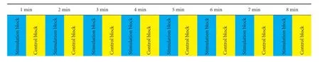
Figure 1 Experimental block design.

Figure 2 Activation during swallowing of primary motor cortex (BA4) and premotor cortex (BA6/8) in the right/left cerebral infarction group and the control group.
Our results showed that in the process of normal swallowing, the extent of cortical activation in the left or right hemisphere was quite different (depending on the side of the injury), but that the intensity was not. The extent of activation in primary oropharyngeal cortex (BA4) was signifcantly larger than other cortical regions, and was greater in the left hemisphere than in the right. Consistent with previous reports, this suggested that the size of the activated area was normal, and that especially in the dominant hemisphere, it was an important factor for implementing normal autonomous swallowing (Regard and Landis, 1997; Jubault et al., 2007; Price, 2010). Similarly, the size of supramarginal gyrus (BA40) activation in the dominant (left) hemisphere was also signifcantly greater than that in the non-dominant one, while that of the supplementary motor area was did not differ across hemispheres, although it was smaller than what we observedfor BA4. However, the activation intensity of these regions was not signifcantly different. These results further illustrated that BA40 in both the supramarginal gyrus and premotor cortex/supplementary motor area were important centers for planning the movements needed for swallowing. Volume of activation of the apply center of dominant hemisphere in a normal state is an important security condition for autonomic swallowing completed accurately and effciently.
Meanwhile, we also observed that both swallowing coordination centers in the posterior lobe of cerebellar cortex were activated in some adults, resulting in smoother and more coordinated swallowing. When the normal independent swallowing began, the size of primary motor cortex activation in the left hemisphere was greater than in the right, the total LI value and activation in the central gyrus, anterior cingulate cortex, and insula were all greater than 10%. The LI value in BA6/8 was signifcantly less than 10% and that in BA40 was approximately 10%. Therefore, initiation and execution of independent swallowing was obviously lateralized (Daniels et al., 2006; Martin et al., 2007; Teismann et al., 2007; Babaei et al., 2012; Li et al., 2014). Thus, while independent swallowing was regulated by the central gyrus, anterior cingulate cortex, insula, and supramarginal gyrus in the dominant hemisphere, it was planned bilaterally.
However, in patients with right recurrent cerebral infarction, the extent of activation in BA4 and BA6/8 on the same side of the lesion decreased, while that of BA40 and BA13 areas was signifcantly different. Additionally, activation volume in BA6/8 contralateral to the lesion was greater, and that in BA4, BA40, and BA13 showed no signifcant differences. Similarly, in those with left recurrent cerebral infarction, activation volume in BA4 and BA6/8 ipsilateral to the lesion were smaller than on the right, and those of BA40 and BA13 showed no signifcant differences. Further, activation volume of BA4 and BA6/8 contralateral to the lesion were larger, and those of BA4, BA40, and BA13 showed no significant differences. Therefore, when the non-dominant hemisphere infarction occurred, activation volume of the regions needed to initiate swallowing (such as BA13) did not differ across hemispheres, but those of regions needed for planning the action (such as BA6/8)were reduced on the side of the lesion and increased on the side opposite the lesion. Activation volume in BA40 showed no significant difference between hemispheres, a smaller region of activation in primary motor cortex (BA4), but no compensatory contralateral increase. In those with a dominant hemispheric infarction, the volume of activation for regions needed to initiate swallowing did not differ across hemispheres, a reduced extent of activation in BA6/8 on the side of the lesion side, and a compensatory increase in activation volume contralaterally. However, activation volume in BA40 was not signifcantly different activation volume in the primary motor cortex (BA4) was reduced on the side of the lesion, and no compensatory contralateral increase was observed.
Our fndings showed that the size of cortical activity was lateralized when normal swallowing occurred, but that the activation intensity of both regions related to swallowing was not. However, activation intensity of the central gyrus, anterior cingulate cortex, insula, and supramarginal gyrus has been shown to be an obvious change in dysphagia patients (Ertelt et al., 2007; Steinhagen et al., 2009; Babaei et al., 2013). We showed that activation intensity in ipsilateral BA4, BA6/8, BA40, and BA13 was reduced in patients with right recurrent cerebral infarction of the non-dominant hemisphere, but the intensity was not obviously different when contralateral central cortex was activated. The activation intensity of BA4, BA6/8, and BA13 were reduced, while that of BA40 was not obviously different when ipsilateral central cortex was activated. Activation intensity of BA4, BA6/8, BA13, and BA40 also had no obvious differences when contralateral central cortex was activated in patients with left recurrent infarction of the non-dominant hemisphere. Therefore activation intensity was not obviously different in ipsilateral swallowing related regions in patients with dysphagia after acute cerebral infarction, but it decreased in ipsilateral swallowing centers and was not generally affected in BA40, a dominant swallowing center.
Pleasant stimulation related to appetite could activate the swallowing centers, such as the anterior insula and the anterior cingulate cortex, and then activate the motor cortex related to planning and execution of swallowing, such as the supramarginal gyrus and premotor area. The activation volume of central cortex is the basis for regular swallowing motor activity. The central regulation of swallowing has obvious advantages in dominant hemisphere laterality. In patients with dysphagia resulting from recurrent acute cerebral infarction, while regions dedicated to planning and execution of swallowing were active, the size of activity was reduced in regions involved in the initiation of movement, while the intensity in the contralateral swallowing regions had no obvious change, nor did that in BA40 area of the dominant side. In our study, non-response bias and investigation bias might have occurred, and we patiently explained this to patients to improve compliance with the examination. Therefore, making individualized rehabilitation programs for dysphagia patients based on the results of this study will promote compensatory reorganization of cortical centers and further improve the effect of rehabilitation treatment. Meanwhile, to fully understand the impact and role of each nerve and biological function of the body, we need to further research the characteristics and compensatory reorganization mechanisms of brain function in a variety of sensory dysfunctions and cognitive impairments. We will expand the sample size in order to make conclusions that can be used for clinical rehabilitation treatment.
Acknowledgments:We would like to thank M.D. Wen-jiang Zhang from Tangshan Vocational Technical College in China for software applications and M.D. Ling-min Meng from Kailuan General Hospital in China for the help in medical statistics. This work gained supports and technical assistance of teachers from Hebei United University in China.
Author contributions:XDY was responsible for the design, organization and coordination of the study, involved in dataprocessing and analysis, responsible for the results analysis and writing the manuscript. LFZ was involved in the design of the study and responsible for the experimental operations, data processing, results analysis and writing the manuscript. SJW was responsible for the choice of subjects, involved in the design of the study, results analysis and writing the manuscript. YSZ, SHW and YJZ were involved in the experimental operations, data processing and results analysis. XJW and LC were involved in the choice of subjects and data processing. LLZ was involved in results analysis and writing the manuscript. All authors approved the final version of the paper.
Conficts of interest:None declared.
Babaei A, Kern M, Antonik S, Mepani R, Ward BD, Li SJ, Hyde J, Shaker R (2010) Enhancing effects of favored nutritive stimuli on cortical swallowing network activity. Am J Physiol Gastrointest Liver Physiol 299:G422-429.
Babaei A, Siwiec RM, Kern M, Douglas Ward B, Li SJ, Shaker R (2013) Intrinsic functional connectivity of the brain swallowing network during subliminal esophageal acid stimulation. Neurogastroenterol Motil 25:992-e779.
Babaei A, Ward BD, Ahmad S, Patel A, Nencka A, Li SJ, Hyde J, Shaker R (2012) Reproducibility of swallow-induced cortical BOLD positive and negative fMRI activity. Am J Physiol Gastrointest Liver Physiol 303:G600-609.
Black-Schaffer RM, Kirsteins AE, Harvey RL (1999) Stroke rehabilitation. 2. Co-morbidities and complications. Arch Phys Med Rehabil 80:S8-16.
Cola MG, Daniels SK, Corey DM, Lemen LC, Romero M, Foundas AL (2010) Relevance of subcortical stroke in dysphagia. Stroke 41:482-486.
D’Esposito M, Deouell LY, Gazzaley A (2003) Alterations in the BOLD fMRI signal with ageing and disease: a challenge for neuroimaging. Nat Rev Neurosci 4:863-872.
Daniels SK, Brailey K, Priestly DH, Herrington LR, Weisberg LA, Foundas AL (1998) Aspiration in patients with acute stroke. Arch Phys Med Rehabil 79:14-19.
Daniels SK, Corey DM, Fraychinaud A, DePolo A, Foundas AL (2006) Swallowing lateralization: the effects of modifed dual-task interference. Dysphagia 21:21-27.
Ertekin C, Aydogdu I (2003) Neurophysiology of swallowing. Clin Neurophysiol 114:2226-2244.
Ertelt D, Small S, Solodkin A, Dettmers C, McNamara A, Binkofski F, Buccino G (2007) Action observation has a positive impact on rehabilitation of motor deficits after stroke. Neuroimage 36 Suppl 2:T164-173.
Ettlin DA, Zhang H, Lutz K, Järmann T, Meier D, Gallo LM, Jäncke L, Palla S (2004) Cortical activation resulting from painless vibrotactile dental stimulation measured by functional magnetic resonance imaging (FMRI). J Dent Res 83:757-761.
Forman SD, Cohen JD, Fitzgerald M, Eddy WF, Mintun MA, Noll DC (1995) Improved assessment of signifcant activation in functional magnetic resonance imaging (fMRI): use of a cluster-size threshold. Magn Reson Med 33:636-647.
Fukunaga A, Uematsu H, Sugimoto K (2005) Influences of aging on taste perception and oral somatic sensation. J Gerontol A Biol Sci Med Sci 60:109-113.
Galovic M, Leisi N, Müller M, Weber J, Abela E, Kägi G, Weder B (2013) Lesion location predicts transient and extended risk of aspiration after supratentorial ischemic stroke. Stroke 44:2760-2767.
Gazzaniga MS, Ivry RB, Mangun GR (2002) Cognitive Neuroscience: the Biology of the Mind. New York: W.W.Norton & Company.
Hamdy S, Mikulis DJ, Crawley A, Xue S, Lau H, Henry S, Diamant NE (1999) Cortical activation during human volitional swallowing: an event-related fMRI study. Am J Physiol 277:G219-225.
Han TR, Paik NJ, Park JW (2001) Quantifying swallowing function after stroke: A functional dysphagia scale based on videofuoroscopic studies. Arch Phys Med Rehabil 82:677-682.
Humbert IA, McLaren DG, Malandraki G, Johnson SC, Robbins J (2011) Swallowing intentional off-state in aging and Alzheimer’s disease: preliminary study. J Alzheimers Dis 26:347-354.
Jubault T, Ody C, Koechlin E (2007) Serial organization of human behavior in the inferior parietal cortex. J Neurosci 27:11028-11036.
Kern M, Birn R, Jaradeh S, Jesmanowicz A, Cox R, Hyde J, Shaker R (2001) Swallow-related cerebral cortical activity maps are not specific to deglutition. Am J Physiol Gastrointest Liver Physiol 280:G531-538.
Komisaruk BR, Mosier KM, Liu WC, Criminale C, Zaborszky L, Whipple B, Kalnin A (2002) Functional localization of brainstem and cervical spinal cord nuclei in humans with fMRI. Am J Neuroradiol 23:609-617.
Kumar S, Wagner CW, Frayne C, Zhu L, Selim M, Feng W, Schlaug G (2011) Noninvasive brain stimulation may improve stroke-related dysphagia: a pilot study. Stroke 42:1035-1040.
Lacourse MG, Orr EL, Cramer SC, Cohen MJ (2005) Brain activation during execution and motor imagery of novel and skilled sequential hand movements. Neuroimage 27:505-519.
Li S, Luo C, Yu B, Yan B, Gong Q, He C, He L, Huang X, Yao D, Lui S, Tang H, Chen Q, Zeng Y, Zhou D (2009) Functional magnetic resonance imaging study on dysphagia after unilateral hemispheric stroke: a preliminary study. J Neurol Neurosurg Psychiatry 80:1320-1329.
Li S, Ma Z, Tu S, Zhou M, Chen S, Guo Z, Gong Q, He L, Huang X, Yao D, Lui S, Yu B, Wang X, Zhou D, He C (2014) Altered resting-state functional and white matter tract connectivity in stroke patients with dysphagia. Neurorehabil Neural Repair 28:260-272.
依照规定,水行政执法机关一旦发现涉水违法行为可能触犯刑法,有义务将相关案件移送至公安、检察等司法机关处理。按照最高检规定,水行政执法机关向公安机关移送案件,公安机关作立案、不立案决定处理,应向同级检察院备案。但对未报送检察机关备案的,没有规定监督和惩罚措施,这使得部分水行政执法机关对应移送的案件持一种消极态度。虽然刑法第402条规定了行政执法机关不移送案件的刑事责任,但该条款的适用取决于检察机关及时发现行政机关在移送案件过程中的渎职行为。事实上在未及时备案又缺乏信息共享的前提下,检察机关很难发现部分水行政执法机关的渎职行为。
Logemann JA (2007) Swallowing disorders. Best Pract Res Clin Gastroenterol 21:563-573.
Lotze M, Montoya P, Erb M, Hülsmann E, Flor H, Klose U, Birbaumer N, Grodd W (1999) Activation of cortical and cerebellar motor areas during executed and imagined hand movements: an fMRI study. J Cogn Neurosci 11:491-501.
Lowell SY, Poletto CJ, Knorr-Chung BR, Reynolds RC, Simonyan K, Ludlow CL (2008) Sensory stimulation activates both motor and sensory components of the swallowing system. Neuroimage 42:285-295.
Malandraki GA, Johnson S, Robbins J (2011a) Functional MRI of swallowing: from neurophysiology to neuroplasticity. Head Neck 33 Suppl 1:S14-S20.
Malandraki GA, Sutton BP, Perlman AL, Karampinos DC (2010) Age-related differences in laterality of cortical activations in swallowing. Dysphagia 25:238-249.
Malandraki GA, Perlman AL, Karampinos DC, Sutton BP (2011b) Reduced somatosensory activations in swallowing with age. Hum Brain Mapp 32:730-743.
Martin R, Barr A, MacIntosh B, Smith R, Stevens T, Taves D, Gati J, Menon R, Hachinski V (2007) Cerebral cortical processing of swallowing in older adults. Exp Brain Res 176:12-22.
Martin RE, Goodyear BG, Gati JS, Menon RS (2001) Cerebral cortical representation of automatic and volitional swallowing in humans. J Neurophysiol 85:938-950.
Martin RE, MacIntosh BJ, Smith RC, Barr AM, Stevens TK, Gati JS, Menon RS (2004) Cerebral areas processing swallowing and tongue movement are overlapping but distinct: a functional magnetic resonance imaging study. J Neurophysiol 92:2428-2443.
Martino R, Pron G, Diamant N (2000) Screening for oropharyngeal dysphagia in stroke: insuffcient evidence for guidelines. Dysphagia 15:19-30.
Martino R, Foley N, Bhogal S, Diamant N, Speechley M, Teasell R (2005) Dysphagia after stroke: incidence, diagnosis, and pulmonary complications. Stroke 36:2756-2763.
Meng NH, Wang TG, Lien IN (2000) Dysphagia in patients with brainstem stroke: incidence and outcome. Am J Phys Med Rehabil 79:170-175.
Michou E, Mistry S, Jefferson S, Singh S, Rothwell J, Hamdy S (2012) Targeting unlesioned pharyngeal motor cortex improves swallowing in healthy individuals and after dysphagic stroke. Gastroenterology 142:29-38.
Murakami T, Hama S, Yamashita H, Onoda K, Kobayashi M, Kanazawa J, Yamawaki S, Kurisu K (2013) Neuroanatomic pathways associated with poststroke affective and apathetic depression. Am J Geriatr Psychiatry 21:840-847.
Ney DM, Weiss JM, Kind AJ, Robbins J (2009) Senescent swallowing: impact, strategies, and interventions. Nutr Clin Pract 24:395-413.
Ogura E, Matsuyama M, Goto TK, Nakamura Y, Koyano K (2012) Brain activation during oral exercises used for dysphagia rehabilitation in healthy human subjects: a functional magnetic resonance imaging study. Dysphagia 27:353-360.
Peck KK, Branski RC, Lazarus C, Cody V, Kraus D, Haupage S, Ganz C, Holodny AI, Kraus DH (2010) Cortical activation during swallowing rehabilitation maneuvers: a functional MRI study of healthy controls. Laryngoscope 120:2153-2159.
Power ML, Hamdy S, Singh S, Tyrrell PJ, Turnbull I, Thompson DG (2007) Deglutitive laryngeal closure in stroke patients. J Neurol Neurosurg Psychiatry 78:141-146.
Price CJ (2010) The anatomy of language: a review of 100 fMRI studies published in 2009. Ann N Y Acad Sci 1191:62-88.
Regard M, Landis T (1997) “Gourmand syndrome”: eating passion associated with right anterior lesions. Neurology 48:1185-1190.
Rogus-Pulia N, Robbins J (2013) Approaches to the rehabilitation of dysphagia in acute poststroke patients. Semin Speech Lang 34:154-169.
Saad ZS, Chen G, Reynolds RC, Christidis PP, Hammett KR, Bellgowan PS, Cox RW (2006) Functional imaging analysis contest (FIAC) analysis according to AFNI and SUMA. Hum Brain Mapp 27:417-424.
Satow T, Ikeda A, Yamamoto J, Begum T, Thuy DHD, Matsuhashi M, Mima T, Nagamine T, Baba K, Mihara T, Inoue Y, Miyamoto S, Hashimoto N, Shibasaki H (2004) Role of primary sensorimotor cortex and supplementary motor area in volitional swallowing: a movement-related cortical potential study. Am J Physiol Gastrointest Liver Physiol 287:G459-G470.
Steinhagen V, Grossmann A, Benecke R, Walter U (2009) Swallowing disturbance pattern relates to brain lesion location in acute stroke patients. Stroke 40:1903-1906.
Szameitat AJ, Shen S, Sterr A (2007) Motor imagery of complex everyday movements. An fMRI study. Neuroimage 34:702-713.
Taylor JK, Fleming GB, Singanayagam A, Hill AT, Chalmers JD (2013) Risk factors for aspiration in community-acquired pneumonia: analysis of a hospitalized UK cohort. Am J Med 126:995-1001.
Teismann IK, Steinstraeter O, Stoeckigt K, Suntrup S, Wollbrink A, Pantev C, Dziewas R (2007) Functional oropharyngeal sensory disruption interferes with the cortical control of swallowing. BMC Neurosci 8:62.
Uğurbil K, Adriany G, Andersen P, Chen W, Gruetter R, Hu X, Merkle H, Kim DS, Kim SG, Strupp J, Zhu XH, Ogawa S (2000) Magnetic resonance studies of brain function and neurochemistry. Annu Rev Biomed Eng 2:633-660.
Wiles CM (1991) Neurogenic dysphagia. J Neurol Neurosurg Psychiatry 54:1037-1039.
Yang EJ, Baek SR, Shin J, Lim JY, Jang HJ, Kim YK, Paik NJ (2012) Effects of transcranial direct current stimulation (tDCS) on post-stroke dysphagia. Restor Neurol Neurosci 30:303-311.
Copyedited by Phillips A, Norman C, Yu J, Yang Y, Li CH, Song LP, Zhao M
*
10.4103/1673-5374.153701
http://www.nrronline.org/
Accepted: 2014-10-28
猜你喜欢
上海建材(2021年4期)2021-02-12
人大建设(2019年8期)2019-11-17
山西教育·招考(2019年8期)2019-09-10
职工法律天地·下半月(2016年5期)2017-05-31
今古传奇·故事版(2016年23期)2017-01-12
环球时报(2016-12-13)2016-12-13
上海故事(2016年12期)2016-12-09
人间(2015年17期)2015-12-30
杂文选刊(2015年4期)2015-03-18
- 中国神经再生研究(英文版)的其它文章
- RAFting the rapids of axon regeneration signaling
- TAM receptors: two pathways to regulate adult neurogenesis
- Synapsing with NG2 cells (polydendrocytes), unappreciated barrier to axon regeneration?
- Targeting the body to protect the brain: inducing neuroprotection with remotely-applied near infrared light
- Novel advancements in threedimensional neural tissue engineering and regenerative medicine
- Functional regeneration of the brain: white matter matters