Brugada phenocopy: emergence of a new clinical entity
2015-12-21 02:52:26,,
实用心电学杂志
2015年4期
, ,
Brugada phenocopy: emergence of a new clinical entity
ByronHGottschalk,DanielDAnselm,AdrianBaranchuk
[Abstract]Brugada phenocopies (BrP) are clinical entities that present with ECG patterns identical to true Brugada syndrome (BrS) but are induced by various clinical conditions. They are characterized by type 1 or type 2 Brugada ECG patterns in precordial leads (V1-V3) that present during an associated underlying condition. Upon resolution of the underlying condition, these ECG patterns normalize. In this study, we reviewed the classification of BrP, methods for differentiating BrP from BrS, and recently discussed etiologies of BrP. In addition, we provided an update on the international online registry for BrP and discussed future directions in BrP research.
[Key words]Brugada ECG pattern; Brugada phenocopy; Brugada syndrome
1Introduction
Brugada syndrome (BrS) is an inherited sudden cardiac death syndrome with autosomal dominant inheri-tance and variable penetrance. Over 70 mutations have been linked to the condition, most of which are found in cardiac sodium channels[1]. About 20-30 percent of patients affected by true BrS will have an identifiable SCN5A mutation[2].The condition is characterized on ECG by ST-segment changes in the right precordial leads (V1-V3) resulting in two defining patterns. The Type 1 (“coved”) Brugada ECG pattern is defined by a high take-off ST-segment elevation(≥2 mm), followed by a slow down-sloping concave or rectilinear ST-segment and a negative symmetric T wave (Fig.1)[1]. The Type 2 (“saddleback”) Brugada ECG pattern presents with a high take-off r′≥2 mm from the isoelectric baseline, followed by a convex ST-segment elevation of ≥0.05 mV with a variable T wave in lead V1and a positive or flat T-wave in lead V2(Fig.2)[1].While these patterns serve as the hallmark of BrS, it is important to note that the changes seen on ECG may be dynamic and are often concealed. In some patients, the typical ECG findings are “unmasked” by certain drugs or febrile states[3-4]. A personal or family history of syncope or aborted sudden cardiac death(SCD) provide additional clues to the BrS diagnosis. In cases where the diagnosis is not clear based on history and ECG changes alone, a positive provocative challenge with a potent sodium channel blocking agent such as ajmaline, procainamide, or flecainide suggests sodium channel dysfunction. BrS is important to identify as it is associated with a high risk for sudden cardiac death in young, otherwise healthy adults[5]. Upon diagnosis, patients should undergo risk stratification to determine their likelihood of malignant ventricular arrhythmias and high-risk patients are often referred for implantable cardiac defibrillator (ICD) therapy to reduce the morbidity and mortality associated with BrS.
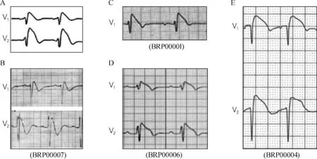
(A) True congenital type 1 Brugada syndrome electrocardiogram shown in comparison to (B) congenital hypokalemic periodic paralysis
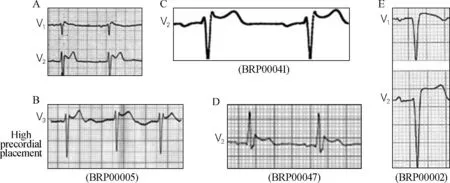
(A) True congenital type 2 Brugada syndrome shown in comparison to (B) congenital pectus excavatum causing mechanical mediastinal
Brugada phenocopies (BrP) are clinical entities with ECG findings identical to either the type 1 or type 2 Brugada ECG patterns yet differ etiologically from true congenital BrS[6-8]. These patient’s ECG findings are associated with an identifiable condition and upon resolution of that condition, the ECG pattern normalizes. Importantly, these patients have a negative sodium channel blocker provocative challenge suggesting a pathophysiological basis other than sodium channel dysfunction[7]. Alternatively, the sodium channel dysfunction may be transient occurring only during the inciting clinical event such as metabolic abnormality or ischemia, although this remains speculative. While the arrhythmogenic properties of BrS are well known along with the risk of sudden cardiac death, not much is known about the natural history of BrP. In an effort to facilitate research into the various etiologies, pathophysiology, and prognosis of the condition, we have developed an online educational portal and international registry for BrP (see www.brugadaphenocopy.com)[8]. This paper reviews previous cases of BrP, the various etiologies that have been identified in BrP, and future directions in research on this relatively new clinical entity.
2Classification of BrP
BrP may be induced under a multitude of clinical circumstances that have been characterized in six distinct etiological groups: (i) metabolic conditions, (ii) mechanical compression, (iii) ischemia, (iv) myocardial and pericardial disease, (v) ECG modulation, and (vi) miscellaneous(Tab.1)[7, 9]. In addition, they are further classified according to the presenting ECG morphology: type 1 BrP if the patient presents with a type 1 Brugada ECG pattern; or type 2 BrP if the patient presents with a type 2 Brugada ECG pattern. Cases of BrP are further sub-classified as A, B, and C(Tab.1). Class A includes true BrP in which all mandatory diagnostic criteria have been including negative provocative challenge with a sodium channel blocking agent. Class B includes cases highly suspected to be BrP; however, the mandatory diagnostic criteria are incomplete. These are cases where a provocative challenge is not possible due to factors such as the patient being deceased or lost to follow-up. Class C includes highly suspected cases of BrP; however, provocative testing was not justified such as in cases with recent surgical right ventricular outflow tract(RVOT) manipulation[10].
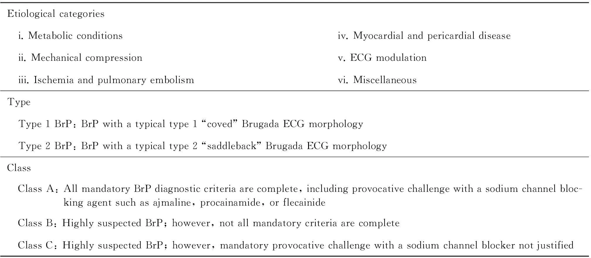
Tab.1 Brugada phenocopy etiological categories and morphological classification system
Adapted from Gottschalk et al[10]
3Differentiating BrP from BrS
Establishing a clear diagnostic distinction between BrP and BrS is of the utmost importance[11-12]. The fact that BrS may occur spontaneously, with no personal or family history of suggestive of the condition and in the absence of known genetic abnormalities makes differentiating between the two conditions challenging[1-2]. Current recommendations for diagnosis and risk stratification of BrS rely on a combination of clinical factors, presenting ECG morphologies, and positive provocative testing with a sodium channel blocking agent. It is this combination of factors that guides the clinical decision of whether to implant an ICD or not[1]. Similarly, the clinical diagnosis of BrP requires clinical factors(an identifiable underlying condition that triggers the ECG pattern in the absence of personal or family history suggestive of BrS), presenting ECG morphologies identical to BrS that normalizes upon resolution of the underlying condition, and a negative provocative test with sodium channel blocking agents[7]. Tab.2 depicts the current recommended systematic diagnostic criteria for BrP.
The importance of this distinction is highlighted by the natural history of the two conditions; while for patients with high risk of BrS who are candidates for treatment with an ICD, the clinical implications of BrP remain unknown. Therefore, current recommendations for treatment of BrP focus on resolving the underlying condition.
3.1Methods
Using Ovid MEDLINE(R) and Ovid OLDMEDLINE(R) from 1947 to May Week 1 2015, EMBASE from 1980 to May Week 1 2015, and PubMed we performed a search using “Brugada Syndrome”MeSH heading combined with keyword searches for “Brugada Phenocopy”“mimicking Brugada”“induced Brugada syndrome”“acquired Brugada”“Brugada-like”“Brugada type”, and “Brugada pattern”. The results were reviewed and case reports were selected according to the following inclusion criteria: (i) the case report was published; (ii) the case report includes a legible ECG showing a Brugada ECG pattern; (iii) the patient in the case does not have true BrS determined by a low clinical probability, absence of a positive provocative test, and/or absence of a positive genetic test for BrS. Case reports were excluded if they reported an example of confirmed (or suspected) underlying BrS being unmasked by drug, fever, or other clinical circumstances.

Tab.2 Brugada phenocopy diagnostic criteria
Adapted from Anselm et al[13]. Abbreviation: ECG, electrocardiogram; RVOT, right ventricular outflow tract; SCN5A, sodium channel voltage-gated type V alpha subunit
3.2Results
Fifty-eight case reports, reporting 71 individual cases of BrP met our inclusion criteria. These cases were classified according to the updated classification of BrP[9]. Tab.3 depicts an updated summary of BrP as of May, 2015. Since the first literature review on BrP published by our group in 2012, 37 cases have been added to the database[7]. These include BrP in the context of myocardial infarction, pulmonary embolism, and intracranial hemorrhage.
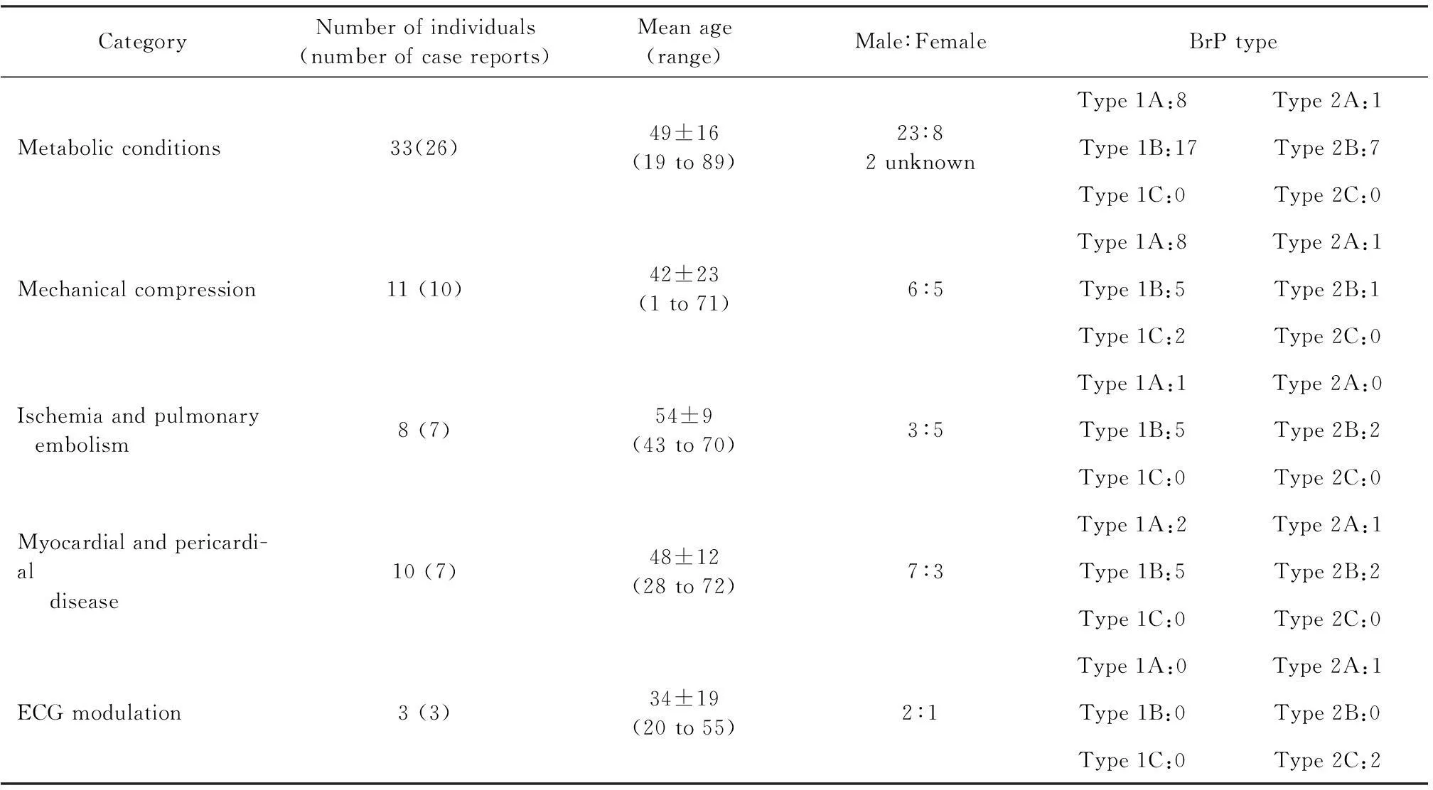
Tab.3 Summary of Brugada Phenocopy Registry
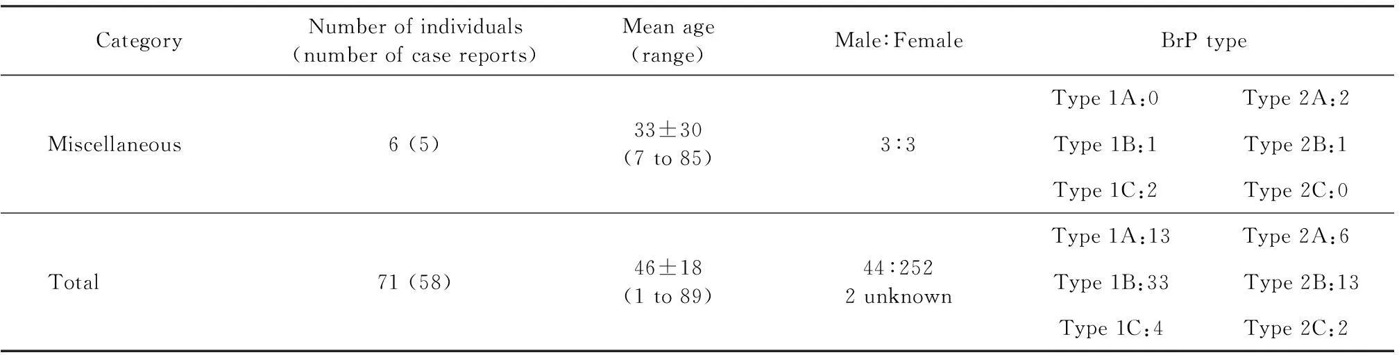
Continued
Quoted from Gottschalk et al[8]
3.2.1BrP:myocardial infarctionAt this time,there are 5 cases of BrP in the context of myocardial ische-mia[14-18]. Of the reports demonstrating BrP in the context of myocardial infarction, 3 were the result of a right coronary artery occlusion and 2 were the result of a left coronary artery occlusion. All 5 of the cases presented without a family history or personal history suggestive of BrS, thus making BrP very likely. Two of the 5 cases was a confirmed type 1A BrP in that all the diagnostic criteria were met[14, 18].
Is ischemia eliciting BrP or unmasking BrS?
Interestingly, ischemia may either induce BrP as previously discussed, or it may result in unmasking of true BrS through modulation of myocardial sodium channels[19-20]. Two interesting case reports demonstrate this phenomenon and illustrate the importance of proper diagnostic investigations in this patient population[18, 21]. Jiang et al[18]described a patient who presented with a type 1 Brugada ECG pattern and elevated troponin Ⅰ levels shortly after radiofrequency catheter ablation as treatment of atrial fibrillation. The patient underwent coronary angiography that revealed a sub-total occlusion in the first diagonal. In this case, we hypothesized that the ST-segment elevations produced after this procedure were likely the result of ischemia[20]. This patient underwent a diagnostic provocative test using a sodium channel blocker for BrS. The result was negative,which coupled with the patient’s lack of personal or family history suggestive of BrS, which was diagnostic for a type 1A BrP due to myocardial ischemia. This case is the second confirmed BrP reported in the context of myocardial ischemia in the international registry.
In contrast to the previously discussed case, Garg et al[21]discussed a particularly interesting case of a patient presenting with a Brugada ECG pattern during an acute coronary event. This patient subsequently underwent a provocative test with ajmaline, which proved to be positive, thereby diagnosing BrS in this patient that was unmasked by ischemia.
These cases highlight the need to investigate the cause of a Brugada ECG pattern. Indeed, the patient described by Garg et al would require risk stratification in order to determine his risk for SCD.
3.2.2BrP:pulmonary embolismTo date, there have been 4 reported cases of BrP in the context of acute pulmonary embolism[22-24]. Three of these cases presented with a type 1 Brugada ECG pattern while the final case presented with a type 2 Brugada ECG pattern. The phenomenon of right precordial ST-elevations caused by moderate to severe pulmonary emboli has been previously reported in the literature[25-28]. In all 4 reported cases of BrP in the context of acute pulmonary embolism, the responsible embolism was either large or massive. A speculated mechanism behind these ECG manifestations is the idea that large emboli results in significant right ventricular strain leading to transient sodium channel dysfunction[13].Zhan et al[24]further postulated that the right ventricular strain results in transmural ischemia of the right ventricle leading to the ST-elevations through a similar mechanism that is found during BrP in the context of right ventricular myocardial infarctions.
3.2.3BrP:neurological diseaseOur group published the first documented case of a confirmed BrP caused by a neurological condition in 2014[29].Briefly, an 85-year-old man was admitted to ICU after syncope complicated by traumatic head injury. A CT scan demonstrated temporo-parietal subarachnoid hemorrhage and 12 hours after the patient was admitted, he developed a type 2 Brugada ECG pattern that resolved within 9 hours. This patient underwent a subsequent CT scan demonstrating an insular hematoma with intraventricular hemorrhage.The patient underwent sodium channel provocation with flecainide that failed to induce a Brugada ECG pattern.He was therefore diagnosed with a type 2A BrP.There are various reports in the literature of cardiac manifestations of neurologic disease[30-32].These complications include ST-segment elevation on ECG in cases of subarachnoid hemorrhage. Indeed, there are multiple reports[30, 33-35]of the relationship between the insular region and ECG abnormalities(including ST-segment elevation).We postulated that our patient’s Brugada ECG manifestations were a result of neurological dysfunction caused by hematoma formation in the insular region of the brain[29].It is likely that resolution of the pressure effect of the hematoma resulted in normalization of the ECG pattern in this patient.To our knowledge,this is the only published case of BrP due to a neurologic disease to date and the only case of its kind registered in our international database.
3.3Future directions
Most reported cases of BrP discussed a single inciting event such as metabolic derangement or myocardial ischemia during which the Brugada ECG pattern was observed. Recently, we advanced the concept of clinical reproduction in BrP where resolution of an underlying condition resulted in normalization of the ECG[36]. The pattern subsequently reappeared when the same underlying condition reoccurred. In short, a young patient with diarrhea was admitted to the hospital following the development of severe hypokalemia(K+1.5 mEq/L). An ECG was performed and demonstrated a type 1 Brugada ECG pattern. After correction of the patient’s metabolic abnormalities, an ECG showed resolution of the Brugada ECG pattern. During the same admission, the patient again became hypokalemic(K+2.6 mEq/L) due to ongoing gastrointestinal losses. The corresponding ECG demonstrated recurrence of the type 1 Brugada ECG pattern that subsequently normalized after correction of the metabolic derangement. This patient had no personal or family history suggestive of BrS and underwent a negative provocative challenge. This is the first case that demonstrated clinical reproducibility of BrP.
Future investigations of BrP must focus on the development of experimental models designed to fully understand the mechanisms behind BrP. Through experimentation, we will begin to understand whether BrP are the result of transient sodium channel dysfunction that is not reproduced by sodium channel blocking agents, or whether the key to the ECG manifestations lies in malfunction of other ion channels. We may find that BrP and BrS are indeed entities along a continuous spectrum of ion channel dysfunction.
3.4Conclusions
The BrP international registry is aimed at furthering knowledge on the etiology, pathophysiology, and natural history of these phenomena. At this point, it is unknown whether BrP is a risk factor for malignant arrhythmias since there have been no studies examining the long-term prognosis of these patients. The question of clinical reproducibility of BrP has been demonstrated through one report thus far; however, we will be looking for more such examples in the future. Questions that remain unanswered include whether development of BrP predisposes patients to malignant arrhythmias under certain clinical conditions, or whether a patient that develops BrP in the context of one etiology is at risk of developing BrP in another etiology. The international database will aid in answering these questions through collating patients, identification of patterns, and assessing the long-term morbidity and mortality of these patients with longitudinal follow-up. Within the first year of the website being operational, it has generated over 2 000 unique visitors resulting in the submission of a number of potential BrP cases. With further awareness of the concept and more widespread use of the term “Brugada phenocopy” in the literature, we expect this number to grow more rapidly. In order to facilitate research on this phenomenon, we encourage other investigators to use the term “Brugada phenocopy” when reporting such cases. In addition, we invite the submission of cases to our online international registry at www.brugadaphenocopy.com.
References
[1] Bayés de Luna A, Brugada J, Baranchuk A, et al. Current electrocardiographic criteria for diagnosis of Brugada pattern:a consensus report[J]. J Electrocardiol, 2012, 45(4):433-442.
[2] Probst V, Wilde AA, Barc J, et al. SCN5A mutations and the role of genetic background in the pathophysiology of Brugada syndrome[J]. Circ Cardiovasc Genet, 2009, 2(6):552-557.
[3] Postema PG, Wolpert C, Amin AS, et al. Drugs and Brugada syndrome patients:review of the literature, recommendations, and an up-to-date website(www.brugadadrugs.org)[J]. Heart Rhythm, 2009, 6(9):1335-1341.
[4] Wilde AA, Antzelevitch C, Borggrefe M, et al. Proposed diagnostic criteria for the Brugada syndrome:consensus report[J]. Circulation, 2002, 106(19):2514-2519.
[5] Antzelevitch C, Brugada P, Borggrefe M, et al. Brugada syndrome:report of the second consensus conference:endorsed by the Heart Rhythm Society and the European Heart Rhythm Association[J].Circulation. 2005, 111(5):659-670.
[6] Anselm DD, Gottschalk BH, Baranchuk A. Brugada phenocopies:consideration of morphologic criteria and early findings from an international registry[J]. Can J Cardiol, 2014, 30(12):1511-1515.
[7] Baranchuk A, Nguyen T, Ryu MH, et al. Brugada phenocopy:new terminology and proposed classification[J]. Ann Noninvasive Electrocardiol, 2012, 17(4):299-314.
[8] Gottschalk BH,Anselm DD,Baranchuk A.Brugada Phenocopy international registry and online educational portal[EB/OL].(2013-08-01)[2015-06-10].www.brugadaphenocopy.com.
[9] Anselm DD, Baranchuk A. Brugada phenocopy:redefinition and updated classification[J]. Am J Cardiol, 2013, 111(3):453.
[10] Gottschalk B, Anselm DD, Baranchuk A. Brugada phenocopy:morphological classification and importance of provocative testing[J]. Ann Noninvasive Electrocardiol, 2014, 19(6):604-605.
[11] Gottschalk BH, Anselm DD, Baranchuk A. Brugada phenocopies are the leading differential diagnosis of Brugada syndrome[J]. Clin Med, 2015, 15(3):308-309.
[12] Gottschalk BH, Anselm DD, Baranchuk A. Brugada syndrome unmasked by ischemia needs full risk evaluation[J]. Intern Emerg Med, 2015, 10(1):111-112.
[13] Anselm DD, Baranchuk A. Brugada Phenocopy in the context of pulmonary embolism[J]. Int J Cardiol, 2013, 168(1):560.
[14] Anselm D, Barbosa-Barros R, de Sousa Belém L, et al. Brugada Phenocopy induced by acute inferior ST-segment elevation myocardial infarction with right ventricular involvement[J].Inn Card Rhythm Manag,2013,4:1092-1094.
[15] Eggebrecht H, Wieneke H, Erbel R. Brugada-like ECG pattern in a patient with isolated right ventricular infarction[J]. Herz, 2009, 34:327.
[16] Nakazato Y,Kurata T,Yamaguchi H.ST segment elevation in the precordial leads mimicking Brugada syndrome[J].Heart,2000,83(2):216.
[18] Jiang C, Wang R, Lu W, et al. A Brugada-like electrocardiogram pattern induced after radiofrequency catheter ablation of atrial fibrillation[J]. Int J Cardiol, 2015, 178:e1-e4.
[19] Di Diego JM, Antzelevitch C. Acute myocardial ischemia:cellular mechanisms underlying ST segment elevation[J]. J Electrocardiol, 2014, 47(4):486-490.
[20] Gottschalk BH,Anselm DD,Baranchuk A.Brugada phenocopy induced by ischemia or Brugada syndrome unmasked by ischemia?[J].Int J Cardiol,2014,177(2):619-620.
[21] Garg P,Nelson T,Sahu J,Sheridan P.Is this ST-elevation because of myocardial ischemia or a Brugada pattern?An interesting case review[J].Intern Emerg Med,2015,10(2):249-250.
[22] Wynne J, Littmann L. Brugada electrocardiogram associated with pulmonary embolism[J]. Int J Cardiol, 2013, 162(2):e32-e33.
[23] Yetkin Ö, Turhan H. Massive pulmonary embolism simulating electrocardiographic pattern of Brugada syndrome[J]. TAPE, 2007, 5:260-264.
[24] Zhan ZQ, Wang CQ, Nikus KC, et al. Brugada phenocopy in acute pulmonary embolism[J]. Int J Cardiol, 2014, 177(3):e153-e155.
[25] Grand A, Taine B, Huret JF, et al. An overlooked electrocardiographic sign of acute embolic cor pulmonale:elevation of the ST segment in right precordial leads[J]. Ann Cardiol Angeiol(Paris), 1985, 34(5):361-362.
[26] Cassin M, Charmet P, Collazzo R, et al. Electrocardiographic aspects simulating acute myocardial infarct in massive pulmonary embolism. Description of 2 cases[J]. G Ital Cardiol, 1986, 16(10):882-885.
[27] Panneerselvam A, Dattatreya PV, Sridhar L, et al. ST-segment elevation in right precordial leads in acute pulmonary embolism[J]. JICC, 2011, 1:112-114.
[28] Zhan ZQ, Wang CQ, Wang ZX, et al. Significance of ST-segment deviation in patients with acute pulmonary embolism and negative T waves[J]. Cardiol J, 2015 May 25. doi:10.5603/CJ.a2015.0023. [Epub ahead of print]
[29] Labadet C, Gottschalk BH, Rivero M, et al. Brugada phenocopy in the context of intracranial hemorrhage[J]. Int J Cardiol, 2014, 177(3):e156-e157.
[30] Baranchuk A, Nault MA, Morillo CA. The central nervous system and sudden cardiac death:what should we know?[J]. Cardiol J, 2009, 16(2):105-112.
[31] Goldberger ZD, Creutzfeldt CJ, Goldberger AL. Catastrophic neurologic syndrome with dramatic ECG changes[J]. J Electrocardiol, 2014, 47(1):80-83.
[32] Saritemur M,Akoz A,Kalkan K,et al.Intracranial hemorrhage with electrocardiographic abnormalities and troponin elevation[J]. Am J Emerg Med, 2013, 31(1):271.e5-271.e7.
[33] Christensen H,Boysen G,Christensen AF,et al.Insular lesions,ECG abnormalities,and outcome in acute stroke[J].J Neurol Neurosurg Psychiatry, 2005, 76(2):269-271.
[34] Koppikar S, Baranchuk A, Guzmn JC, et al. Stroke and ventricular arrhythmias[J].Int J Cardiol,2013,168(2):653-659.
[35] Svigelj V, Grad A, Tekavcic I,et al. Cardiac arrhythmia associated with reversible damage to insula in a patient with subarachnoid hemorrhage[J].Stroke,1994,25(5):1053-1055.
[36] Genaro NR, Anselm DD, Cervino N, et al. Brugada phenocopy clinical reproducibility demonstrated by recurrent hypokalemia[J]. Ann Noninvasive Electrocardiol, 2014, 19(4):387-390.
Brugada拟表型:一个新的临床实体
ByronHGottschalk,DanielDAnselm,AdrianBaranchuk蒋祖勋 译并审校
[摘要]Brugada拟表型(Brugada phenocopy, BrP)是临床实体,它与真正的Brugada综合征(Brugada syndrome, BrS)具有相同的心电图波形,但前者是由各种临床状况诱发的。在某种相关的潜隐状况下,BrP的特征表现为心前区导联V1~V3呈1型或2型Brugada心电图波形。随着潜隐状况的纠正,这些心电图波形恢复正常。本研究中,我们回顾了BrP的分类、与BrS鉴别的方法及有关BrP病因的最新研究结果。此外,我们提供了BrP国际在线注册的更新数据,并讨论了BrP未来的研究方向。 表3Brugada拟表型注册的类别个体数量(病例报道的数量)平均年龄(范围)男性:女性BrP类型代谢状况33(26)49±16(19~89)23:82例未知Type1A:8Type2A:1Type1B:17Type2B:7Type1C:0Type2C:0机械压迫11(10)42±23(1~71)6:5Type1A:8Type2A:1Type1B:5Type2B:1Type1C:2Type2C:0缺血和肺动脉栓塞8(7)54±9(43~70)3:5Type1A:1Type2A:0Type1B:5Type2B:2Type1C:0Type2C:0心肌和心包疾病10(7)48±12(28~72)7:3Type1A:2Type2A:1Type1B:5Type2B:2Type1C:0Type2C:0心电图调制3(3)34±19(20~55)2:1Type1A:0Type2A:1Type1B:0Type2B:0Type1C:0Type2C:2其他6(5)33±30(7~85)3:3Type1A:0Type2A:2Type1B:1Type2B:1Type1C:2Type2C:0
[关键词]Brugada心电图波形; Brugada拟表型; Brugada综合征
1引言
Brugada综合征(Brugada syndrome, BrS)是一种遗传心源性猝死综合征,具有常染色体显性遗传和可变的外显率。超过70种基因突变与这种状况有关,大多数突变发现在心脏钠通道中[1]。在真正的BrS患者中,约20%~30%可检测出SCN5A基因突变[2]。这种状况的特征性心电图表现为在右心前区导联V1~V3上出现ST段改变,产生两个典型的波形。1型(穹窿型)Brugada心电图波形定义为迅速上升的ST段抬高(≥2 mm),紧接着一个较慢的下斜凹面或直线ST段,以及负性对称T波[图1:各种1型Brugada拟表型(Brugada phenocopy, BrP)的比较;(A) 真正的先天性1型BrS心电图;(B) 先天性低钾周期性瘫痪(1B型BrP);(C) 急性下壁ST段抬高型心肌梗死伴右室受累(1A型BrP);(D) 并行高钾、低钠及酸中毒(1A型BrP);(E) 急性肺动脉栓塞(1B型BrP)。图下数字是BrP国际注册的身份编号。][1]。2型(马鞍型)Brugada心电图波形表现为从等电位基线出现一个迅速上升的r′≥2 mm,紧接着一个凸面ST抬高(≥0.05 mV),伴V1导联变化的T波和V2导联正向或扁平的T波[图2:各种2型BrP的比较;……
登录APP查看全文