
单椎体压缩骨折 PKP 术后原椎体引起背痛复发因素的探讨
2016-05-12 12:03吴叶卜国云卢祥商卫林孙玉鹏侯树勋
中国骨与关节杂志 2016年1期
吴叶 卜国云 卢祥 商卫林 孙玉鹏 侯树勋
单椎体压缩骨折 PKP 术后原椎体引起背痛复发因素的探讨
吴叶 卜国云 卢祥 商卫林 孙玉鹏 侯树勋
作者单位:100048 北京,解放军总医院第一附属医院全军骨科研究所、北京市骨科植入医疗器械工程技术研究中心(吴叶、卢祥、商卫林、孙玉鹏、侯树勋);300222 天津医院 (卜国云)
【摘要】目的 探讨骨质疏松性单节段椎体骨折经皮椎体后凸成形术 (percutoneous kyphoplasty,PKP)后,责任椎体引起背部疼痛复发的原因,为预防和临床治疗提供参考。方法 笔者所在单位同一组医生自 2006 年 1 月至 2009 年 12 月共收治因骨质疏松引起单节段椎体压缩骨折而行 PKP 的患者共 52 例,其中 28 例术后随访>12 个月,其中男 11 例,女 17 例;年龄 68~86 岁,平均 (74.13±4.18) 岁。随访时间 12~34 个月,平均(18.50±7.26) 个月。按照是否复发背部疼痛分为 A、B 组:术后未复发背部疼痛为 A 组 (n=20),术后复发背部疼痛为 B 组 (n=8)。所有患者均记录年龄、性别、症状出现的时间,采用相同手术方法 (PKP),记录骨水泥注入量 (filling PMMP volume,FPV)。手术前、后及复查时均采用视觉模拟评分 (visual analogue scale,VAS)评估疼痛程度;通过骨密度 (bone mineral density,BMD) 比较手术前后及随访时 BMD 的 T 评分;通过 X 线、MRI 检查比较手术前后及随访时骨折椎体的压缩率 (compression rate,CR)、压缩的恢复率或丢失率,比较手术前后及随访时骨折椎体的后凸角度 (kyphotic angle,KA) 和后凸角度恢复率或丢失率;比较术后及随访时骨水泥 (甲基丙烯酸甲酯) 与椎体上下终板间距离 (postoperation PMMP to endplate distance,PPED) 的改变。对所有数据进行统计学分析。结果 背部疼痛复发患者原手术椎体均有不同程度的再骨折或塌陷。A、B 组 FPV 有差异,但无统计学意义。A、B 组 CR 术后均明显增加,两组差异无统计学意义;末次随访时 B 组 CR 恢复率为 (17.4±9.3) %,A 组为 (24.1±11.1) %,两组间差异有统计学意义 (P<0.05);KA 呈现同样的改变。A、B 两组手术前 VAS 评分分别为 (8.8±0.6) 分、(8.6±0.5) 分;术后分别为 (2.1±0.6) 分、(2.2±0.8) 分,与术前比较差异有统计学意义 (P<0.05);B 组末次随访时 VAS 评分为 (7.5±1.2) 分,与 B 组术后及 A 组末次随访 (3.1±0.7) 分比较,差异有统计学意义 (P<0.05)。A 组骨水泥上缘至上终板距离术后与末次随访比较,差异无统计学意义 (P>0.05);术后 A 组骨水泥下缘至下终板距离平均为 (2.5±0.6) mm,末次随访时为 (2.4± 0.7) mm,与术后比较差异无统计学意义 (P>0.05);术后 B 组骨水泥上缘至上终板距离平均为 (3.5±0.4) mm,末次随访时为 (2.7±0.9) mm,与术后比较差异有统计学意义 (P<0.05),术后 B 组骨水泥下缘至下终板距离平均为 (4.5±0.6) mm;末次随访时 (3.3±0.4) mm,与术后比较差异有统计学意义 (P<0.05)。结论 手术椎体再骨折或塌陷是引起单椎体压缩骨折 PKP 术后背痛复发的重要原因,适量的 FPV 可能有利于减少手术椎体再骨折或塌陷。适时监测 CR、KA、T 评分、PPED 的变化可以为预防复发背部疼痛提供参考。
【关键词】骨质疏松性骨折;骨折,压缩性;椎体后凸成形术;背痛;复发
经皮椎体后凸成形术 (percutaneous kyphoplasty,PKP) 是治疗原发性骨质疏松引起胸腰段椎体压缩骨折的主要手段,既可以即刻缓解患者背痛,又可以较好地恢复椎体的高度,防止后凸畸形的形成,治疗结果较为满意[1-3]。但是,临床上经常发现有些胸腰段压缩性骨折患者采用 PKP 治疗后,短期内疗效满意,后期却再次复发较为严重的腰背痛。引起复发背部疼痛的原因不一,文献中对于非手术椎体发生骨折引发背部疼痛的报道较多[4-6],而对于手术椎体再次引起背部疼痛的报道较少。笔者对所在单位2006 年 1 月至 2009 年 12 月采用 PKP 治疗的部分患者进行回顾性分析,探讨手术椎体再次引起严重背痛的原因,为预防和临床治疗提供参考。
资料与方法
一、入组和排除标准
1.入组标准:(1) 骨质疏松引起的单节段胸腰段椎体压缩骨折;(2) 手术方法均为经双侧椎弓根行球囊扩张 PKP、椎体内填充物为同一种骨水泥聚甲基丙烯酸甲酯 (polymethyl methacrylate,PMMA);(3) 术后没有出现手术相关并发症者;(4) 没有再次创伤病史;(5) 术后持续较为规律的抗骨质疏松治疗;(6) 随访时间>12 个月。
2.排除标准:(1) 肿瘤或长期使用激素等原因引起的病理性骨折;(2) 多节段骨折;(3) 爆裂骨折;(4) 有神经损害症状。
二、一般资料
2006 年 1 月至 2009 年 12 月,我科同一组医生共行 PKP 治疗单节段胸腰椎骨折患者 52 例,符合入组标准并获得随访者 28 例,其中男 11 例,女17 例。按照是否复发背部疼痛分为 A 组 (术后未复发背部疼痛) 和 B 组 (术后复发背部疼痛)。A 组20 例,平均年龄 (72.54±3.66) 岁,B 组 8 例,平均年龄 (76.13±6.87) 岁。A、B 两组患者出现胸腰段背部剧烈疼痛至行 PKP 手术的时间分别为 (7.2± 0.8) 天和 (6.4±0.3) 天,平均随访时间分别为(20.2±10.4) 个月和 (16.8±5.4) 个月。
三、手术方法
所有患者采用相同体位及麻醉,相同穿刺方法,在 C 型臂或 G 型臂机监测下完成手术,均采用经双侧椎弓根行球囊扩张的方法手术,空腔填充物为 PMMA,填充时骨水泥黏稠度基本一致。记录填充 PMMA 总量 (filling PMMP volume,FPV)。
四、复发背部疼痛的定义
骨质疏松引起胸腰段椎体压缩骨折,行球囊扩张 PKP 的患者,术后背部相同部位再次出现视觉模拟评分 (visual analogue scale,VAS)>6 分的持续疼痛定义为复发背痛。
五、临床观察指标
所有患者均记录年龄、性别、背部疼痛症状持续的时间。手术前、后及复查时均采用 VAS 评估疼痛程度。通过 BMD-T 评分 (双能 X 线骨密度仪行L1~3骨密度测定,计算 T 值) 比较手术前、后及随访时骨密度情况。
六、影像学测量及计算
由同一组医生通过 UNITE 影像系统进行测量:
骨折前椎体前缘正常高度=(骨折椎体上位椎体前缘高度+骨折椎体下位椎体前缘高度)/2;
骨折椎体压缩高度=骨折前椎体前缘正常高度-骨折椎体实际测量前缘高度;
骨折椎体压缩率 (compression rate,CR)=(骨折椎体压缩高度/骨折前椎体前缘正常高度)×100%。
骨折椎体后凸成角 (kyphotic angle,KA) 为骨折椎体上位椎体下缘水平线与骨折椎体下位椎体上缘水平线夹角;骨髓内与椎体上下终板间距离(postoperation PMMP to endplate distance,PPED) 为骨水泥上下缘与上下终板间的最小距离。
七、统计学分析
结 果
A、B 两组在年龄、症状持续时间、随访时间上差异无统计学意义 (P>0.05)。A、B 两组 FPV 分别为 (5.63±1.41) ml、(6.27±0.72) ml,两组之间虽然有一定的差异,但无统计学意义 (P>0.05)。A、B 两组 CR 术后均明显减少,两组 CR 恢复率差异无统计学意义 (P>0.05);末次随访时 B 组 CR 恢复率为 (17.4±9.3) %,A 组为 (24.1±11.1) %,两组间差异有统计学意义 (P<0.05),B 组末次随访 CR 为(32.0±10.6) %,与术后比较差异有统计学意义 (P<0.05)。A、B 两组术前 KA 分别为 (17.1±6.3) °、(21.4±5.9) °,A 组术后 KA 为 (11.8±5.3) °,B 组为 (16.3±4.5) °,两组间差异有统计学意义 (P< 0.05),B 组末次随访时 KA 为 (22.2±5.8) %,与术后比较差异有统计学意义 (P<0.05) (表 1)。A、B 两组手术前、后及末次随访的 VAS 评分见表 2。A、B 两组术后及末次随访时骨水泥上下缘至上下终板间距离见表 3。手术前、后及随访时骨密度有差异,但差异无统计学意义 (P>0.05) (表 4)。典型病例见图 1、2。
表 1 A、B 两组手术前、后及末次随访的影像学测量指标比较(±s,°)Tab.1 The data on image measuring between Group A and B pre-,post-operatively and final follow-up (±s,°)

表 1 A、B 两组手术前、后及末次随访的影像学测量指标比较(±s,°)Tab.1 The data on image measuring between Group A and B pre-,post-operatively and final follow-up (±s,°)
注:CR,骨折椎体压缩率;KA,骨折椎体后凸成角Notice:CR,compression rate; KA,kyphotic angle
项目 组别 术前 术后 末次随访CR A 组 40.5±11.3 24.1±11.1 25.1±11.2 B 组 38.8±13.6 17.4± 9.3 32.0±10.6 KA A 组 17.1± 6.3 11.8± 5.3 11.8± 5.3 B 组 21.4± 5.9 16.3± 4.5 22.2± 5.8
表 2 A、B 两组手术前、后及末次随访的背痛 VAS 评分比较(±s,分)Tab.2 A comparison between Group A and B pre-,post-operatively and final follow-up (±s,point)

表 2 A、B 两组手术前、后及末次随访的背痛 VAS 评分比较(±s,分)Tab.2 A comparison between Group A and B pre-,post-operatively and final follow-up (±s,point)
注:a与术前比较,差异有统计学意义 (P < 0.05);b与末次随访比较,差异有统计学意义 (P < 0.05);c与 A 组末次随访比较,差异有统计学意义(P < 0.05)Notice:aA comparison with preoperatively,statistically significant (P < 0.05);baComparison with the last follow-up,statistically significant (P < 0.05);caComparison with the last follow-up in Group A,statistically significant (P < 0.05)
组别 术前 术后 末次随访A 组 8.8±0.6 2.1±0.6a 3.1±0.7 B 组 8.6±0.5 2.2±0.8ab 7.5±1.2c
表 3 手术后及末次随访时骨水泥上下缘至上下终板间距离(±s,mm)Tab.3 The distance from PMMA margin to upper and lower endplate postoperatively and final follow-up (±s,mm)

表 3 手术后及末次随访时骨水泥上下缘至上下终板间距离(±s,mm)Tab.3 The distance from PMMA margin to upper and lower endplate postoperatively and final follow-up (±s,mm)
注:a末次随访与术后骨水泥距上终板距离比较,差异有统计学意义 (P < 0.05)。b末次随访与术后骨水泥距下终板距离比较,差异有统计学意义(P < 0.05)Notice:aThe distance from PMMA margin to upper end-plate postoperatively and final follow-up,statistically significant (P < 0.05);bThe distance from PMMA margin to lower end-plate postoperatively and final follow-up,statistically significant (P < 0.05)
项目 A 组 B 组骨水泥上缘与上终板距离术后 2.2±0.8 3.5±0.4末次随访 2.1±0.7 2.7±0.9a骨水泥下缘与下终板距离术后 2.5±0.6 4.5±0.6末次随访 2.4±0.7 3.3±0.4b
表 4 A、B 两组手术前、后及末次随访的骨密度 (L1~3) 比较(T 评分,±s)Tab.4 BMD (L1-3) between Group A and Group B pre-,postoperatively and final follow-up (T-score,±s)
组别 术前 术后 末次随访A 组 -3.59±0.78 -3.79±0.45 -3.65±0.71 B 组 -3.98±0.88 -4.00±0.31 -3.77±0.63

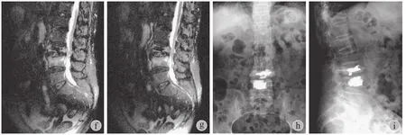
图 1 患者,姜某 a~b:第 1 次骨折,L3椎体骨折术前;c~d:第 1 次骨折术后;e~g:第 2 次骨折,L4椎体骨折术前;h~i:第 2 次骨折术后Fig.1 Patient Jiang a-b:the first fracture,L3fracture preoperatively; c-d:the first fracture postoperatively; e-g:the second fracture,L4fracture preoperatively; h-i:the second fracture postoperatively
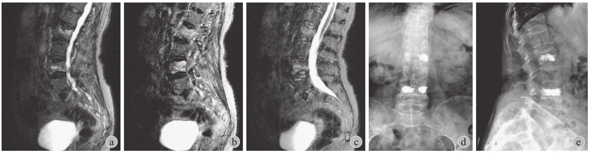
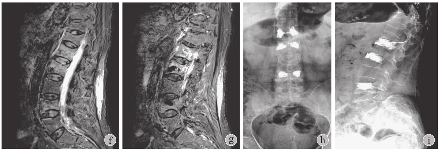
图 2 患者,张某 a~c:第 1 次骨折,L2和 L4椎体骨折术前;d~e:第 1 次骨折术后;f~g:第 2 次骨折,L2椎体骨折术前;h~i:第2 次骨折术后Fig.2 Patient Zhang a-c:The first fracture,L2,L4fracture preoperatively; d-e:The first fracture postoperatively; f-g:The second fracture,L2fracture preoperatively; h-i:The second fracture postoperatively
讨 论
随着人口老龄化,骨质疏松性椎体压缩骨折的患者逐渐增加。Lieberman 等[7]报道每年大约有400 万人发生骨质疏松性椎体压缩骨折。邱贵兴等[8]认为,骨质疏松引起骨折有多种危险因素,而低骨密度是最重要的危险因素。尚伟等[9]对北京老年骨质疏松性骨折与再骨折进行分析,也强调低骨密度是再骨折发生的重要因素。
对于骨质疏松引起的脊柱骨折,传统的手术治疗主要是采用脊柱内固定,对于高龄患者来说,该方法受到麻醉及创伤较大等多方面的限制,手术风险高,很多患者不愿意或不能接受。为了避免内固定带来的高风险,目前临床通常采用经皮椎体成形术 (percutaneous vertebroplasty,PVP) 或 PKP 手术,这种手术侵袭性小,局麻下可以完成,安全性高,可以为几乎所有骨质疏松性椎体压缩骨折患者所接受,正因为如此,PVP 或 PKP 技术已经得到广泛临床应用。
PKP 治疗的原理有以下 3 方面:(1) PMMA 等注入椎体后可以在短时间内凝固,在骨折椎体内产生支撑作用,即刻部分或完全恢复骨折部位的高度,重建脊柱稳定,防止骨折部位异常活动及微小再骨折的发生;(2) 椎体高度恢复可使脊柱重心后移,改善脊柱生物力学的变化,减少肌肉和韧带损伤引起的疼痛;(3) PMMA 注入时产生 70 ℃ 左右的热效应,可以破坏骨折部位的痛觉感受器。
尽管 PKP 治疗骨质疏松性椎体压缩骨折在临床得到广泛引用,但近年来研究发现,部分患者在手术后一定时期内再次出现背部剧烈疼痛。文献报道,引起 PKP 治疗后背部疼痛复发的原因主要是非手术椎体发生骨折或骨折椎体的再骨折或塌陷所致[10]。
Lo 等[11]研究 PVP 治疗后发生非治疗椎体骨折者占 6.8%,这些骨折患者在背部再次出现较为严重的疼痛,其中骨折椎体相邻椎体骨折者占 57.1%,约61.1% 发生在胸腰段。Tseng 等[12]有类似的报道,其发现相邻椎体骨折较其它椎体骨折发生更早,且发生其它椎体骨折的时间平均在术后 11.6 个月。有些作者报道的比例更高。Syed 等[13]报道原发性骨质疏松患者术后 1 年以上发生非手术椎体骨折约占 20.6%,长期服用激素患者发生非骨折椎体骨折的比例是原发性骨质疏松者的 1.84 倍。Kanis 等[14]报道长期使用激素者发生非手术椎体骨折的比例为37%。申勇等[15]通过回顾性分析 PVP 术后发生非手术椎体骨折病例认为,非手术椎体发生骨折可以引起背部严重疼痛,发生原因主要是与骨密度减低有关的自然病程,而患者性别、体重、手术部位及手术入路对椎体再骨折没有明显影响。
除了非手术椎体问题引起复发背痛外,也有文献报道了手术椎体本身问题再次引起严重的背部疼痛。Kim 等[16]研究了骨质疏松性椎体压缩骨折手术椎体再骨折的影响因素,其认为手术椎体再骨折可导致背痛复发。Heo 等[17]报道 PKP 或 PVP 治疗骨质疏松性椎体压缩骨折后 2 年,骨折椎体容易发生骨折椎体的形态变化,引起复发的背部疼痛。Heo 等[18]在另外一篇文章报道骨折椎体的再次塌陷,认为骨折椎体塌陷是引起复发背痛的重要原因。
笔者研究发现,骨质疏松性椎体压缩骨折采用PKP 治疗后常规应该进行正规的抗骨质疏松治疗,通过定期复查 X 线片,测量 CR、KA、PPED 等变化,可以早期判断治疗椎体是否发生再骨折或塌陷。所有 PKP 术后患者,随访 12 个月以上可以发现手术椎体有不同程度的高度丢失,但一般患者只主诉背部存在不适,有轻微疼痛,VAS 均<4 分,服用 NSAID 类药物可以缓解,而真正复发背部剧烈疼痛的患者较少,一旦出现复发背痛,应该考虑非手术椎体骨折或手术椎体发生较为严重的再骨折或塌陷。
笔者研究发现,治疗椎体再骨折和骨水泥下沉最重的 1 例,术后 15 个月开始出现手术部位疼痛,开始服用美洛昔康可以缓解,服用后 1 周疼痛加重,卧床翻身困难,复查发现原手术部位有压痛和叩击痛,X 线片及 MRI 见手术椎体再次骨折和塌陷。骨折椎体上位椎体前缘高度 21 mm,下位 22 mm,骨折后测量椎体前缘高度 11 mm,压缩高度 10.5 mm,CR 49.9%;术后 CR 20%,恢复率29.9%;随访时 CR 69.6%,丢失 49.6%。同时该患者填充骨水泥显著下沉,骨水泥下缘突破骨折椎体下位终板。该患者术后没有创伤病史,一直坚持抗骨质疏松治疗。但是,从该患者 X 线片可见,PKP 后填充 PMMA 过于饱满,这样 PMMA 承担了负重的主体,可能形成一种应力遮挡,对骨折椎体下终板造成过大的压力,最后 PMMA 突破下终板;同时,骨折椎体非骨水泥区骨质长期处于休闲状态,导致骨质疏松进一步发展,最后椎体前缘稍受力即发生再次骨折。
笔者认为,PKP 手术时 PMMA 的填充应该适量,不能一味追求恢复骨折椎体的高度而过多填充,操作过程中注意球囊扩张压力不能过大,以免在骨折椎体内造成过大的空腔。适当的球囊扩张,可以使骨折椎体得到一定的复位,同时可保持球囊上部骨密度适当,尽可能保持完整的上终板,这样在填充 PMMA 后,压力传递过程中上终板和骨组织可以将重力通过 PMMA 和周围的骨组织向下终板传递,从而避免 PMMA 产生过大的应力遮挡作用,较好地防止椎体再骨折和 PMMA 过多下沉,避免再次发生严重的背部疼痛。引起治疗椎体再骨折或塌陷的主要原因除患者本身骨质疏松进行性加重外,术中扩张球囊的压力、PMMA 填充量与再骨折或塌陷有直接关系。
由于本研究的患者例数少,有些患者术后因为其它疾病服用了较多的药物,活动量也不完全一样,虽然患者没有明显的创伤病史,引起手术椎体再骨折或塌陷的因素不能完全确定,因此,要说明引起手术椎体再骨折或塌陷的原因,尚需要对许多相关因素进行进一步的分析。
参考文献
[1]Bouza C,López T,Magro A,et al.Efficacy and safety of balloon kyphoplasty in the treatment of vertebral compression fractures:a systematic review.Eur Spine J,2006,15(7):1050-1067.
[2]Taylor RS,Fritzell P,Taylor RJ.Balloon kyphoplasty in the management of vertebral compression fractures:an updated systematic review and meta-analysis.Eur Spine J,2007,16(8):1085-1100.
[3]Voggenreiter G.Balloon kyphoplasty is effective in deformity correction of osteoporotic vertebral compression fractures.Spine,2005,30(24):2806-2012.
[4]Frankel BM,Monroe T,Wang C.Percutaneous vertebral augmentation:an elevation in adjacent-level fracture risk in kyphoplasty as compared with vertebroplasty.Spine J,2007,7(5):575-582.
[5]Korovessis P,Zacharatos S,Repantis T,et al.Evolution of bone mineral density after percutaneous kyphoplasty in fresh osteoporotic vertebral body fractures and adjacent vertebrae along with sagittal spine alignment.J Spinal Disord Tech,2008,21(4):293-298.
[6]Pflugmacher R,Schroeder RJ,Klostermann CK.Incidence of adjacent vertebral fractures in patients treated with balloon kyphoplasty:two years’ prospective follow-up.Acta Radiol,2006,47(8):830-840.
[7]Lieberman IH,Dudeney S,Reinhardt MK,et al.Initial outcome and efficacy of “kyphoplasty” in the treatment of painful osteoporotic vertebral compression fractures.Spine,2001,26(14):1631-1638.
[8]邱贵兴,陈宾,翁习生,等.老年骨质疏松骨折主要部位的骨折阈值测定研究.中华医学杂志,2005,85(16):1113-1116.
[9]尚伟,余卫,徐苓,等.北京老年骨质疏松性椎体初发骨折与再骨折相关性分析.中华医学杂志,2005,85(2):84-87.
[10]李亮,聂艳,姜保恩,等.经皮椎体成形术后手术椎体再发骨折的相关因素分析.实用骨科杂志,2014,20(4):339-343.
[11]Lo YP,Chen WJ,Chen LH,et al.New vertebral fracture after vertebroplasty.Trauma,2008,65(6):1439-1445.
[12]Tseng YY,Yang TC,Tu PH,et al.Repeated and multiple new vertebral compression fractures after percutaneous transpedicular vertebroplasty.Spine,2009,34(18):1917-1922.
[13]Syed MI,Patel NA,Jan S,et al.Symptomatic refractures after vertebroplasty in patients with steroid-induced osteoporosis.AJNR Am J Neuroradiol,2006,27(9):1938-1943.
[14]Kanis JA,Johansson H,Oden A,et al.A meta-analysis of prior corticosteroid use and fracture risk.J Bone Miner Res,2004,19(6):893-899.
[15]申勇,刘法敬,张英泽,等.骨质疏松椎体压缩性骨折PVP术后非手术椎体骨折的相关因素分析.中国脊柱脊髓杂志,2010,20(12):975-979.
[16]Kim YY,Rhyu KW.Recompression of vertebral body after balloon kyphoplasty for osteoporotic vertebral compression fracture.Eur Spine J,2010,19(11):1907-1912.
[17]Heo HD,Cho YJ,Sheen SH,et al.Morphological changes of injected calcium phosphate cement in osteoporotic compressed vertebral bodies.Osteoporos Int,2009,20(12):2063-2070.
[18]Heo DH,Chin DK,Yoon YS,et al.Recollapse of previous vertebral compression fracture after percutaneous vertebroplasty.Osteoporos Int,2009,20(3):473-480.
(本文编辑:王萌)
.作者须知 Instruction for authors.
An analysis of recurrent back pain induced by primary single vertebral compression fracture after percutaneous kyphoplasty
WU Ye,BU Guo-yun,LU Xiang,SHANG Wei-lin,SUN Yu-peng,HOU Shu-xun.Orthopaedic Institute,the first Affiliated Hospital of PLA General Hospital,Beijing,100048,PRC
【Abstract】Objective To explore the reasons of recurrent back pain induced by the osteoporosis single vertebral compression fracture after percutaneous kyphoplasty (PKP) surgery and to provide clinical and preventable guiding.Methods In the authors’ hospital,52 patients experienced PKP executed by the same group of doctors from January 2006 to December 2009 due to osteoporosis single vertebral compression fracture.In the 52 patients,28 patients (male,11; female,12; age 68 to 86 years,averaged 74.13 ± 4.18 years) were followed up for more than 12 months.The duration of the follow-up was from 12 to 34 months (averaged 18.50 ± 7.26 months).The patients were divided into 2 groups depending on the recurrent back pain (Group A:recurrent pain,n = 20; Group B:no pain,n = 8).Age,gender,and the duration of symptoms were recorded.Patients underwent PKP and the filing PMMPbook=63,ebook=68volume (FPV) was recorded.Preoperatively,postpostoperatively and in the follow-up,visual analogue scale (VAS) was employed to evaluate the pain.T score was applied to compare the bone mineral density (BMD).Compression rate (CR),recovery rate of the compression or lost rate of the recovery of the fracture were compared on plain films and MRI images.Kyphosis angle (KA),recovery or lost rate of the kyphosis angle were compared on plain films and MRI images.The distance from PMMP to end-plate was compared on plain films.The data were analyzed statistically.Results The primary surgical vertebrae presented refracture or collapse in patients with recurrent back pain.There were differences between Group A and B in FPV but not on statistics.CR increased both in Group A and B without statistical differences postoperatively,but the recovery rate in Group B was (17.4 ± 9.3) % compared with that (24.1 ± 11.1) % in Group A with statistical differences (P < 0.05),and the same trend was seen on KA.The preoperative VAS score were (8.8 ± 0.6),(8.6 ± 0.5) in Group A and B respectively comparing those were (2.1 ± 0.6),(2.2 ± 0.8) postoperatively with statistical differences (P < 0.05).The VAS was (7.5 ± 1.2) at the final follow-up in Group B compared with that in Group B postoperatively and that in Group A at the final follow-up (3.1 ± 0.7) with statistical significances (P < 0.05).In Group A,the distance from the upper end-plate to PMMP were (2.2 ± 0.8) mm postoperatively and (2.1 ± 0.7) mm at the final follow-up without statistical differences (P > 0.05).The distance from the lower end-plate to PMMP were (2.5 ± 0.6) mm postoperatively and (2.4 ± 0.7) mm at the final follow-up without statistical differences (P > 0.05).In Group B,the distance from the upper end-plate to PMMP were (3.5 ± 0.4) mm postoperatively and (2.7 ± 0.9) mm at the final follow-up with statistical differences (P < 0.05).The distance from the lower end-plate to PMMP were (4.5 ± 0.6) mm postoperatively and (3.3 ± 0.4) mm at the final follow-up with statistical differences (P < 0.05).Conclusions The surgical vertebrae refracture or collapse are important reasons for recurrent back pain after PKP for single vertebral compression fracture.Detection of the changes of CR,KA,T-score and PPED can provide guiding for the prevention of recurrent back pain.
【Key words】Osteoporotic fractures; Fractures,compression; Kyphoplasty; Back pain; Recurrence
(收稿日期:2015-02-09)
DOI:10.3969/j.issn.2095-252X.2016.01.014
中图分类号:R683.2
猜你喜欢
中国伤残医学(2022年14期)2022-12-23
祝您健康(2022年1期)2022-01-10
中老年保健(2021年2期)2021-08-22
世界最新医学信息文摘(2021年12期)2021-06-09
科学之谜(2020年4期)2020-06-19
保健与生活(2019年23期)2019-09-10
意林·全彩Color(2018年9期)2018-10-12
中国实用医药(2016年29期)2016-12-26
大家健康(2016年4期)2016-12-23
华夏医学(2016年4期)2016-12-12
