多层螺旋CT三维血管成像对腹部血管疾病的诊断价值
2016-05-14 17:40:38陈志林
中外医疗
2016年6期
陈志林
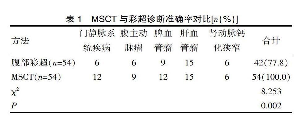
[摘要] 目的 探讨多层螺旋CT三维血管成像对腹部血管疾病的诊断价值。 方法 整群选取2014年5月—2015年8月该院收治的疑似腹部血管性疾病的54例患者纳入该研究,采用多层螺旋CT腹部血管成像扫查(MSCT)方法进行肝脏动脉期与门脉期增强扫描,对比腹部彩超与MSCT的检测结果。 结果 多层螺旋CT三维成像对于腹部大、中、小血管形态改变可予以清晰显示,对于腹部脏器肿瘤其他表现也可同时展开观察,诊断准确率为100.0%(54/54),彩超诊断准确率为77.8%(42/54),多层螺旋CT对于腹部血管性疾病检出率远高于腹部彩超(P<0.05)。 结论 多层螺旋CT腹部血管成像扫查属于无创检查技术的一种,其对于腹部血管疾病患者血管走行以及血管与周围脏器之间的关系可展开多角度乃至多方位观察,有效支持了临床诊断工作。
[关键词] 腹部血管疾病;多层螺旋CT;数字减影血管造影;诊断价值
[中图分类号] R814.42 [文献标识码] A [文章编号] 1674-0742(2016)02(c)-0185-02
Value of Multi-slice Spiral CT Three-dimensional Angiography in Diagnosis of Abdominal Vascular Diseases
CHEN Zhi-lin
Jiangbei Hospital District, Zhongda Hospital Affiliated to Southeast University, Nanjing, Jiangsu Province, 210044 China
[Abstract] Objective To observe the value of multi-slice spiral CT three-dimensional angiography in diagnosis of abdominal vascular diseases. Methods 54 cases of patients suspected with abdominal vascular diseases admitted and treated in our hospital from May 2014 to August 2015 were selected as the research object, and their livers in?hepatic artery phase?and polal vein phase were given enhancement scanning by the method of multi-slice spiral CT angiography in the abdomen, the detection results of abdominal Doppler ultrasound and MSCT were compared. Results The morphological changes of large, medium and small micrangium in the abdomen could be clearly showed by multi-slice spiral CT three-dimensional angiography, at the same time, the other manifestations of abdominal viscera tumor could be observed, and the diagnostic accuracy rate of it was 100.0%(54/54)and of Doppler ultrasound was 77.8%(42/54), the detection rate of multi-slice spiral CT for abdominal vascular diseases was much higher than that of Doppler ultrasound(P<0.05). Conclusion Multi-slice spiral CT angiography in the abdomen belongs to one of non-invasive examination technologies, which can make a multi-angle and multi-azimuth observation of vessel arranged and the correlation between the blood vessel and peripheral viscera in patients with abdominal vascular diseases, and it effectively supports the clinical diagnosis.
[Key words] Abdominal vascular diseases; Multi-slice spiral CT; Digital substraction angiograph; Diagnostic value
随着影像技术的快速发展,多层螺旋CT技术是影像学领域的一大突破,特别是16层CT的研发与应用。多层螺旋CT容积扫描速度更快,且Z轴分辨率更高,对于腹部血管三维重组图像质量有明显的提升,进而推动了CT血管造影成像技术的进步[1]。该文以数字减影血管造影检查技术为对照,总结分析了2014年5月—2015年8月该院收治的54例疑似腹部血管性疾病患者的多层螺旋CT三维血管成像结果,以便于对腹部血管疾病做出直观、准确的评价,现报道如下。
1 资料与方法
1.1 一般资料
整群选取2014年5月—2015年8月该院收治的疑似腹部血管性疾病的54例患者纳入该研究,其中男30例,女24例;年龄40~68岁,平均年龄(56.7±5.2)岁。
1.2 方法
检测仪器为飞利浦生产的MX-16型16层螺旋CT,常规腹部平扫联合增强扫描。其中扫描层厚设定为1.25 mm,转速为0.5 s/360°,螺距比为1.75∶1,管电压设定为120 kV,管电流在平扫以及增强扫描下分别设定为200 mA、350~400 mA,扫描持续10~15 s。……
登录APP查看全文