Studies on the Interaction of Perfluorononanoic Acid with Human Serum Albumin by Multi-Spectroscopic,Molecular Docking and Isothermal Titration Calorimetry Techniques
2016-06-05 14:58HUTaoyingHUANGFangZHOUShanshanLIUYing
光谱学与光谱分析 2016年12期
HU Tao-ying,HUANG Fang,ZHOU Shan-shan,LIU Ying,2*
1. College of Life and Environmental Sciences,Minzu University of China,Beijing 100081,China 2. Beijing Engineering Research Center of Food Environment and Public Health,Minzu University of China,Beijing 100081,China
Studies on the Interaction of Perfluorononanoic Acid with Human Serum Albumin by Multi-Spectroscopic,Molecular Docking and Isothermal Titration Calorimetry Techniques
HU Tao-ying1,HUANG Fang1,ZHOU Shan-shan1,LIU Ying1,2*
1. College of Life and Environmental Sciences,Minzu University of China,Beijing 100081,China 2. Beijing Engineering Research Center of Food Environment and Public Health,Minzu University of China,Beijing 100081,China
Perfluorononanoic acid (PFNA) is the third most frequently detected in serum among all perfluoroalkyl acids (PFAAs) which is a kind of toxic emerging environmental contaminant. The influence of PFNA on the conformation and even function of human serum albumin (HSA) is still just at the beginning of research. The attempt of this paper was to completely elucidate the interaction mechanism of PFNA with HSA by means of multi-spectroscopic,molecular docking and isothermal titration calorimetry (ITC) techniques. The inner filter effect of all fluorescence data in the paper was eliminated to get accurate binding parameters. The results showed that the fluorescence of HSA was quenched by PFNA through a combined quenching procedure of dynamic and static quenching. Through site marker competitive experiments,subdomain IIA of HSA had been assigned to possess the high-affinity binding site of PFNA. Furthermore,molecular docking reconfirmed that PFNA was bound in subdomain IIA mainly through polar force,hydrophobic interaction and halogen-bond,and the calculated free energy was -26.54 kJ·mol-1which indicated that the PFNA molecule exhibited large binding affinity towards HSA. The thermodynamic characterizations of two different classes of binding sites by ITC displayed that the first class with a higher affinity constant was dominated by an enthalpic contribution due to electrostatic interactions and halogen-bond,whereas the second class with a lower affinity constant was preponderated by hydrophobic interaction. The three-dimensional fluorescence revealed that the conformation of HSA was changed and the hydrophobicity of the Trp and Tyr residues microenvironment increased after formation of PFNA-HSA complex. The alterations of the protein secondary structure were quantitatively calculated from circular dichroism (CD) spectroscopy with reduction ofα-helix content about 14.3%,β-sheet 5.3%,β-turn 3.5%,and augment in random content from 14.4% to 37.5%. Above results revealed that the binding of PFNA with HSA can alter the secondary structure of HSA,further probably affecting HSA physiological function. The results can provide insights with the binding mechanism of PFNA with HSA and salient biophysical and biochemical clues on elucidating the transport and distribution of PFNA in vivo.
Perfluorononanoic acid; Human serum albumin; Multi-spectroscopic techniques; Molecular docking; Isothermal titration calorimetry
Introduction
Human serum albumin (HSA) is a well-known target because of its availability of hydrophobic pockets inside the network and flexibility to adapt its shape. Over recent decades,many researches have centered on the interaction of HSA with various small molecules,such as drugs,metals,dyes,environmental hormone,pesticide and fertilizer,etc.[1],which will alter the distribution,free concentration,metabolism and elimination of the small molecule and consequently affect the levels of its activity and toxicity in organism[2].
Perfluorononanoic acid (PFNA,structure shown in Fig.1) is a representative of the synthetic perfluoroalkyl acids (PFAAs) which are composed of hydrophobic and hydrophilic functional group[3],and widespread used as surfactants in firefighting foams,food packaging,polymer additives and water- and stain-resistant coatings,which are one large class of the active ingredients receiving comparatively little attention but used in large amounts throughout the world[4]. Numerous reports have shown that PFNA was the third most frequently detected in serum after perfluorooctane sulfonate (PFOS) and perfluorooctanoic acid (PFOA)[5]. In recent years,most interaction studies of PFAAs with proteins have been focused on the eight carbon atoms of PFOS and PFOA[6]. However,toxicological researchers demonstrated that compared with PFOS and PFOA,PFNA was more likely to accumulate and express reproductive toxicity,hepatotoxicity and immunotoxicity[7]in vivo. Thus,the binding of PFNA to HSA affected conformation,physiological function and activity of HSA,and hindered the transport of endogenous materials. By far,only few trials have been reported on the binding of PFNA proteins[8],and they were insufficient on the binding mechanism,such as lacking the specific information about binding sites,mainly driving force,changes for conformation and secondary structure of protein,which are of great importance for perfectly demonstrating the interaction mechanism of PFNA with proteins.
In this paper,the binding mechanism and thermodynamic characterization of PFNA-HSA interactions at molecular level is elucidated by multi-spectroscopic,molecular docking and isothermal titration calorimetry (ITC) approaches.
1 Experimental
1.1 Materials
PFNA (Shanghai Aijie Biological Technology Co.,Ltd.,China) was dissolved and diluted to 1.0×10⁻³mol·L-1with ultrapure water,2.0×10-5mol·L-1HSA (Sigma,USA) working solution was prepared. The stock solutions of phenylbutazone and ibuprofen were prepared to 1.0×10⁻³mol·L-1. Phosphate buffer solution and 1.0 mol·L-1NaCl solution were used. All reagents were of analytical reagent grade and ultrapure water was used throughout the experiment.
1.2 Methods
The methods and setting of parameters on fluorescence spectroscopy,UV-Vis spectroscopy,site marker competitive experiments,circular dichroism spectroscopy and molecular modeling are referred to literature [9].
The steady state fluorescence anisotropy measurements were performed on an F-4500 spectrophotometer. The excitation wavelength was 295 nm in order to selectively excite the tryptophan residues of HSA,with slit widths of 5 nm for both excitation and emission. The steady-state anisotropy valuerwas defined as[10]:
(1)
Where,IVVandIVHare the intensities obtained with the excitation polarizer oriented vertically and the emission polarizer oriented in vertical and horizontal directions,respectively. TheGwas the correction factor for detector sensitivity to the polarization direction of the emission and defined as
(2)
Where,Irefers to the similar parameters as mentioned above for the horizontal position of the excitation polarizer.
1.3 Isothermal titration calorimetry
ITC of PFNA with HSA was performed using a Model Nano-ITC 2G biocalorimetry instrument (TA,USA) at 298 K. The direct analysis of ITC data curves for PFNA binding to HSA allowed the determination of binding stoichiometry (Ni) and enthalpy change (ΔHi) in theith class of binding site according to Eq.(3)[11]
(3)
whereQ(j) is the heat evolved afterjth injection,Mtthe total concentration of the protein,V0the active cell volume,andθithe fraction of sites occupied by PFNA. The heat released fromjth injection ΔQ(j) for an injection volumedVjis then given by the following equation[12]
(4)
Results were analyzed with either availability of one or two binding sites by NanoAnalyze v3.1.2 provided with the manufacturer.
2 Results and discussions
2.1 Steady state fluorescence
Fig.1 showed the fluorescence emission spectra of HSA with various amounts of PFNA. The maximum intensity of HSA in absence of PFNA was observed at 351 nm with excitation at 280 nm,and further increasing in PFNA concentration caused a concentration dependent quenching of intrinsic fluorescence of HSA accompanied with a blue shift in the maxima from 351 to 347 nm. This suggested that there were interactions between PFNA and HSA,which was responsible for quenching the fluorescence of HSA.
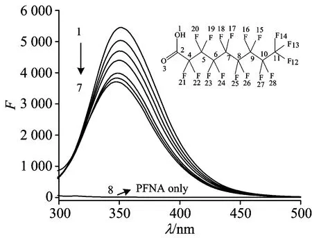
Fig.1 Fluorescence emission spectra of PFNA-HSA system,the inset is the structure of PFNA
cHSA=2.0×10⁻⁶mol·L-1;cPFNA(×10-6mol·L-1)(1—7): 0,1.0,2.0,3.0,4.0,5.0,6.0; curve 8:cPFNA=1.0×10⁻⁶mol·L-1;T=298 K
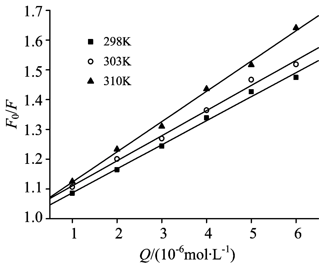
Fig.2 The Stern-Volmer plots for the PFNA-HSA system at different temperatures
cPFNA(×10-6mol·L-1)(1—7): 0,1.0,2.0,3.0,4.0,5.0,6.0;cHSA=2.0×10⁻⁶mol·L-1
2.2 Mechanisms of fluorescence quenching
Stern-Volmer equation is used to explain the quenching mechanism[9]
(5)
WhereF0,F,Ksv,[Q] andτ0are seen literature [9],kqis the bimolecular quenching rate constant,and its maximum scattering collision quenching constant is 2.0×10¹⁰L·mol-1·s-1. The Stern-Volmer plots are shown in Fig.2,Ksvandkqat corresponding temperatures are listed in Table 1.
Table 1 Stern-Volmer quenching constants for interaction of PFNA with HSA at different temperatures
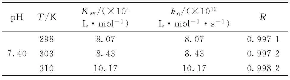
pHT/KKsv/(×104L·mol-1)kq/(×1012L·mol-1·s-1)R2988.078.070.99717.403038.438.430.997231010.1710.170.9982
There was a good linear dependence betweenF0/Fand [Q]PFNA,Ksvvalues increased with increasing temperature,revealing the occurrence of dynamic quenching interaction between PFNA and HSA. Furthermore,the values ofkqwhich were all greater than the upper limit of 2.0×10¹⁰L·mol-1·s-1indicated that the static quenching may exist between them. Besides,the fluorescence spectra of HSA (Fig.1) not only decreased gradually with increasing PFNA but also obtained a blue shift (4 nm),manifesting a decrease in polarity of the microenvironment around the Trp residues after binding of PFNA with HSA. It was probably due to the growth of the compact structure of hydrophobic subdomain IIA where Trp-214 is placed. This phenomenon also reconfirmed that probable quenching mechanism of the intrinsic fluorescence of HSA was initiated by PFNA-HSA complex formation.
Fluorescence anisotropy provides useful information on the changes in orientation of a small molecule upon binding with macromolecules. The anisotropy values (r) of PFNA (5.0×10⁻⁶mol·L-1) at three different HSA concentrations (0,1.0,5.0×10-6mol·L-1) were 0,0.103 and 0.126,respectively.rof a small molecule may vary from 0 (randomly oriented molecule) to 0.4 when there is no rotation or restricted motion of molecule. This augment in the anisotropy values with the addition of HSA deduced that a reduction of rotation freedom resulted from the complex formation between PFNA and HSA. In a word,the quenching mechanism of PFNA-HSA system should be a combined quenching process (including dynamic and static quenching)[13].
2.3 Site marker competitive experiments
Two site marker phenylbutazone (site Ⅰ in the subdomain ⅡA) and ibuprofen (site Ⅱ in the subdomain ⅢA) were used to identify bonding sites. The fluorescence of the complex was remarkably affected in presence of phenylbutazone,but remained invariant with ibuprofen. The result showed PFNA was to be bound to site Ⅰ in the subdomain ⅡA of HSA.
2.4 Molecular docking
In order to further determine the preferred binding sites of PFNA on HSA (PDB: 2BXN),molecular docking was carried out using AutoDock. The calculated free energy was -26.54 kJ·mol-1and the best energy ranked results are shown in Fig.3. The results showed that PFNA bound at subdomain ⅡA (site Ⅰ) [Fig.3(a)],which was consistent with the results observed in the site marker competitive experiments. PFNA molecule was surrounded by the hydrophobic chains and amino acid residues,such as Arg 218,Leu 219,Phe 223,Leu 260,Ala 261,Ile 264,Ser 287 and Val 293. Thus we can conclude that PFNA was able to fit well within the hydrophobic cavity of subdomain IIA. The docking result showed the existence of polar,hydrophobic interactions and halogen-bond between PFNA and HSA (Table 2). For example,O1 and O3 of PFNA interacted with Arg 222 through polar force. Hydrocarbon alkyl chains on Leu 219 and Ile 290 residues interacted with PFNA through hydrophobic interaction. Furthermore,it can be seen that there were four specific halogen-bonds of PFNA with Arg 257,Ser 287,Ile 290 and Ala 291 residues of HSA considering the distance between donor and acceptor atoms from 2.6 to 4.0,which played a crucial role in binding of PFNA to HSA.
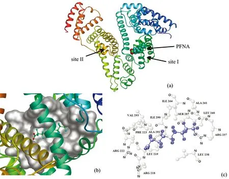
Fig.3 (a) The binding site of PFNA on HSA. HSA is shown in cartoon and PFNA is represented using spheres; (b) Enlarged binding mode between PFNA and HSA. HSA is shown in cartoon,the interacting side chains of HSA are displayed in surface mode and PFNA is represented using balls and sticks; (c) Molecular modeling of the interaction between PFNA and HSA. The atoms of PFNA are blue

Table 2 The distances and driving forces between the PFNA atoms and the atoms of residues obtained by molecular docking
2.5 Isothermal titration calorimetry measurement
To explore the binding procedure and main driving force of PFNA to HSA,a representative calorimetric titration profile of PFNA with HSA is shown in Fig.4(a),the exothermicity and endothermicity of calorimetry peaks suggested that there were more than one binding process for PFNA-HSA interaction. Fig.4(b) shows the integrated heat profile after eliminating dilution heat of PFNA into buffer solution,the solid smooth line represents the best fit of experimental data using the standard nonlinear least-squares regression binding model which two classes of binding sites fitted well to calorimetric data. The thermodynamic parameters are the average of three independent experiments for the interaction of PFNA with HSA obtained from ITC (Table 3). From Table 3 can be seen that for the binding of PFNA to HSA,the number of second-class binding site (N2=9.95) on HSA molecules is higher than correspondingN1(3.33),whereas the second-class binding constant (Ka,2=4.58×10⁴L·mol-1) is much smaller thanKa,1(22.71×10⁴L·mol-1). Moreover,the binding events only occurred in subdomain IIA of HSA according to the results of site marker competitive experiments and molecular docking.
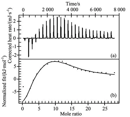
Fig.4 (a) ITC titration profile of PFNA HSA binding; (b) Integrated heat profile of the calorimetric titration shown in panel A. The solid line represents the best nonlinear least-squares fit to the independent binding sites model
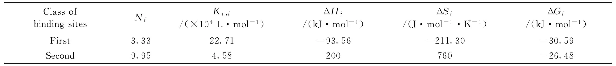
Table 3 Binding constants and relative thermodynamic parameters of PFNA-HSA at 298 K
As evident in Table 3,for the first binding site of this system,the both negative enthalpy (ΔH1) and entropy (ΔS1) changes indicated that this binding was an exothermic and entropy decreasing process. This can be explained by two factors: (1) the hydrated PFNA molecules should lose some water molecules when they approached to the binding sites and,simultaneously,the hydration layers on the surface of HSA molecules were partly destroyed. Both dehydration processes were endothermic and entropy increasing. (2) Directly electrostatic interaction of dipole groups of PFNA with peptide sections of HSA molecules,which caused exothermic effects and negative contribution to entropy. The experimentally negative enthalpy and entropy changes indicated that Factor (2) was evidently stronger than Factor (1) for this type of binding. Since seventeen electrophilic F-groups in PFNA can attract strongly the lone pair electrons of polar side groups of peptide chain,and partly PFNA molecules which were negatively charged under physiological condition interacted with the positively charged amino acids of HSA surface. Thus the strong electrostatic interactions gave rise to the strong negative values of enthalpic and entropic changes. Because the first class of binding was entropically opposed but enthalpically favored,the negative change of Gibbs free energy (ΔG1) was due to the contribution of heat effect and the process was mainly driven by enthalpy.
The positive values of both ΔH2and ΔS2indicated that the second binding was an endothermic and entropy increasing process. This phenomenon can be explained by considering following reasons: firstly,when a PFNA molecule (partly) inserted itself into a hydrophobic cavity of HSA molecule formed by folding and twisting of peptide chain,the hydrophobic interaction between PFNA molecule and the cavity would cause a decrease in both the enthalpy and entropy. Secondly,the hydrophobic interaction led to some water molecules transferring into bulk solution from the hydrophobic cavity at the binding sites of HSA molecule,which was also exothermic but entropy increasing. Thirdly,accompanying the inserting of PFNA molecule into hydrophobic cavity,the original iceberg structure surrounding PFNA molecule was destroyed,which was endothermic and made a major positive contribution to the entropy. Because of the experimentally positive ΔH2and ΔS2,i.e.,entropy effect resulted in the negative change of Gibbs free energy (ΔG2),the second-class binding was entropy driven process. On the other hand,hydrophobic interaction between PFNA and HSA played a key role for this class of binding process as corroborated by the positive entropy changes (ΔS2) at complex formation.
2.6 Three-dimensional fluorescence spectra

Fig.5 Three-dimensional fluorescence spectra for HSA and PFNA HSA complexcHSA=2.0×10⁻⁶ mol·L-1,cPFNA=2.0×10⁻⁶ mol·L-1; T=298 K
2.7 Circular dichroism spectra
Further evidence of conformational changes of HSA upon addition of PFNA was confirmed by CD spectroscopy (Fig.6). It was apparently observed that the CD bands of HSA were at 209 and 222 nm. The binding of PFNA to HSA caused only a decrease in negative band intensity without any significant shift of the peaks which implied that HSA had predominantlyα-helix in nature even after binding to PFNA. The secondary structural contents of HSA were expressed in terms of mean residue ellipticity (MRE) according to literature [14],the calculated results exhibited that PFNA caused decrease inα-helical content of HSA from 55.6% in free HSA to 41.3% at a molar ratio of PFNA to HSA of 15∶1,β-sheet from 13.5% to 8.2%,β-turn from 16.5% to 13.0%,and increase in random content of HSA from 14.4% to 37.5%. From above results,it was apparent that the binding of PFNA to HSA led to a secondary structure change of HSA with the loss of helical stability.

Fig.6 The CD spectra of HSA in the absence and presence of PFNA
cHSA=2.0×10⁻⁶mol·L-1; molar ratiosnPFNA∶nHSAfrom 1 to 3: 0∶1,10∶1,15∶1
3 Conclusions
The results indicated that PFNA could quench the intrinsic fluorescence of HSA through static and dynamic quenching process. It was worthy noted that the binding site was located in the hydrophobic pocket of subdomain IIA according to the competitive binding experiment and molecular docking studies. Furthermore,molecular docking offered a molecular level explanation with the ability to estimate the participation of specific chemical groups and their interactions in complex stabilization. The ITC results showed that binding occurred at two different classes of binding sites. It also demonstrated that both electrostatic and hydrophobic interactions were presented in the formation of PFNA-HSA complex,the former being more important when PFNA bound to the first class of binding site,whereas hydrophobic forces were generally predominant in the second class of site,as seen from entropy increases. As further revealed by three-dimensional fluorescence and CD spectra,PFNA caused microenvironmental and conformational changes of HSA. Investigation of PFNA-HSA interaction has a great significance for thoroughly understanding the interaction process of PFNA-HSA,the relationship of structure and function of HSA,and the chemical essence of the interaction between biomacromolecule and ligand.
[1] Yang B J,Hao F,Li J R,et al. Food Chem. Toxicol.、2014,65: 227.
[2] Deng F Y,Dong C Y,Liu Y. Mol. Biosyst.、2012,8(5): 1446.
[3] Brieger A,Bienefeld N,Hasan R,et al. Toxicol. in Vitro、2011,25(4): 960.
[4] Christian G D,Thomas A T. Environ. Health Persp.、1999,107: 907.
[5] Kärrman A,Harada K H,Inoue K,et al. Environ. Int.、2009,35(4): 712.
[6] Qin P F,Liu R T,Pan X R,et al. J. Agric. Food Chem.、2010,58(9): 5561.
[7] Ohmori K,Kudo N,Katayama K,et al. Toxicology、2003,84(2-3): 135.
[8] MacManus-Spencer L A,Tse M L,Hebert P C,et al. Anal. Chem.、2010,82(3): 974.
[9] Hu T Y,Liu Y. J. Pharm. Biomed. Anal.、2015,107: 325.
[10] Molina-Bolívar J A,Galisteo-González F,Ruiz C C,et al. J. Lumin.、2014,56: 141.
[11] Sun X J,Xu X Y,Liu M,et al. J. Solution Chem.、2010,39(1): 77.
[12] Cheema M A,Taboada P,Barbosa S,et al. J. Chem. Thermodyn.、2009,41(4): 439.
[13] Li J H,Wang S M. J. Chem. Thermodyn.、2013,58: 206.
[14] Matei I,Hillebrand M. J. Pharm. Biomed. Anal.、2010,51(3): 768.
*通讯联系人
O657.3
A
光谱法联合分子对接和等温滴定微量热法研究全氟壬酸与人血清白蛋白的相互作用
胡涛英1、黄 芳1、周珊珊1,2、刘 颖1,2*
1. 中央民族大学生命与环境科学学院、北京 100081 2. 中央民族大学北京市食品环境与健康工程技术研究中心、北京 100081
全氟壬酸(PFNA)是在血清中检测到第三多的全氟烷酸类(PFAAs)新型有毒环境污染物。目前PFNA对人血清白蛋白(HSA)结构甚至是功能的影响还处于起步阶段、借助于多光谱、分子对接和等温滴定微量热(ITC)技术研究了PFNA和HSA相互作用的结合机理。所有荧光数据均进行了内滤光校正以获得更准确的结合参数。荧光结果表明PFNA通过动静态猝灭方式可以猝灭HSA的内源荧光。取代实验和分子对接结果表明、PFNA主要通过极性键、疏水力和卤素键键合在HSA亚域ⅡA疏水腔中、最佳对接自由能为-26.54 kJ·mol-1、表明PFNA分子与HSA有较大的结合亲和力。ITC表明两者的结合属于两类结合位点模型并给出了相应的热力学参数:第一类结合位点有较大的亲和力、属于焓驱动、静电力和卤键作为主要驱动力; 第二类结合位点亲和力较小、主要驱动力是疏水力。三维荧光光谱揭示PFNA与HSA生成复合物后、可以改变HSA的构象、引起Trp和Tyr残基微环境疏水性增强。圆二色谱(CD)定量测定了HSA与PFNA作用前后的二级结构含量:α-螺旋、β-折叠和β-转角含量分别降低14.3%、5.3%和3.5%、无规卷曲含量从14.4%增加到37.5%。以上结果表明、PFNA与HSA的结合可以改变HSA的二级结构、进而可能影响HSA的生理功能。结果阐述了PFNA与HSA相互作用机理、并且为PFNA在体内的运输和分配提供了可靠的生物物理和生物化学的相关依据。
全氟壬酸; 人血清白蛋白; 光谱法; 分子对接; 等温滴定微量热法
2015-06-08、
2015-10-29)
2015-06-08; accepted:2015-10-29
The National Natural Science Foundation of China (21177163),111 Project B08044,Special Guidance Fund of Building World First-class Universities (Disciplines) and Characteristic Development of Minzu University of China (2016,ydzxxk201619),Coordinate Development of First-Class and First-Class University Discipline Construction Funds (10301-0150200604),The Academic Team Construction Project of Minzu University of China (2015MDTD25C&13C),First-class Universities and First-class Discipline Construction Transitional Funds Under Special Funding (10301-01404031,2015),2015MDTD08C
10.3964/j.issn.1000-0593(2016)12-4141-07
Biography:HU Tao-ying,(1989—),Master of College of Life and Environmental Science,Minzu University of China e-mail: hty0945020@163.com *Corresponding author e-mail: liuying4300@163.com
猜你喜欢
新闻传播(2018年11期)2018-08-29
新闻传播(2018年13期)2018-08-29
造纸化学品(2017年1期)2017-01-21
新闻传播(2016年9期)2016-09-26
化工生产与技术(2016年5期)2016-03-13
化工生产与技术(2016年5期)2016-03-13
中国人兽共患病学报(2016年6期)2016-01-30
中国塑料(2015年3期)2015-11-27
分析化学(2015年7期)2015-07-30
新闻传播(2015年7期)2015-07-18