
全球近10年无绿藻病例报道文献的回顾
2016-12-12 10:05曾炫皓章强强
中国真菌学杂志 2016年5期
曾炫皓 章强强
(复旦大学附属华山医院皮肤科,上海 200040)
·综述·
全球近10年无绿藻病例报道文献的回顾
曾炫皓 章强强
(复旦大学附属华山医院皮肤科,上海 200040)
该文系统性地回顾分析了人类无绿藻病在近10 a来的发病率及其趋势,并对该病的流行病学、临床表现、诊断标准、治疗方案及其预后的新特点进行了总结。全球至今已报道190例无绿藻病,约33%的无绿藻病例在近10 a内发生;系统性无绿藻发病率在近10 a病例数与皮肤型无绿藻病基本持平。我国大陆地区总共报道有9例无绿藻病,大约占我国 (包括港澳台地区)已有报道的无绿藻病例总数的50%。无绿藻病的诊断主要依靠标本的真菌学及组织病理学检查。两性霉素B及其脂质体仍然是该病治疗最常用且最有效的药物。提示无绿藻病在近10 a中发病率明显上升,且系统性无绿藻发病率显著升高。
无绿藻病;中型无绿藻;小型无绿藻
[Chin J Mycol,2016,11(5):310-315]
无绿藻病是由无绿藻感染引起的一种罕见的疾病,在全球多个区域均有分布,全球至今已有190例文献报道,其中我国报道19例。近10 a来无绿藻病发病率有增高的趋势,大量的新病例在近年报道。本文对无绿藻病的流行病学、临床表现以及诊断和治疗的新特点进行综合性分析。
1 病原学分类
无绿藻是一种条件致病菌,常存在于污水,土壤,植物,生牛奶,以及哺乳动物 (如猫、狗、牛、羊)身上,并能寄居在人类的指甲、皮肤、呼吸道、消化道等部位[1],只有在创伤或机体免疫功能下降时,无绿藻才入侵致病。然而,目前也发现免疫正常且无创伤等诱因的原因不明的无绿藻病报道[2-6]。无绿藻在生物学上处于真核生物 (Eukaryota)、绿色植物界 (Vwidiplantae)、绿藻门 (Chlorophyta)、Trebouxlophyceae纲、绿藻目 (Chlorellales)、绿藻科 (Chlorellaceae)、无绿藻属 (Prototheca)[7]。已知的包括大型无绿藻 (Protothecastagnora)、中型无绿藻 (Protothecazopfii)、小型无绿藻 (Protothecawickerhamii)、Protothecalmea、Protothecablaschkeae以及Protothecacutis6个种。近几年尤其多见于器官移植术后患者[8]。无绿藻病一旦发生系统性感染,治愈率低[1]。
2 流行病学
导致人和动物致病的通常认为是大型无绿藻,中型无绿藻和小型无绿藻[9]。人类无绿藻病仅与中型无绿藻以及小型无绿藻有关,并且以小型无绿藻感染更为多见[1,10]。无绿藻病自1952年被报道至今,除外南极洲的各个大陆板块都有过病例报道,尤其多见于温暖潮湿的地区[11]。Todd等[11]撰写的综述中提到截止2012年7月全球已经有160例无绿藻病报道,并且其中1/3为最近5 a的新增病例。
如表1所示,通过Pubmed检索到的近10 a的52个病例中,男女比约为1∶1,平均发病年龄为64岁,年龄最大的81岁[12],年龄最小的仅6个月[13]。60岁以上的33例,占比最大。39岁以下人群无绿藻病多表现为系统性无绿藻病,而皮肤型无绿藻病更多见于60岁以上的老年患者。除外8例中型无绿藻感染,其余病例均为小型无绿藻感染。近10 a报道的系统性无绿藻病患者常长期服用糖皮质激素,或有器官移植病史。同时,免疫功能正常患者发生的系统性无绿藻病4例[2,5-6]。近10 a报道的病例中,皮肤型无绿藻病治愈率为32.00%,死亡率为8.00%;系统性无绿藻病的治愈率为32.00%,死亡率为24.00%。
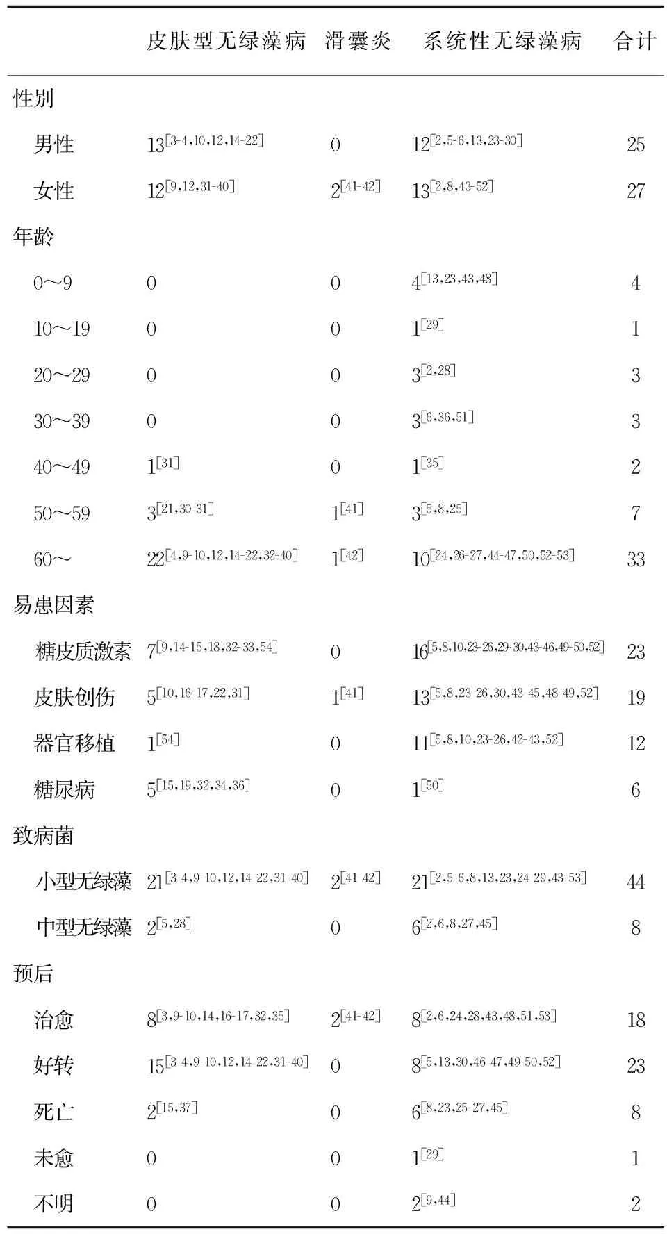
表1 近10 a无绿藻病发病分布情况
我国已有19例报道[3,7,55],其中大陆12例,台湾6例,香港1例。我国大陆地区近10 a报道的无绿藻病共有9例,其中表现为皮肤型无绿藻病的有7例,3例为小型无绿藻感染[3,36],1例为中性无绿藻感染[34],3例未明确到种[55-57];表现为系统性无绿藻病有2例,1例为中型无绿藻感染[6],1例为小型无绿藻感染[28],并且患者的体液及细胞免疫指标都正常。除此以外,Zak等[13]报道了波兰的首例人类无绿藻病,表现为以脑炎为特点的系统性无绿藻病;Takano等[45]报道了日本首例由2型中型无绿藻感染所致的皮肤无绿藻病;Leni等[29]在印度报道了首例由小型无绿藻感染所致的系统性无绿藻病;Jae等[4]报道了韩国第1例由中型无绿藻感染所致的皮肤无绿藻病。
3 临床表现
3.1 临床表现概述
无绿藻病临床上常见有3个类型:皮肤及皮下组织感染;滑膜炎以及纤维组织炎;系统性感染。其中又以皮肤及皮下组织感染最常见,此种类型多见于免疫力正常的人群,目前的观点认为皮肤无绿藻病的病因多和暴露部位的创伤有关,一般表现为在创伤数周后发生的炎症和皮损。皮肤损害表现最常见为红斑、丘疹、硬结节、斑块、脓疱,也可以发生浅表溃疡、疣状增生、疱疹样损害。其发病部位多为身体的暴露部位,如头面部和四肢末端,常表现为局限型病灶[1]。无绿藻甲病较为少见,已有2例报道[38],主要表现为甲变色、增厚。皮肤型无绿藻病常由于缺乏显著的临床特点而被误诊,确诊时感染的时间一般都较长。此外,皮肤型无绿藻病如果不及时治疗,可以转变为系统性无绿藻病,近10 a已有3例相关报道[29,45-46]。近几年还报道过角膜移植后由小型无绿藻引起的角膜炎[5]和表现为四肢蜂窝织炎样皮损的无绿藻病[44]。此外,近年来由于医院创伤性治疗导致的无绿藻病也有报道[8,46,49-50]。
系统性的无绿藻感染多见于由各种原因造成自身免疫力低下的人群,临床表现为多系统损害,特点是病情发展迅速、全身症状显著、死亡率高。近10 a国外报道的系统性无绿藻感染共22例,其中7例死亡,但1例尸检已经无法检测到无绿藻感染[15]。相关病例报道显示,糖尿病、系统性红斑狼疮等自身免疫性疾病、器官移植、干细胞移植、慢性肾功能衰竭、长期应用糖皮质激素或免疫抑制剂、HIV感染,以及恶性肿瘤等是系统性无绿藻患者免疫力低下最常见的原因。另外,2012年Meijia-Otero等[50]报道了1例患有SLE和糖尿病的女患者在住院期间应用belimumab导致的小型无绿藻院内感染,同年Min Z等[51]报道了1例使用rituximab治疗SLE期间发生的小型无绿藻感染,2010年Gaur S等[49]报道了1例在移除Hickman导管时发现的小型无绿藻感染,此前均未见以上原因导致的无绿藻感染。
无绿藻性滑膜炎及纤维组织炎多发生在免疫正常的患者,较为少见。近10 a来仅报道2例[41-42],1例为患者在接受静脉曲张注射治疗后,出现右脚背的肿胀,MRI显示跖骨到楔状骨的皮下组织肿胀伴骨损害迹象,作拇长伸肌穿刺后提示为滑膜炎,穿刺液培养发现小型无绿藻生长。另1例71岁女性患者是小型无绿藻感染导致的滑囊炎,表现为左手鹰嘴部肿胀疼痛[42]。系统性无绿藻病患者鹰嘴部也有可能发生滑囊炎,近10 a有2例相关的报道[29,58]。
3.2 老年患者无绿藻病临床表现
尽管各个年龄段都有无绿藻病发病的报道,但老年人无绿藻病的发病率最高。60岁以上的老年患者病例数约占近10 a报道病例的65%,具体为皮肤型无绿藻病22例[4,9-10,12,14-22,32-40],滑囊炎型无绿藻病1例[42],系统性无绿藻病10例[24,26-27,44-47,50,52-53],除外4例为中型无绿藻[4,27,34],其余均为小型无绿藻感染。皮肤损害都是老年无绿藻病最常见的临床表现,并且老年系统性无绿藻病全部继发于机体免疫功能受损后。文献报道的10例60岁以上系统性无绿藻病中,死亡3例[26-27,45],其中2例确认死于无绿藻感染,1例尸检发现经治疗后已经无法检测到无绿藻[15]。国内近10 a报道的60岁以上老年无绿藻病共有4例,均表现为皮肤型无绿藻病,且经过治疗后病情都得到了好转[3,6,36,55]。
3.3 中年患者无绿藻病临床表现
44~59岁的中年人无绿藻感染国内外近10 a共有9例报道,约占17.65%左右。其中4例表现为系统性无绿藻病[5,8,25,49],4例表现为皮肤型无绿藻病[21,30-31,36],1例表现为无绿藻所致的滑膜炎[41]。除外2例系统性无绿藻病由中型无绿藻引起[6,8],其余均为小型无绿藻感染。4例系统性无绿藻病均发生在患者器官或造血干细胞移植后,其中2例预后为死亡,另外2例分别为患者造血干细胞移植后在血培养时发现的中型无绿藻感染[8]和肾移植后患者在肾周脓肿后发现的小型无绿藻感染[25]。
3.4 青年患者无绿藻病临床表现
14~43岁的年龄段无绿藻感染较少见,近10 a仅有5例相关报道,预后均为治愈。其中2型中型无绿藻感染的脑膜炎2例[2],SLE患者伴发的系统性无绿藻病1例[51],中型无绿藻波多黎各变种所致淋巴结炎1例[6],小型无绿藻引起的脑膜炎1例[28]。尤其值得关注的是这些病例经检查免疫功能都正常。国内吴绍熙等[57]报道的1例18岁女性表现为面部皮肤无绿藻病。
3.5 儿童患者无绿藻病临床表现
儿童无绿藻病近10 a共有5例报道,均表现为小型无绿藻感染所致的系统性无绿藻病。分别是3岁美国男孩在干细胞移植后发生的系统性无绿藻病,表现为发热、中性粒细胞减少、梗阻性黄疸,后由于移植物抗宿主反应导致的肾衰竭死亡[23];4岁中国女孩在肝移植后发生的系统性无绿藻病,表现为持续的咳嗽,呼吸困难,食欲下降,颜面部和上肢的红色斑块和小脓疱,经过治疗后痊愈[43];2岁墨西哥女孩在未经正规处理的颏下脓肿切开后发生的播散性无绿藻感染,经治疗后好转[48];波兰6个月的男婴的无绿藻中枢神经感染,经治疗后痊愈[13];10岁印度男孩以颜面部和躯干和前臂簇集性的红色丘疹,鹰嘴处滑囊炎及巨脾为显著表现的系统性无绿藻病,预后为未愈[29]。近10 a我国 (包括港澳台地区)未见儿童无绿藻病报道。
4 诊 断
无绿藻病缺乏特异性的临床表现,患者首诊常常被误诊,因而无绿藻病被确诊时常常已是患者患病数个月甚至数年以后。目前用于检查无绿藻病的诊断手段可以分为实验室检查、影像学检查、微生物学检查、组织病理学检查以及分子生物学检测[1]。无绿藻病诊断更推荐将微生物学检查和组织病理学检查联合[51]。无绿藻的鉴定主要基于菌落形态、镜下结构,生长温度试验、对糖和醇的同化利用等。未形成孢子囊孢子的无绿藻与皮炎芽生菌、新生隐球菌、副球孢子菌、耶氏肺孢子菌等病原体形态上相似,尤其需要鉴别,上述几种病原体孢子囊的大小的不同是鉴别它们的的一个重要区分点。特征性的内孢子是无绿藻属鉴定的一个重要依据[7]。相比传统的表型鉴定方法,分子生物学检测具有快速、敏感、特异性高的特点,特别是对于变种、亚种的鉴定具有更大的优势[11]。
5 治 疗
无绿藻病的治疗尚无标准方案。国外一线治疗最常使用两性霉素B或其脂质体、伏立康唑、伊曲康唑。具体药物常根据药敏试验的结果进行选择。近10 a来,对49例无绿藻感染病例药敏试验结果如表2所示。从中可以看出对两性霉素B及其脂质体敏感的菌株最多,其次为各种唑类药物,而对其他常见抗真菌药物敏感的菌株较少。
表2 近10 a无绿藻病药敏试验敏感药物汇总
Tab.2 The summary sheet of drug sensitivity test of prototheca in last decade

小型无绿藻中型无绿藻合计伊曲康唑15[3,9,16,18-19,28-30,33,35,39,41,45,52-53]2[4,6]17酮康唑2[38,52]02伏立康唑11[14,19,24-25,32,36-37,46,49,52]011氟康唑3[3,10,17]03卡泊芬净1[49]01两性霉素B及其脂质体23[10,15,18-19,23-25,36-41]4[4,6,27,34]27特比萘芬02[31,45]2多西环素2[35,49]02
无绿藻病的药物治疗需要较长时间的坚持,48例病例中预后为好转或治愈的患者治疗时间都在6个月以上,但是两性霉素B的副作用常常导致患者无法耐受,因而有多例的治疗方案选择了短时间的两性霉素B治疗后以唑类药物长期治疗[4,9,30,33,35,46,49]。此外,Nanako Yamada等[16]采用口服伏立康唑结合患者皮损处55℃热敷的治疗方法,并取得了较好的疗效。Grzesiak B等[59]利用从牛奶中分离出的8株中型无绿藻菌株进行药敏试验后发现百里香、丁香、以及肉桂精油能有效抑制中型无绿藻,Bouari C等[60]以BALB/c小鼠为载体动物进行实验后发现洋薄荷精油能够有效治疗中型无绿藻菌株感染所致的皮肤型无绿藻病,这些植物精油也许能够用作无绿藻病治疗的辅助手段。
6 小 结
全球无绿藻病的发病率有明显的上升趋势,近1/3的无绿藻病例在近10 a内被报道。近几年国内外报道的免疫功能正常的系统性无绿藻病病例,进一步证实了系统性无绿藻病并非一定发生于免疫缺陷患者。以往认为无绿藻病的感染大多与皮肤的创伤有关,但近10 a来的病例报道中,也有大量患者没有明确的皮肤创伤病史。同时从近10 a的病例报道看,系统性无绿藻病病例数与皮肤型无绿藻病基本持平,呈现明显的升高趋势,这提示我们在诊断中要重视无绿藻系统性感染的可能性。系统性无绿藻病病例数的增多,和由各种原因所导致的免疫缺陷患者的增多有直接联系。从近10 a来的病例报道来看,患有糖尿病、长期服用糖皮质激素、器官移植或造血干细胞移植的患者 (尤其是60岁以上的老年患者)以及其他各种免疫功能下降的患者,如果暴露部位皮肤发生了慢性、无痛性的红斑、丘疹、硬结节、溃疡,且经验性治疗无效时,一定要警惕无绿藻感染的可能性。上述类型的患者如果发生了难以治疗的系统性感染,更要警惕系统性无绿藻病的可能。
[1] Mayorga J,Barba-Gomez JF,Verduzco-Martinez AP,et al.Protothecosis[J].Clin Dermatol,2012,30(4):432-436.
[2] Hench J,Roschanski N,Hewer E,et al.Granulomatous Encephalitis:Protothecosis Excluded[J].Histopathology,2016.doi:10.1111/his.13020.[Epub ahead of print]
[3] Zhang Q,Li L,Yuli K,et al.A case of cutaneous protothecosis mimics eczema[J].Mycopathologia,2015,179(1-2):163-166.
[4] Seok JY,Lee Y,Lee H,et al.Human cutaneous protothecosis:report of a case and literature review[J].Korean J Pathol,2013,47(6):575-578.
[5] Solky AC,Laver NM,Williams J,et al.Prototheca wickerhamii infection of a corneal graft[J].Cornea,2011,30(10):1173-1175.
[6] Zhang Q,Weng X,Li L,et al.An unusual case of granulomatous lymphadenitis due toProtothecazopfiivar.portoricensis in an immunocompetent man in China[J].Int J Infect Dis,2010,14 Suppl 3:e32-e35.
[7] 章强强.中国无绿藻病发病的现状分析及其诊治[J].实用皮肤病学杂志,2013,6(02):65-67.
[8] Macesic N,Fleming S,Kidd S,et al.Protothecosis in hematopoietic stem cell transplantation:case report and review of previous cases[J].Transpl Infect Dis,2014,16(3):490-495.
[9] Woo JY,Suhng EA,Byun JY,et al.A case of cutaneous protothecosis in an immunocompetent patient[J].Ann Dermatol,2016,28(2):273-274.
[10] Ramirez I,Nieto-Rios JF,Ocampo-Kohn C,et al.Protothecal bursitis after simultaneous kidney/liver transplantation:a case report and review[J].Transpl Infect Dis,2016,18(2):266-274.
[11] Todd JR,King JW,Oberle A,et al.Protothecosis:report of a case with 20-year follow-up,and review of previously published cases[J].Med Mycol,2012,50(7):673-689.
[12] Hightower KD,Messina JL.Cutaneous protothecosis:a case report and review of the literature[J].Cutis,2007,80(2):129-131.
[13] Zak I,Jagielski T,Kwiatkowski S,et al.Protothecawickerhamiias a cause of neuroinfection in a child with congenital hydrocephalus.First case of human protothecosis in Poland[J].Diagn Microbiol Infect Dis,2012,74(2):186-189.
[14] Kwong JC,Ward PB,Johnson PD.Cutaneous protothecosis in a patient with hypogammaglobulinemia[J].Med Mycol Case Rep,2013,2(1):132-133.
[15] Gaitanis G,Nomikos K,Zioga A,et al.Multifocal cutaneous protothecosis in a patient with myelodysplastic syndrome[J].Hippokratia,2012,16(1):95.
[16] Yamada N,Yoshida Y,Ohsawa T,et al.A case of cutaneous protothecosis successfully treated with local thermal therapy as an adjunct to itraconazole therapy in an immunocompromised host[J].Med Mycol,2010,48(4):643-646.
[17] Humphrey S,Martinka M,Lui H.Cutaneous protothecosis following a tape-stripping injury[J].J Cutan Med Surg,2009,13(5):273-275.
[18] Carneiro FP,Moraes MA,Rebelo AM,et al.Cutaneous protothecosis:case report[J].Rev Soc Bras Med Trop,2007,40(4):466-468.
[19] Sheikh-Ahmad M,Goldstein S,Potasman I.Protothecawickerhamiihand infection successfully treated by itraconazole and voriconazole[J].J Travel Med,2006,13(5):321-323.
[20] Curbelo A,Pankey GA.A man presenting with nodules on hands and elbows[J].Clin Infect Dis,2009,48(8):1114-1115,1160-1161.
[21] Pimentel CL,Alegre M,Dalmau J,et al.Erythematous papules on the leg[J].Arch Dermatol,2006,142(7):921-926.
[22] Dalmau J,Pimentel CL,Alegre M,et al.Treatment of protothecosis with voriconazole[J].J Am Acad Dermatol,2006,55(5 Suppl):S122-S123.
[23] Sykora T,Horakova J,Buzzasyova D,et al.Protothecal peritonitis in child after bone marrow transplantation:case report and literature review of paediatric cases[J].New Microbes New Infect,2014,2(6):156-160.
[24] Narita M,Muder RR,Cacciarelli TV,et al.Protothecosis after liver transplantation[J].Liver Transpl,2008,14(8):1211-1215.
[25] Bandaranayake TD,Paniz MA,Peaper DR,et al.Protothecawickerhamiialgaemia:an emerging infection in solid organ transplant recipients[J].Transpl Infect Dis,2015,17(4):599-604.
[26] Mohd TR,Sabaratnam P,Salleh MA,et al.Characterization ofProtothecawickerhamiiisolated from disseminated algaemia of kidney transplant patient from Malaysia[J].Mycopathologia,2012,173(2-3):173-178.
[27] Van den Bossche D,De Bel A,Hendrickx M,et al.Galactomannan enzymatic immunoassay cross-reactivity caused byProtothecaspecies[J].J Clin Microbiol,2012,50(10):3371-3373.
[28] Zhang QQ,Zhu LP,Weng XH,et al.Meningitis due to Prototheca wickerhamii:rare case in China[J].Med Mycol,2007,45(1):85-88.
[29] Mathew LG,Pulimood S,Thomas M,et al.Disseminated protothecosis[J].Indian J Pediatr,2010,77(2):198-199.
[30] Fong K,Tee SI,Ho MS,et al.Cutaneous protothecosis in a patient with previously undiagnosed HIV infection[J].Australas J Dermatol,2015,56(3):e71-e73.
[31] Gandham NR,Vyawahare CR,Chaudhaury N,et al.Onychoprotothecosis:An uncommon presentation of protothecosis[J].Indian J Med Microbiol,2015,33(3):435-437.
[32] Yun CH,Jeong JH,Ryu HR,et al.Cutaneous protothecosis responds rapidly to voriconazole[J].Int J Dermatol,2015,55(12):1373-1377.
[33] Silva PC,Costa ESS,Lima RB,et al.Cutaneous protothecosis--case report[J].An Bras Dermatol,2013,88(6 Suppl 1):183-185.
[34] Zhang QQ,Li L,Zhu LP,et al.Cutaneous protothecosis in patient with diabetes mellitus and review of published case reports[J].Mycopathologia,2012,173(2-3):163-171.
[35] Srisuttiyakorn C,Sindhuphak W.Cutaneous protothecosis:a case report from Thailand[J].Int J Dermatol,2012,51(11):1340-1342.
[36] Lu S,Xi L,Qin W,et al.Cutaneous protothecosis:two new cases in China and literature review[J].Int J Dermatol,2012,51(3):328-331.
[37] Okazaki C,Wakusawa C,Chikama R,et al.A case of cutaneous protothecosis in a polyarteritis nodosa patient and review of cases reported in Japan[J].Dermatol Online J,2011,17(9):2.
[38] Zaitz C,Miranda GA,de Sousa VM,et al.Onychoprotothecosis:report of the first case in Brazil[J].Int J Dermatol,2006,45(9):1071-1073.
[39] Zaitz C,Godoy AM,Colucci FM,et al.Cutaneous protothecosis:report of a third Brazilian case[J].Int J Dermatol,2006,45(2):124-126.
[40] Jeunon T,Fantin-Ribeiro A,Jeunon-Sousa MA,et al.Erythematous plaque and shallow ulcers on right arm and forearm[J].Int J Dermatol,2009,48(11):1171-1173.
[41] Lee JS,Moon GH,Lee NY,et al.Case report:protothecal tenosynovitis[J].Clin Orthop Relat Res,2008,466(12):3143-3146.
[42] Knox J,Coloe SV,Perera C,et al.Protothecawickerhamiiolecranon bursitis successfully treated with adjunctive systemic itraconazole[J].Pathology,2015,47(4):388-391.
[43] Tan RM,Aw MM,Quak SH,et al.Pulmonary protothecosis in a pediatric liver transplant patient[J].J Pediatric Infect Dis Soc,2014,3(3):e31-e34.
[44] Kovalyshyn I,Brankov N,Fernandez AP.Erythematous and edematous plaques on the bilateral extremities in an immunocompromised patient.Protothecosis[J].Int J Dermatol,2016,55(2):e59-e61.
[45] Takano M,Hoshi S,Nagai K,et al.The first case of human protothecosis caused byProtothecazopfiiin Japan[J].J Infect Chemother,2014,20(10):647-649.
[46] Figueroa CJ,Camp BJ,Varghese GI,et al.A case of protothecosis in a patient with multiple myeloma[J].J Cutan Pathol,2014,41(5):409-413.
[47] Murata M,Ito T,Nagae K,et al.Disseminated protothecosis manifesting with multiple,rapidly-progressing skin ulcers:successful treatment with amphotericin B[J].Eur J Dermatol,2015,25(2):208-209.
[48] Tello-Zavala MC,Mercado-Lara A,Gomez-Hernandez N,et al.Disseminated protothecosis in a Mexican child[J].Pediatr Infect Dis J,2013,32(12):e476-e477.
[49] Gaur S,Marrin C,Barnes RA.Disseminated protothecosis following traumatic Hickman line removal in a patient with leukaemia[J].Med Mycol,2010,48(2):410-412.
[50] Mejia-Otero C,Singh S,Arias UL,et al.A rare case of prototheca algaemia in a patient with systemic lupus erythematosus and recent belimumab infusion[J].Case Reports Immunol,2012,2012:754901-754902.
[51] Min Z,Moser SA,Pappas PG.Protothecawickerhamiialgaemia presenting as cholestatic hepatitis in a patient with systemic lupus erythematosus:A case report and literature review[J].Med Mycol Case Rep,2012,2:19-22.
[52] Mcmullan B,Muthiah K,Stark D,et al.Protothecawickerhamiimimicking yeast:a cautionary tale[J].J Clin Microbiol,2011,49(8):3078-3081.
[53] Perez MC,Camba M,Tinajas A,et al.Protothecawickerhamiiperitonitis in patients on peritoneal dialysis[J].Nefrologia,2007,27(1):81-82.
[54] Waine B.Protothecosis:a lethal infection due to an achlorophylous algae[J].Clin Infect Dis,2014,59(12):187-197.
[55] 于长平,田洪青,周桂芝,等.皮肤无绿藻病1例[C]//中国中西医结学会皮肤性病专业委员会.全国中西医结合皮肤性病学术年会.南昌:中国中西医结学会皮肤性病专业委员会,2014:146.
[56] 虞胜镭,陈澍.无绿藻感染一例[J].中华传染病杂志,2008,26(11):700.
[57] 吴绍熙,吕桂霞,姜祎群,等.无绿藻病[J].临床皮肤科杂志,2007,36(2):127.
[58] Satoh K,Ooe K,Nagayama H,et al.Protothecacutissp.nov.,a newly discovered pathogen of protothecosis isolated from inflamed human skin[J].International Journal of Systematic & Evolutionary Microbiology,2009,60(Pt 5):1236-1240.
[59] Grzesiak B,Glowacka A,Krukowski H,et al.Theinvitroefficacy of essential oils and antifungal drugs againstProtothecazopfii[J].Mycopathologia,2016,181(7-8):609-615.
[60] Bouari C,Bolfa P,Borza G,et al.Antimicrobial activity ofMenthapiperitaandSaturenjahortensisin a murine model of cutaneous protothecosis[J].J Mycol Med,2014,24(1):34-43.
[本文编辑] 卫凤莲
Human Protothecosis:literature review of previously published cases in the last decade
ZENG Xuan-hao,ZHANG Qiang-qiang
(DepartmentofDermatology,HuaShanHospital,FudanUniversity,Shanghai200040,China)
We systematically review all the published cases in the last decade to realize the newest incidence and trends of protothecosis and comprehensively analyze the new character of protothecosis in epidemiology,clinical manifestations,therapy and prognosis.The amount of protothecosis cases since the first case had been reported was 190,and nearly 33% of the cases were reported in last decade.In recent years,the cases of disseminated protothecosis almost equals cutaneous protothcosis.In last decade,9 cases had been reported in mainland which conclude half of total number of protothcosis in China.The diagnose of protothecosis still relied on mycology and histopathology examination.Amphotericin B and its liposome were still the most commonly used and effective antifungal to cure protothcosis.These showed the rising incidence of protothcosis or disseminated protothecosis worldwide.
Protothecosis;Protothecazopfii;Protothecawickerhamii
国家自然科学基金 (81573056)
曾炫皓,男 (汉族),硕士研究生在读.E-mail:969358560@qq.com
章强强,E-mail:zhangqq8@163.com
R 519.5
A
1673-3827(2016)11-0310-06
2016-07-25
猜你喜欢
临床误诊误治(2021年12期)2021-12-04
中国新闻周刊(2021年9期)2021-03-29
作文评点报·低幼版(2020年25期)2020-07-23
浙江农林大学学报(2019年4期)2019-07-24
中国真菌学杂志(2018年2期)2018-05-17
中国感染与化疗杂志(2018年3期)2018-01-20
中国男科学杂志(2016年5期)2016-12-01
小品文选刊(2016年21期)2016-11-27
右江医学(2014年1期)2014-03-22
右江医学(2014年1期)2014-03-22
