
肿瘤特异性成像中分子探针验证模型的建立及评估
2017-03-30 10:20霍天龙杨硕李天然康钰赵赟赟杜湘珂
中国医疗设备 2017年3期
霍天龙,杨硕,李天然,康钰,赵赟赟,杜湘珂
1.北京大学人民医院 放射科,北京100044;2.解放军95医院 放射科,福建 莆田 351100
肿瘤特异性成像中分子探针验证模型的建立及评估
霍天龙1,杨硕1,李天然2,康钰1,赵赟赟1,杜湘珂1
1.北京大学人民医院 放射科,北京100044;2.解放军95医院 放射科,福建 莆田 351100
目的建立同一个体裸鼠肿瘤受体阳性和阴性双瘤动物模型。方法选取 SSTR2 阳性表达细胞系人BEL-7402细胞系和SSTR2阴性表达细胞系人 A549细胞系,进行细胞培养,选取对数生长期细胞,消化和悬浮后,分别移植到同一裸鼠各一侧腋窝(移植细胞悬液浓度在 5×10⁶个/0.2 mL/侧以上),制作皮下移植瘤双瘤模型,观察成瘤效果,对成瘤效果进行分析和总结。结果成功进行了2种细胞的细胞培养,采用腋窝作为注射部位,成功制造了BEL-7402/A549双瘤模型。其中BEL-7402成瘤率在80%以上(24/30),接种细胞的量足够即可很好地成瘤;A549细胞浓度在5×106个/0.2 mL以上成瘤率略低,约73%(22/30),增加细胞数量和补充注射可以部分提高成瘤率。最终双侧成瘤大小相差在1 cm以内的占32%(8/22)。组织学检查显示,与裸鼠正常肝脏相比,裸鼠肝脏移植肿瘤明显呈恶性形态,细胞核大,多见病理性核分裂像,双核或多核多见;肿瘤组织可见明显坏死区和 大量血管分布,提示生长迅速和微血管较多。A549移植瘤和BEL-7402移植瘤大体形态不同,但微观形态同BEL-7402一样,与人肿瘤形态一致。结论同一裸鼠个体可以成功接种不同肿瘤,掌握好两种肿瘤的生长差别,可以制作出大小相差在允许范围的合适的双瘤模型,为肿瘤特异性成像奠定基础。
分子影像学;动物模型;裸鼠移植瘤;双瘤模型;分子探针
引言
恶性肿瘤是当今威胁人类健康的最大顽疾之一,其中尤以晚期肿瘤的疗效最差。因此,现阶段治疗恶性肿瘤的关键在于早期诊断,而分子影像学即是早期诊断的希望。分子影像学立足于从细胞和分子水平显示病变,是一种特异性的、全新的活体显像方法。分子影像学可使我们从整体上了解疾病的发生、发展,在原位揭示生化改变,减少有创的检查,对疾病进行早期检测、鉴别定性以及治疗评价[1]。
合适的肿瘤模型是完成肿瘤早期成像和特异性成像的基础。以往研究仅在同一个体单纯构建目标肿瘤受体阳性动物模型,同时利用不同个体构建目标受体阴性肿瘤作为对照,因此这种对照方式存在内在缺陷,效果欠理想。若能在同一个体同时构建目标受体阳性肿瘤和对应的目标受体阴性肿瘤模型则是解决问题的关键,它可以有效地克服不同个体在不同时间和生理状态下分子探针分布偏移,从而客观和直观验证分子探针特异性,完成肿瘤受体特异性显像。有鉴于此,本研究选择高表达SSTR2受体的人BEL-7402阳性细胞系和不表达SSTR2的人A549细胞系进行同一裸鼠个体双侧移植瘤模型制作,科学直观地完成受体定量特异性显像,为分子探针特异性和肿瘤早期特异性成像奠定基础。60%~70%时适时冻存。
(1)冻存程序:将欲冻存细胞消化、悬浮后,用15 mL离心管低速离心(800 r/min)7 min,收集细胞,吸去上清;用1.5 mL冻存液吹打细胞,将其完全移入将冻存管,旋紧管口;将冻存管放入冰箱,4℃约40 min;置于普通冰箱上层冰盒(约-20℃),30~60 min;-30℃放置30 min左右;-70℃或-80℃过夜;做好标记后,将冻存管放入冻存格内,然后放入液氮罐。
(2)冻存液配方:小牛血清89%,DMSO(分析纯,Sigma-2650)10%,双抗1%。
1.2 双瘤模型建立
BEL-7402和A549肿瘤细胞传代、生长到一定数量,取对数生长期细胞,用0.25%的胰酶(含0.04%的EDTA,欣惠泽奥生物科技有限公司)消化、离心、调整细胞数,使其达到5×10⁶个/0.2 mL以上。选4周龄雄性Balb/c裸小鼠(北京大学医学部动物培育中心提供,全部实验小鼠都在无特定病原体环境下饲养,体重15~18 g),于其双腋窝部皮下分别注射肿瘤细胞约5×10⁶个/0.2 mL,进行模型构建。考虑到二者成瘤速度不同,预实验掌握好时间差,尽量使HCC移植瘤和对照瘤相差不大(直径最好在1 cm左右)。选取二者体积相近的成功模型进行后续实验。
1 材料和方法
1.1 细胞培养
BEL-7402人肝癌细胞系来源于北京大学基础医学院细胞生物学系,A549人肺癌细胞系来源于北京大学人民医院呼吸科实验室。使用前,解冻、复苏,将细胞离心收集,除去冻存液后,在超净台(CJ-IS,天津泰斯特仪器有限公司)中,用含 10%小牛血清(北京金泽奥生物科技有限公司)和双抗(青霉素和链霉素各100 U/mL)的DMEM低糖完全培养液(北京博奥图瑞生物科技有限公司)1.5 mL重新悬浮细胞,将悬液完全吸入75 cm2Corning培养瓶(北京天悦基因生物科技有限公司),补足培养液后,置于37℃,5%CO2孵箱(北京迪索公司)中培养。待细胞长到
2 结果
2.1 细胞培养
状态良好的培养细胞在倒置显微镜下观察,形态为典型肿瘤细胞形态,排列紧密,核大,核浆比高,多呈多边形,边界清楚,内含颗粒,折光较强,见图1。

图1 BEL-7402(a)和A549(b)细胞培养图片
2.2 双瘤模型建立
采用腋窝作为注射部位,成功制造了BEL-7402/A549双瘤模型,见图2。其中BEL-7402成瘤率在80%以上(24/30)。接种细胞的量足够(浓度在5×106个/0.2 mL以上)即可很好地成瘤。A549细胞浓度在5×10⁶个/0.2 mL以上成瘤率略低,约73%(22/30),增加细胞数量和补充注射可以部分提高成瘤率最终双侧成瘤大小相差在1 cm以内的占32%(8/22)。
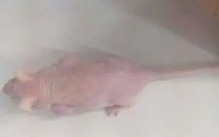
图2 双瘤模型鼠
2.3 组织学检查
扫描结束后,处死裸鼠,提取肿瘤组织,一半组织低温保存,备用;另一半组织用10%Formaldehyde 固定,梯度酒精脱水后,石蜡包埋切片,梯度酒精复水,常规HE染色观察,观察肿瘤形态和血管密度。
组织切片(HE染色)显示,肿瘤明显呈恶性形态,细胞核大,多见病理性核分裂像,双核或多核多见;肿瘤组织可见明显坏死区和大量血管分布,提示生长迅速和微血管较多,见图3~4。
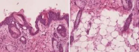
图3 A549肿瘤组织病理图片(HE 200×)

图4 肿瘤组织病理图片(HE 200×)
3 讨论
分子影像学的内涵决定了其必定能在肿瘤的早期诊断和鉴别定性方面发挥不可替代的作用。分子影像学的特点和基本任务就是利用分子生物学的基础研究成果,围绕病变发生发展过程中相关特异性分子,用化学的和生物学的方法构建特异性分子探针,用影像学手段在分子水平进行成像,从而达到对疾病进行早期诊断和鉴别定性的目的。
生长抑素是一种存在于人脑、下丘脑、胃肠、胰腺等部位内分泌细胞中的活性多肽,具有广泛的生理作用,能抑制生长激素、消化道激素等激素的分泌,抑制胰腺和胃的外分泌,作为中枢神经内神经递质,还可调节正常细胞和肿瘤细胞的分化增殖,具有抗增殖作用。生长抑素在体内是通过与生长抑素受体(Somatostatin Receptor,SSTR)结合而发挥生理功能的。生长抑素受体有五种亚型(Subtype),分别称为SSTR1-5。正常情况下,SSTR表达很少。与正常组织相比,肿瘤组织SSTR表达显著增多。大多数肿瘤如肝细胞癌主要表达SSTR2或SSTR5亚型[1]。
能模拟和复制人类疾病的动物模型是研究肿瘤成像的关键。经典的人肝癌细胞系BEL-7402表达高SSTR2,用此细胞系构建的裸鼠皮下移植瘤模型,可以很好地模拟人肝癌,并具有相应的生物学特性。针对SSTR2构建分子探针可以完成肿瘤特异性成像。实际情况下,分子探针是否具有特异性是能否检出特定肿瘤的关键,因此特异性探针的验证方法非常重要。
目前一般有3种方法,即探针分子乱序对比、特异性阻断实验和双瘤模型。其中双瘤模型实施难度较大,除选择合适的受体阳性/阴性细胞系配对外,掌握和平衡二者的的成瘤模式也非常关键。但最能科学、直观验证分子探针特异性的方法就是双瘤模型。Weissleder等[2]利用转基因技术构建了转铁蛋白受体(TfR)阳性和阴性双瘤模型,证明双瘤模型是可行的。因此,选取SSTR2表达阴性的肿瘤细胞系和SSTR2表达阳性的肿瘤细胞系,分别移植到裸鼠体内,可构建裸鼠双瘤模型。研究证明,人BEL-7402肝癌细胞系高表达SSTR2[3],且移植瘤成功后,肿瘤组织亦高表达SSTR2。Singh等[4]用RT-PCR方法证明,人非小细胞肺癌细胞系(NSCLC cell lines)A549、H460和H1299 SSTR2表达阴性;国内赵荣等[5]用Western blot方法也证明A549细胞不表达SSTR2。因此,选用高表达SSTR2的人肝癌BEL-7402细胞系和不表达SSTR2的人非小细胞肺癌A549细胞系在同一裸鼠个体制造双肿瘤模型,根据SSTR2表达不 同,分子探针结合量会出现明显差别,因此同一个体双侧构建双瘤模型是一种极好的方法,通过不同的增强效果可以直观地验证分子探针特异性,完成肝癌的特异性成像,为肝癌的早期诊断和鉴别诊断提供新方法[6-10]。
合适的动物模型对验证探针是否有效非常关键[11-14]。双瘤模型凭借在裸鼠腋窝双侧种植SSTR2不同表达程度的肿瘤,可以“直观地”看到由于SSTR2表达不同而产生的双侧肿瘤的显像差异,从而可靠地评价分子探针是否有作用[15-16]。了解两种细胞系不同的成瘤速度和成瘤特点,掌握二者成瘤规律,以便分别在不同时间开始移植肿瘤,最后达到在特定实验时间窗内二肿瘤体积差别在一个合理范围,有利于成像。
[1] Reubi JC,Waser B,Schaer JC,et al.Somatostatin receptor sst1-sst5 expression in normal and neoplastic human tissues using receptor autoradiography with subtype-selective ligands[J].Eur J Nucl Med Mol Imaging,2001,28(7):836-846.
[2] Weissleder R,Moore A,Mahmood U,et al.In vivo magnetic resonance imaging of transgene expression[J].Nat Med, 2000,6:351-355.
[3] Huo T,Du X,Zhang S,et al.Gd-EDDA/HYNIC-RGD as an MR molecular probe imaging integrin ανβ3receptor-expressed tumor-MR molecular imaging of angiogenesis[J].Eur J Radiol,2010,73:420-427.
[4] Singh SP,Han L,Murali R,et al.SSTR2-based reporters for assessing gene transfer into non-small cell lung cancer: evaluation using an intrathoracic mouse model[J].Hum Gene Ther,2011,22: 55-64.
[5] Zhao R,Yang W,Wang Z,et al.Treatment of transplanted tumor of lung adenocarcinoma A549 transfected by human somatostatin receptor subtype 2 (hsstr2) gene with 188Re-RC-160[J].Nucl Med Biol,2010,37(8):977-987.
[6] Hwang YF,Hwang SL,Kwan AL,et al.Differentiation among metastatic brain tumors,radiation necroses,and brain abscesses using proton magnetic resonance spectroscopy[J].Kaohsiung J Med Sci,2004,20(9):437-442.
[7] Nath K,Agarwal M,Ramola M,et al.Role of diffusion tensor imaging metrics and in vivo proton magnetic resonance spectroscopy in the differential diagnosis of cystic intracranial mass lesions[J].Magn Reson Imaging,2009,27(2):198-206.
[8] Mishra AM,Gupta RK,Jaggi RS,et al.Role of diffusion-weighted imaging and in vivo proton magnetic resonance spectroscopy in the differential diagnosis of ring-enhancing intracranial cystic mass lesions[J].J Comput Assist Tomogr,2004,28(4):540-547.
[9] Lindskog M,Spenger C,Jarvet J,et al.Predicting resistance or response to chemotherapy by proton magnetic resonance spectroscopy in neuroblastoma[J].J Natl Cancer Inst,2004, 96(19):1457-1466.
[10] Shah N,Gibbs J,Wolverton D,et al.Combined diffuse optical spectroscopy and contrast-enhanced magnetic resonance imaging for monitoring breast cancer neoadjuvant chemotherapy: a case study[J].J Biomed Opt,2005,10(5):051503.
[11] Kwock L,Smith JK,Castillo M,et al.Clinical role of proton magnetic resonance spectroscopy in oncology: brain,breast,and prostate cancer[J].Lancet Oncol,2006,7(10):859-868.
[12] Bolan PJ,Nelson MT,Yee D,et al.Imaging in breast cancer: Magnetic resonance spectroscopy[J].Breast Cancer Res,2005, 7(4):149-152.
[13] Tse GM,Yeung DK,King AD,et al.In vivo proton magnetic resonance spectroscopy of breast lesions: an update[J].Breast Cancer Res Treat,2007,104(3):249-255.
[14] Shukla-Dave A,Hricak H,Kattan MW,et al.The utility of magnetic resonance imaging and spectroscopy for predicting insignificant prostate cancer: an initial analysis[J].BJU Int, 2007,99(4):786-793.
[15] Saito K,Kaminaga T,Muto S,et al.Clinical efficacy of proton magnetic resonance spectroscopy (1H-MRS) in the diagnosis of localized prostate cancer[J].Anticancer Res,2008,28(3B): 1899-1904.
[16] Hara T,Kondo T,Hara T,et al.Use of 18F-choline and 11C-choline as contrast agents in positron emission tomography imaging-guided stereotactic biopsy sampling of gliomas[J].J Neurosurg,2003,99(3):474-479.
Establishment and Evaluation of Molecular Probe Verification Model in Brain Tumor Specific Imaging
HUO Tian-long1, YANG Shuo1, LI Tian-ran2, KANG Yu1, ZHAO Yun-yun1, DU Xiang-ke1
1.Department of Radiology, Peking University People’s Hospital, Beijing 100044, China; 2.Department of Radio-logy, the 95thHospital of PLA, Putian Fujian 351100, China
ObjectiveTo establish a dual-tumor animal model in one nude mouse which positively and negatively expresses a specific receptor respectively.MethodsThe human hepatocellular carcinoma cell line BEL-7402 (positively expressed SSTR2) and the human lung cancer cell line A-549 (negatively expressed SSTR2) were selected and cultured respectively. Then cells in logarithmic growth phase were digested and suspended for transplantation to the bilateral axillary fossa of the same nude mouse (the concentration of the cells suspended were over 5×10⁶/0.2 mL), so as to establish the dual-tumor epidermal xenograft nude mouse model. The effects of the tumor transplantation were observed and analyzed.ResultsThe two kinds of cell lines were successfully cultured; the BEL-7402/A549 dual-tumor nude mouse xenograft model was successfully established in the bilateral axillary fossa of one nude mouse. The tumor formation rate of BEL-7402 cell line was above 80% (24/30), and it was easy to form the tumor if the cell number was large enough. Whereas the tumor formation rate of A549 cell line was relatively low, which was about 73% (22/30) over a concentration of 5×10⁶/0.2 mL, and the success rate of tumor formation could be increased via cells augmentation or secondary injection. The number of the bilateral tumor whose size difference was within 1 cm was accounted for 32% (8/22). The histological examination showed that the hepatocellular carcinomas dissected from the tumor-bearing nude mice were typically malignant form compared with normal mice liver, the cell nucleus was large, pathologic mitosis were commonly seen with dual or multi nucleus. The obvious necrosis and abundant vessels were seen in tumors which suggested that the tumor grew fast and the micro-vessels were rich. Although the general form of the A549 transplantation tumors were different from the BEL-7402 transplantation tumors, the micro-morphology form of the two tumors were consistent with the corresponding tumors of human beings.ConclusionThe different tumor of cell lines could be inoculated to the same nude mouse to successfully develop the dual-tumor models. To well-control the growth difference of the two kindsof tumors could establish the size-proper tumor models, so as to lay a foundation for tumor specificityimaging.
molecular imaging; animal model; nude mouse xenograft; dual-tumor model; molecular probe
R81;R445.2
A
10.3969/j.issn.1674-1633.2017.03.003
1674-1633(2017)03-0010-04
2017-01-13
高等学校博士学科点专项科研基金新教师类(2011000 1120083);国家自然科学基金面上项目(81372363)。
霍天龙,副主任医师,主要研究方向为分子影像学。
通讯作者邮箱:huotianlong@bjmu.edu.cn
猜你喜欢
现代仪器与医疗(2022年4期)2022-10-08
健康体检与管理(2022年4期)2022-05-13
中国生殖健康(2020年2期)2021-01-18
装备制造技术(2020年1期)2020-12-25
浙江医学(2020年19期)2020-10-20
中国临床保健杂志(2020年3期)2020-06-08
中国生殖健康(2018年2期)2018-01-12
北京航空航天大学学报(2017年2期)2017-11-24
医学研究杂志(2015年9期)2015-07-01
癌变·畸变·突变(2015年4期)2015-02-27
