膝关节退行性骨关节病诊断中MRI的应用价值研究
2017-05-30 10:48:04胡静宜张杰韩菲菲
影像技术
2017年2期
胡静宜 张杰 韩菲菲
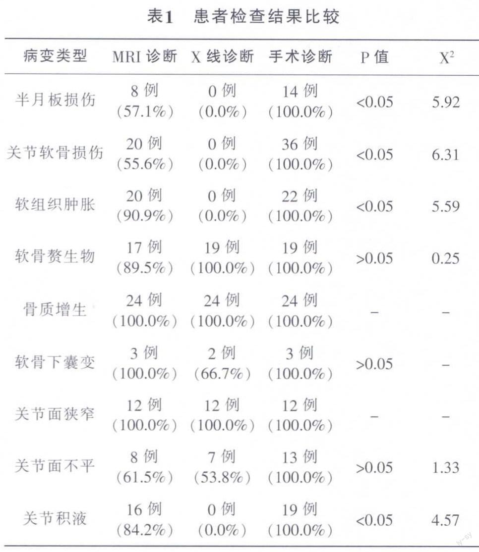
摘要:目的:对核磁共振成像在膝关节退行性骨关节病中的诊断价值进行研究、分析。方法:抽选55例膝关节退行性骨关节病患者,患者入院后先后予以X线、核磁共振成像检查。结果:两种检查方式在软骨赘生物、软骨下囊变、关节面狭窄、关节面不平中的诊断率比较无明显差异,P>0.05。结论:核磁共振成像在膝关节退行性骨关节病中具有较高的诊断准确率。
关键词:膝关节退行性骨关节病;核磁共振成像;诊断价值
中图分类号:R684;R445.2 文献标识码:B DOI:10.3969/j.issn.1001-0270.2017.02.06
Abstract: Objective: The diagnostic value of magnetic resonance imaging in knee joint degene-rative joint disease were studied. Methods: Both examinations in cartilage excrescence, subchondral cystic degeneration, articular surface narrow, uneven articular surface of diagnosis rate is no significant difference, P>0.05. Results: Both examinations in cartilage excrescence, subchondral cystic degeneration, articular surface narrow, uneven articular surface of diagnosis rate is no significant difference, P>0.05. Conclusion: Magnetic resonance imaging has a high diagnostic accuracy in the degenerative joint disease of the knee joint.
Key Words: Knee Degenerative Joint Disease; Magnetic Resonance Imaging; Diagnostic Value
膝關节退行性骨关节病在骨科中的发病率较高,患者临床中多以关节疼痛、关节功能活动受限为主要表现。对于患者来说,尽早诊断、尽早治疗能够促进疾病更好的康复[1]。膝关节退行性骨关节病可采用不同的方式进行诊断,核磁共振成像是临床中常用的影响诊断方式,我院在2015年4月-2016年5月间,抽选了55例膝关节退行性骨关节病患者,对核磁共振成像在膝关节退行性骨关节病中的诊断价值进行研究、分析。
1 资料与方法
1.1 一般资料
抽选55例膝关节退行性骨关节病患者,将其作为研究对象,患者均为2015年4月-2016年5月间入院治疗的人员。患者年龄为57-69岁,患者平均年龄为(63.5±6.8)岁,男性34例,女性21例,患者病程平均为(9.5±4.7)个月。患者均存在膝关节功能活动受限表现,有41例患者关节疼痛明显。所有患者均无风湿性疾病,无痛风、强直性脊柱炎表现。患者无脏腑功能衰竭表现,无血液系统疾病。
1.2 一般方法
1.2.1 对照组
患者采用X线检查,患者仰卧位或坐位,拍摄正位片和侧位片,参数设定:电压为50-60kV,电流为100mA,时间为20-30ms。
登录APP查看全文