
眼眶肌锥内肿瘤MSCT及磁共振诊断影像学表现
2019-04-21 13:36:25张强
中外医学研究
2019年30期
张强

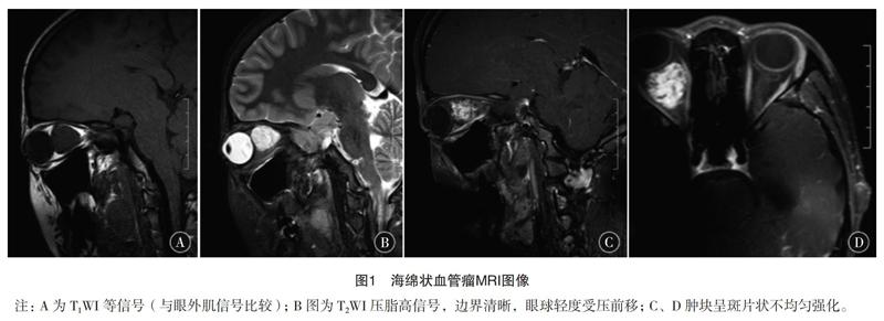

【摘要】 目的:探討眼眶肌锥内肿瘤多层螺旋CT(MSCT)及磁共振(MRI)诊断影像学表现。方法:选择2017年2月-2018年12月疑似眼眶肌锥内肿瘤患者90例作为对象,所有患者均经手术检查最终确诊,手术前均完成MSCT、MRI检查,分析不同影像检查检出率及影像学表现;绘制ROC曲线,分析MSCT、MRI在眼眶肌锥内肿瘤患者中的诊断效能。结果:疑似眼眶肌锥内肿瘤患者90例经手术检查确诊76例,患者经MRI确诊73例,检出率为96.05%(2例误诊,1例漏诊);MSCT确诊60例,检出率为78.95%(3例误诊,13例漏诊);MRI检查检出率高于MSCT检查(P<0.05);MSCT用于在眼眶肌锥内肿瘤患者中表现为类圆形占位病变,边界相对清除,中立于表现为软组织密度;强化明显,动态扫描可呈渐进性强化;而MRI检查患者多表现为T1WI低信号,T2WI高信号,边界不清,增强扫描后患者为中高强化,具有渐进性强化特征;ROC曲线结果表明,MSCT联合MRI在眼眶肌锥内肿瘤中的诊断敏感性、特异性,均高于单一MSCT、MRI检查(P<0.05)。结论:MSCT及MRI用于眼眶肌锥内肿瘤诊断均能获得良好的效果,但是二者联合检测诊断敏感性、特异性更高,为临床诊疗提供影像学依据。
【关键词】 多层螺旋CT 磁共振 眼眶肌锥内肿瘤 诊断效果 影像学表现 ROC曲线
doi:10.14033/j.cnki.cfmr.2019.30.030 文献标识码 B 文章编号 1674-6805(2019)30-00-03
[Abstract] Objective: To investigate the diagnostic imaging findings of multi-slice spiral CT (MSCT) and nuclear magnetic resonance (MRI) in the orbital tumors of the orbital muscle. Method: A total of 90 patients with suspected orbital intracondylar tumors from February 2017to December 2018 were selected. All patients were diagnosed by surgical examination. MSCT and MRI were performed before surgery. The detection rate of different imaging examinations was analyzed. Imaging findings, ROC curves were drawn to analyze the diagnostic efficacy of MSCT and MRI in patients with orbital tumors. Result: Ninety-six patients with suspected orbital conus tumors were diagnosed by surgical examination in 76 cases. The patients were diagnosed by MRI in 73 cases. The detection rate was 96.05% (2 cases were misdiagnosed and 1 case missed diagnosis). 60 cases were diagnosed by MSCT. The detection rate was 78.95% (3 cases were misdiagnosed, 13 cases were missed). The detection rate of MRI was higher than that of MSCT (P<0.05). MSCT was used for round-shaped lesions in the orbital tumor of the orbital muscle. Relative clearance, neutral in the performance of soft tissue density. Enhanced, dynamic scanning can be progressively enhanced. While MRI examination showed more T1WI low signal, T2WI high signal, unclear boundary, after enhanced scan the patient was moderately high, with progressive enhancement characteristics. ROC curve results showed that the diagnostic sensitivity and specificity of MSCT combined with MRI in the orbital conus tumor were higher than single MSCT and MRI (P<0.05). Conclusion: MSCT and MRI can be used for the diagnosis of orbital intracondylar tumors. However, the combination of the two methods is more sensitive and specific, and provides imaging evidence for clinical diagnosis and treatment.
[Key words] MSCT MRI Intraorbital tumor of the orbital muscle Diagnostic effect Imaging findings ROC curve
First-authors address: Nanping Peoples Hospital, Nanping 353000, China
眼眶肌锥内肿瘤是临床上常见的疾病,且多数患者为原发病变,由于眶内组织胚层多样,导致眼眶肌锥内肿瘤组织分类相对复杂、种类繁多,再加上该疾病的发生率与年龄存在较大的差异,导致临床诊断、鉴别难度较大[1-2]。目前,临床上对于眼眶肌锥内肿瘤以手术组织检查为主,虽然能帮助患者确诊,但是检查具有一定的风险性、创伤性,导致患者诊断依从性、耐受性较差[3]。MSCT是眼眶肌锥内肿瘤患者常用的影像学检查方法,具有扫描时间短、分辨率高等特点,能在短时间内对身体进行较长范围、不间断数据采集,可获得大量的信息,能提高图像质量[4];磁共振(MRI)是在外磁场作用下自旋能级发生列变,不仅能发现早期肿瘤,亦可确定病灶部位、大小等[5-6]。因此,本文以疑似眼眶肌锥内肿瘤患者作为对象开展研究,探讨MSCT及MRI在眼眶肌锥内肿瘤中的诊断效果及影像学表现,报道如下。……
登录APP查看全文
