埃兹蛋白在乳腺癌中的表达及其临床意义
2019-12-05 08:41王晓娇练孝春刘正人
医学信息 2019年21期
王晓娇 练孝春 刘正人

摘要:目的 通過检测埃兹蛋白在浸润性乳腺癌、乳腺导管内癌及正常乳腺组织中的表达,探究埃兹蛋白在乳腺癌组织中的表达的临床意义。方法 随机选取南昌大学第一附属医院40例浸润性乳腺癌患者及32例乳腺导管内癌患者,另外选择同期来我院健康体检者22例。通过免疫组化链霉亲和素-过氧化物酶法(S-P)法分别检测埃兹蛋白在22例正常乳腺组织、32例乳腺导管内癌组织和40例浸润性乳腺癌组织中的表达,参照病理结果进行分析。结果 埃兹蛋白在40例浸润性乳腺癌的阳性表达率为72.50%、在32例乳腺导管内癌的阳性表达率为40.63%、在正常乳腺组织中的阳性表达率13.64%,浸润性乳腺癌中埃兹蛋白的表达高于其他两种组织,差异具有统计学意义(P<0.05);埃兹蛋白在G3期乳腺癌组织中的表达高于在G1~G2期乳腺癌组织中的表达,差异具有统计学意义(P<0.05);埃兹蛋白在淋巴结转移患者中的阳性表达率高于在无淋巴结转移的患者中的阳性表达率,差异统计学意义显著(P<0.01);埃兹蛋白在<40岁的患者中的阳性表达率低于在年龄≥40岁的患者中的表达率,差异统计学意义显著(P<0.01);埃兹蛋白在PR阳性患者中的阳性表达率低于在PR阴性患者中的表达率,差异具有统计学意义(P<0.05);乳腺癌患者中埃兹蛋白的表达与组织学分级、腋窝淋巴结转移及年龄呈正相关(P<0.05),与PR呈负相关(P<0.05);埃兹蛋白在浸润性润腺癌中的表达与乳腺癌临床分期、脉管侵犯、肿瘤大小、患者月经状态以及Her-2、ER、Ki-67的表达无相关性(P>0.05)。结论 埃兹蛋白在乳腺癌和正常乳腺组织中的表达不同,对判断乳腺肿瘤性质具有重要参考意义;此外,埃兹蛋白能够帮助判断乳腺癌的转移潜能和预后,可能启发乳腺癌新的靶向治疗。
关键词:乳腺癌;埃兹蛋白;侵袭;转移;免疫组化
中图分类号:R737.9 文献标识码:A DOI:10.3969/j.issn.1006-1959.2019.21.027
文章编号:1006-1959(2019)21-0088-04
Abstract:Objective To investigate the clinical significance of ezrin expression in breast cancer tissues by detecting the expression of ezrin in invasive breast cancer, intraductal carcinoma of the breast and normal breast tissue. Methods 40 patients with invasive breast cancer and 32 patients with intraductal carcinoma of the breast in the First Affiliated Hospital of Nanchang University were randomly selected. In addition, 22 healthy subjects were enrolled in our hospital. The expression of ezrin in 22 normal breast tissues, 32 breast ductal carcinoma tissues and 40 invasive breast cancer tissues was detected by immunohistochemical streptavidin-peroxidase (S-P) method. The analysis was performed with reference to the pathological results. Results The positive expression rate of ezrin in 40 cases of invasive breast cancer was 72.50%, the positive expression rate in 32 cases of breast ductal carcinoma was 40.63%, and the positive expression rate in normal breast tissue was 13.64%.The expression of ezrin in invasive breast cancer was higher than that in the other two tissues,the difference was statistically significant (P<0.05); the expression of ezrin in G3 breast cancer tissues was higher than that in G1~G2 breast cancer tissues, the difference was statistically significant (P<0.05); The positive expression rate of ezrin in patients with lymph node metastasis was higher than that in patients without lymph node metastasis, the difference was statistically significant (P<0.01); The positive expression rate of ezrin in patients <40 years old was lower than that in patients aged 40 years or older, the difference was statistically significant (P<0.01); the positive expression of ezrin in PR positive patients the rate was lower than that in PR-negative patients,the difference was statistically significant (P<0.05). The expression of ezrin in breast cancer patients was positively correlated with histological grade, axillary lymph node metastasis and age (P<0.05),negative correlation with PR (P<0.05); expression of ezrin in invasive adenocarcinoma and clinical stage of breast cancer, vascular invasion, tumor size, patient's menstrual status and Her-2, ER, Ki-67 there was no correlation in expression (P>0.05). Conclusion The expression of ezrin in breast cancer and normal breast tissue is different, which has important reference significance for judging the nature of breast tumor. In addition, ezrin can help determine the metastatic potential and prognosis of breast cancer, and may inspire new targets of breast cancer treatment.
Key words:Breast cancer;Ezrin;Invasion;Metastasis;Immunohistochemistry
乳腺癌(breast cancer)是女性恶性肿瘤中最常见的一种,侵袭和转移是影响乳腺癌患者生存和预后的重要因素[1]。多项研究表明,埃兹蛋白与多种肿瘤的发生发展及预后有着密切的联系[2]。然而,目前对埃兹蛋白促进肿瘤细胞发生发展的机制尚不明确。本研究通过免疫组化检测埃兹蛋白在浸润性乳腺癌、乳腺导管内癌及正常乳腺组织中的表达,参照病理分析埃兹蛋白对浸润性乳腺癌发生发展中所起的作用,旨在探索其在乳腺癌中的临床意义,现在报告如下。
1材料与方法
1.1材料来源 随机选取南昌大学第一附属医院收治的第一次手术且术前均未经新辅助化疗及内分泌等其他治疗的,有完整的临床病理和随访资料的乳腺肿瘤患者72例,均为女性,另外选择同期来我院健康体检者22例。40例浸润性乳腺癌患者年龄28~72岁,平均年龄(41.55±11.58)岁;32例乳腺导管内癌患者年龄28~72岁,平均年龄(43.09±11.45)岁;22例健康体检者年龄28~73歲,平均年龄(46.64±12.34)岁。浸润性乳腺癌按照肿瘤TNM 分期Ⅰ~Ⅳ期分别为 4例、24例、9例和3例。
1.2试剂 埃兹蛋白单克隆抗体购自美国Immunoway公司,一抗浓度配比为1∶100。S-P试剂盒、DBA试剂盒购买自福州迈新生物技术有限公司。
1.3方法 结合研究期间患者的病理结果,采用免疫组化链霉亲和素-过氧化物酶法(S-P法)分别检测埃兹蛋白在浸润性乳腺癌、乳腺导管内癌和正常乳腺组织中的表达,并根据肿瘤TNM分期分级。S-P法按试剂盒说明书进行,以试剂盒内的阳性片为阳性对照,用 PBS液代替一抗为阴性对照。
1.4结果判定 定位:在20倍光学显微镜下(埃兹蛋白阳性通常表达在细胞膜和细胞浆,呈黄色或棕黄色颗粒),观察不同乳腺组织埃兹蛋白染色的情况和分布。定性:计算阳性细胞百分率:随机选择10个高倍视野,计数1000个肿瘤细胞/张切片。百分率≥10%,定为阳性片(+);百分比<10%,定为阴性片(-)。根据Mathew[3]等推荐的标准进行分级:-:无表达;+:<50%为细胞阳性表达或者染色较浅;++:50%~70%阳性表达且深染;+++:>70%且深染。
1.5统计学方法 采用SPSS 21.0软件处理数据。计数资料采用(n)表示,行?字2检验或Fisher确切概率法,相关性分析采用线性趋势检验。P<0.05为差异有统计学意义,P<0.01为差异统计学意义显著。
2结果
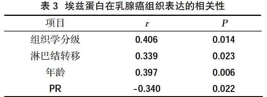
2.1埃兹蛋白在不同乳腺组织中的表达 浸润性乳腺癌组织中埃兹蛋白表达阳性的细胞主要表现为细胞质内棕黄色或棕褐色颗粒弥漫性分布(图1),埃兹蛋白在浸润性乳腺癌组织中的阳性表达率为72.50%(29/40);埃兹蛋白在乳腺导管内癌组织中主要以棕黄色或棕褐色颗粒弥散分布在细胞质中,少部分呈浅黄色染色分布在导管上皮细胞膜上(图2),其在乳腺导管内癌组织中的阳性表达率为40.63%(13/32);埃兹蛋白在正常乳腺组织主要呈浅黄色分布于乳腺导管上皮细胞膜上,其在正常乳腺组织的阳性表达率为13.64%(3/22)。浸润性乳腺癌、乳腺导管内癌及正常乳腺组织中的埃兹蛋白阳性表达率逐渐降低,浸润性乳腺癌中埃兹蛋白的表达与其他两种组织比较,差异具有统计学意义(P<0.05),见表1。
存在恶性程度高、易复发和转移等特点,Her-2基因扩增是乳腺癌患者总体生存率及复发的负性预测因子[19],其预测价值高于激素受体状态和淋巴结转移等多数其他预后因子。埃兹蛋白的表达与这些因素具有相关性说明埃兹蛋白可能与乳腺癌的复发、转移以及预后相关。已有研究显示年轻乳腺癌比年长乳腺癌预后差[20],本研究与之不同,本次结果显示40岁及以上患者的埃兹蛋白阳性率高于40岁以下患者。因一般年轻乳腺癌具有高淋巴转移率、高组织学分级、高临床分期、高三阴性比例及肿块大的特点,预期设想实验结果埃兹蛋白的表达与年龄呈负相关,与实际结果存在差异,这可能与实验样本量少及乳腺癌病理分型有关。
总之,不同的生理状态下埃兹蛋白生物学功能也不同,其异常过度表达,既能抑制乳腺恶性肿瘤细胞的凋亡,同时还可增加细胞的转移能力。埃兹蛋白对判断乳腺肿瘤的良恶性以及指导治疗乳腺癌具有重大意义。此外,埃兹蛋白能够帮助判断乳腺癌的转移潜能和预后,可能启发新的乳腺癌靶向治疗。当然,埃兹蛋白在乳腺癌中的作用机制需更深层次的研究。
参考文献:
[1]兰柯,李文翰,徐子森,等.卡培他滨在治疗转移性乳腺癌中作用的Meta分析[J].中国临床药理学与治疗学,2015,11(4):425-429.
[2]刘宏,张阳德.Ezrin蛋白与肿瘤转移关系[J].中国现代医学杂志,2009,19(19):952.
[3]于兴燕,于东红.Ezrin 蛋白在肿瘤中的研究进展[J].医学综述,2015,36(23):4278-4280.
[4]孔界男,周宪春,韩龙哲,等.甲胎蛋白阴性肝细胞癌中埃兹蛋白(ezrin)和SIX1蛋白表达水平的临床病理意义[J].细胞与分子免疫学杂志,2016,14(2):236-239.
[5]Penchev VR,Chang YT,Begum A,et al.Ezrin Promotes Stem Cell Properties in Pancreatic Ductal Adenocarcinoma[J].Mol Cancer Res,2019,17(4):929-936.
[6]Xie YH,Li LY,He JZ,et al.Heat shock protein family B member 1 facilitates ezrin activation to control cell migration in esophageal squamous cell carcinoma[J].Int J Biochem Cell Biol,2019(112):79-87.
[7]Slik K,Kurki S,Korpela T,et al.Ezrin expression combined with MSI status in prognostication of stage Ⅱ colorectal cancer[J].PLoS One,2017,12(9):e0185436.
[8]李運华,刘绍华,欧阳坚,等.乳腺癌组织Ezrin、VEGF表达变化及其与临床分期的关系[J].山东医药,2017,57(20):58-59.
[9]马小斌,王西京,刘小旭,等.Ezrin在乳腺癌发生发展中的表达及意义[J].现代肿瘤医学,2008,16(3):362-364.
[10]Li N,Kong J,Lin Z,et al.Ezrin promotes breast cancer progression by modulating AKT signals [J].Br J Cancer,2019,120(7):703-713.
[11]Ghaffari A,Hoskin V,Turashvili G,et al.Intravital imaging reveals systemic ezrin inhibition impedes cancer cell migration and lymph node metastasis in breast cancer[J].Breast Cancer Res,2019,21(1):12.
[12]方军.Ezrin蛋白表达与乳腺癌临床病理特征的相关性研究[J].检验医学与临床,2014,11(8):1048-1049.
[13]Antelmi E,Cardone RA,Greco MR,et al.ss1 integrin binding phosphorylates ezrin at T567 to activate a lipid raft signalsome driving invadopodia activity and invasion[J].PLoS One,2013,8(9):e75113.
[14]Mak H,Naba A,Varma S,et al.Ezrin phosphorylation on tyrosine 477 regulates invasion and metastasis of breast cancer cells[J].BMC Cancer,2012(12):82.
[15]Jarvinen TA,Pelto-Huikko M,Holli K,et al Estrogen receptor beta is coexpressed with ERalpha and PR and associated with nodal status, grade, and proliferation rate in breast cancer[J].Am J Pathol,2000,156(1):29-35.
[16]Itoh M,Iwamoto T,Matsuoka J,et al.Estrogen receptor(ER)mRNA expression and molecular subtype distribution in ER-negative/progesterone receptor-positive breast cancers[J].Breast Cancer Res Treat,2014,143(2):403-409.
[17]Aleskandarany MA,Green AR,Benhasouna AA,et al.Prognosis value of proliferation assay in the luminal,HER2-positive,and triple-negative biologic classes of breast cancer[J].Breast Cancer Res,2012(14):R3.
[18]Falck AK,Bendahl PO,Chebil G,et al.Biomarker expression and St Gallen molecular subtype classification in primary tumours,synchronous lymph node metastases and asynchronous relapses in primary breast cancer patients with 10 years' follow-up[J].Breast Cancer Res Treat,2013,140(1):93-104.
[19]黄小娥.Her-2蛋白表达与乳腺癌预后关系的相关性研究[J].中国现代医生杂志,2011,35(10):39-40.
[20]秦颖,张同先.青年乳腺癌临床病理特点分子分型及预后分析[J].中国肿瘤临床,2014,14(4):231-236.
收稿日期:2019-7-26;修回日期:2019-8-9
编辑/王朵梅
猜你喜欢
浙江医学(2020年9期)2020-07-01
浙江中西医结合杂志(2019年4期)2019-05-05
浙江医学(2019年2期)2019-01-23
右江医学(2016年4期)2017-01-05
法制与社会(2016年35期)2016-12-26
中国现代医生(2016年25期)2016-11-19
人民论坛(2016年14期)2016-06-21
中国继续医学教育(2015年1期)2016-01-06
中国当代医药(2014年34期)2014-12-31