结节性甲状腺肿和甲状腺癌的CT诊断与鉴别诊断分析
2020-08-09 08:56:59杨尊现
中外医疗
2020年15期
杨尊现
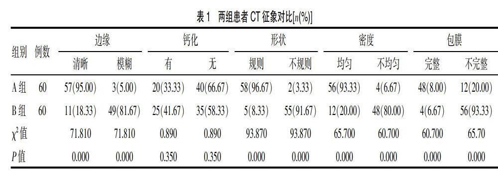
[摘要] 目的 分析结节性甲状腺肿、甲状腺癌的CT诊断影像特征与鉴别诊断。方法 方便选取2018年8月—2019年8月期间于该院接受诊治的60例结节性甲状腺肿患者设为A组,同时将同期收治的60例甲状腺癌患者设为B组,两组均接受CT检查。观察两组CT征象與病灶情况。结果 A组有20例患者(33.33%)有钙化表现,而B组有25例患者(41.67%)有钙化表现,两组在钙化表现比较上差异无统计学意义(χ2=0.890,P>0.05)。同时,A组边缘清晰患者例数多于B组(95.00%>18.33%,χ2=71.810,P=0.000),A组形状规则患者例数多于B组(96.67%>8.33%,χ2=93.870,P=0.000),A组密度均匀患者例数多于B组(93.33%>20.00%,χ2=65.700,P=0.000),A组包膜完整患者例数多于B组(80.00%>6.67%,χ2=65.70,P=0.000),以上两组差异有统计学意义(P<0.05)。A组60例患者病灶表现为局限性,且病灶边缘光滑,可见清晰的周围脂肪间隙,平扫密度不均。B组60例患者病灶表现为低密度,且平扫密度不均,增强扫描后强化程度较正常甲状腺实质低。结论 结节性甲状腺肿、甲状腺癌的CT诊断表现具有特征性,临床可依据掌握上述两种不同类型疾病的CT诊断表现,以判断并鉴别患者的病情。
[关键词] 结节性甲状腺肿;甲状腺癌;CT
[Abstract] Objective To analyze the CT diagnostic imaging features and differential diagnosis of nodular goiter and thyroid cancer. Methods Convenient select sixty patients with nodular goiter who were treated in the hospital from August 2018 to August 2019 were set as group A, and 60 patients with thyroid cancer who were treated in the same period were set as group B. Both groups received CT examination. Observe the CT signs and lesions of the two groups. Results Twenty patients (33.33%) in group A had calcification, and 25 patients (41.67%) in group B had calcification. There was no statistically significant difference in calcification between the two groups (χ2=0.890, P>0.05). At the same time, the number of patients with clear edges in group A was more than that in group B (95.00%>18.33%, χ2=71.810,P=0.000), and the number of patients with regular shape in group A was more than that in group B (96.67%> 8.33%, χ2=93.870,P=0.000). The number of patients with uniform density was more than that of group B (93.33%> 20.00%, χ2=65.700,P=0.000), and the number of patients with intact capsule in group A was more than that of group B (80.00%> 6.67%, χ2=65.70,P=0.000). The difference between the two groups was statistically significant (P<0.05). In group A, the lesions of 60 patients were localized, and the edges of the lesions were smooth. Clear peripheral fat spaces were visible, and the density of plain scanning was uneven. The lesions of 60 patients in group B showed low density and uneven scanning. The intensity of enhancement after enhanced scanning was lower than that of normal thyroid parenchyma. Conclusion The CT diagnosis of nodular goiter and thyroid cancer is characteristic. Based on the CT diagnosis of the above two different types of diseases, the clinical condition can be judged and differentiated.
[Key words] Nodular goiter; Thyroid cancer; CT
在甲状腺疾病中,结节性甲状腺肿、甲状腺癌是两种不同的类型,即前者为良性病变,而后者属于甲状腺恶性肿瘤[1]。CT是临床上检查甲状腺疾病的主要手段之一,虽然该检查具有密度分辨率高、高效精确等特点,但实际检查中以上两种类型的患者影像表现相似度较高[2],不仅鉴别难度大,而且患者的病情极易误诊或漏诊。因此,该院对2018年8月—2019年8月期间收治的60例结节性甲状腺肿、60例甲状腺癌患者CT诊断情况进行深入分析,并对比与总结两者的CT诊断征象,为临床医护人员今后在诊断中提供参考,现报道如下。
1 资料与方法
1.1 一般资料
方便选取该院接受诊治的60例结节性甲状腺肿、60例甲状腺癌作为该次研究对象。纳入标准:①120例患者均于该院均经手术、病理证实病情;②患者知情该次研究,已阅读此次研究相关资料。排除标准:①因合并认……
登录APP查看全文