
头穴围刺对缺血再灌注大鼠脑组织血管生成素-2和血管内皮细胞生长因子表达的影响
2020-10-21 05:38:24师帅栾凯迪李虹霖王玉珏夏昆鹏张金朋马祺昊孙琦月佟晓薇韩盛旺
世界中医药
2020年8期
师帅 栾凯迪 李虹霖 王玉珏 夏昆鹏 张金朋 马祺昊 孙琦月 佟晓薇 韩盛旺
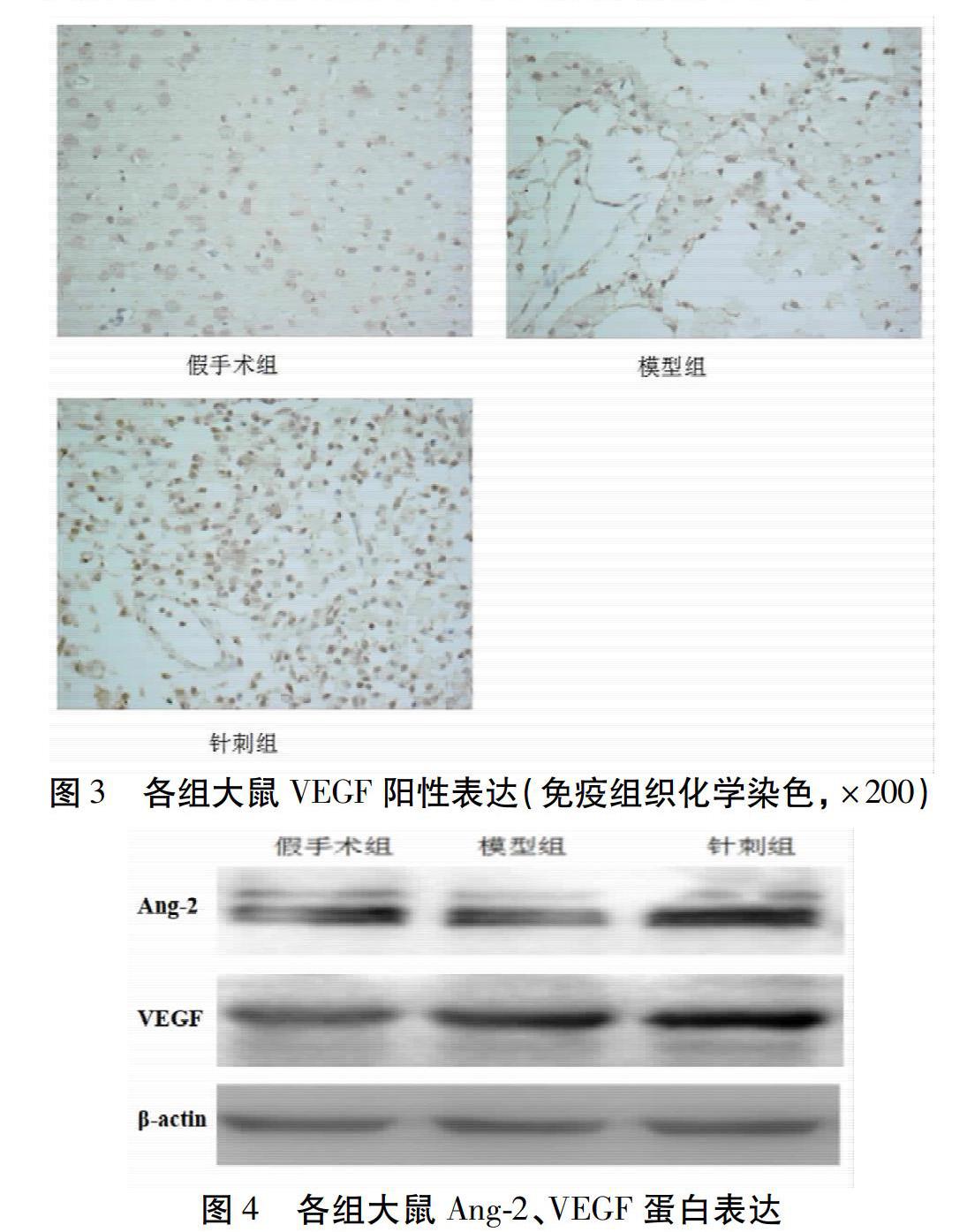
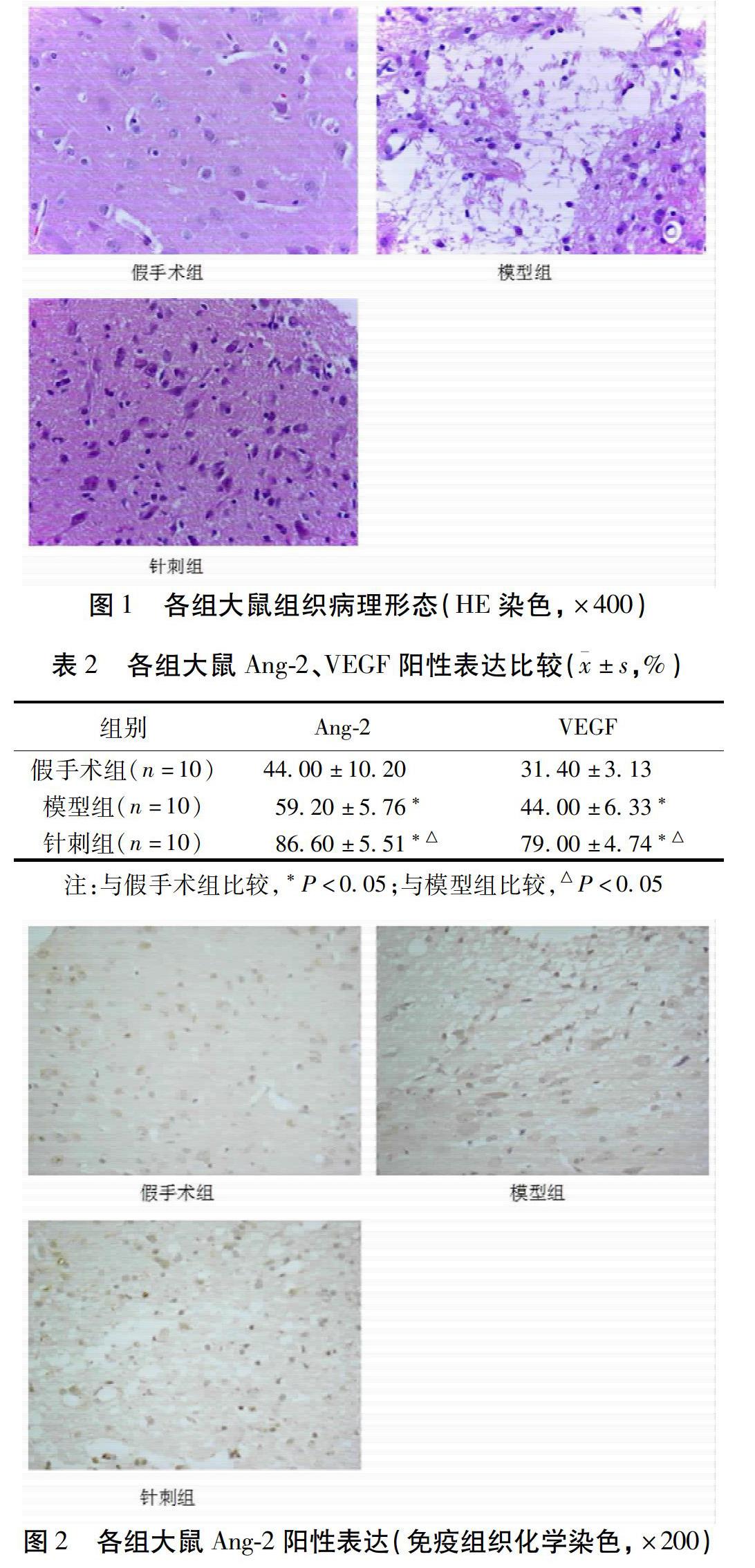
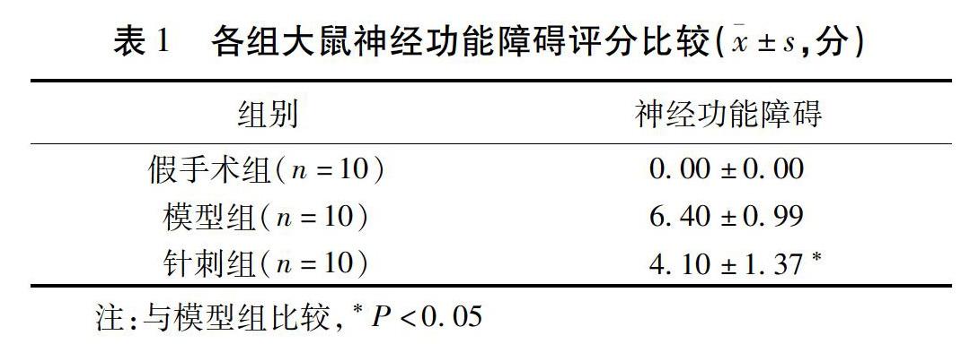
摘要 目的:觀察头穴围刺对缺血再灌注大鼠血管生成素-2(Ang2)和血管内皮细胞生长因子(VEGF)表达的影响。方法:选取30只健康雄性Wistar大鼠随机分为假手术组、模型组和围刺组,参照Zea-Longa报道的线栓法制备大鼠脑缺血再灌注模型,围刺组给予头穴围刺治疗,3组分别于21 d取大鼠脑组织,采用HE染色观察大鼠脑组织缺血结构的变化;免疫组织化学观察大鼠脑缺血阳性细胞表达情况;采用Western检测Ang-2、VEGF蛋白表达水平。结果:针刺21 d后mNSS评分围刺评分优于模型组;与模型组比较,大鼠脑组织病理细胞数减少,差异有统计学意义(P<0.05);针刺21 d后与模型组比较,大鼠脑组织缺血区域Ang-2、VEGF阳性表达明显增加,差异有统计学意义(P<0.05);针刺21 d后与模型组比较,大鼠脑组织Ang-2、VEGF蛋白增多,差异有统计学意义(P<0.05)。结论:头穴围刺可上调大鼠缺血再灌注区脑组织Ang-2、VEGF蛋白的表达,促进梗死区血管新生,帮助脑缺血再灌注损伤后的脑神经修复,降低神经功能缺损程度。
关键词 头穴围刺;脑缺血再灌注;神经功能缺损;促血管生成素-2;血管内皮生长因子;脑神经修复;血管新生
Effects of Scalp Acupoint Encircling Needling on the Expression of Ang-2 and VEGF in Brain Tissue of Rats with Ischemia-Reperfusion Injury
SHI Shuai1,2,LUAN Kaidi1,LI Honglin1,2,WANG Yujue1,2,XIA Kunpeng1,2,ZHANG Jinpeng1,2,Ma Qihao1,SUN Qiyue 1,TONG Xiaowei1,HAN Shengwang1,2
(1 The Second Affiliated Hospital of Heilongjiang University Of Chinese Medicine,Heilongjiang,Harbin 150001,China; 2 Heilongjiang University Of Chinese Medicine,Harbin 150000,China)
Abstract Objective:To observe the effects of scalp acupoint encircling needling on the expression of Ang-2 and VEGF in rats with ischemia-reperfusion injury.Methods:A total of 30 healthy male Wistar rats were randomly divided into a sham operation group,a model group and an encircling needling group,with 10 rats in each group.Rat cerebral ischemia-reperfusion model was prepared by thread embolization method reported by Zea-Longa.The encircling needling group was given scalp point encircling needling.The brain tissues of each group were taken from 21 days in 3 groups,and the changes of cerebral ischemic structure were observed by HE staining.Immunohistochemistry was used to observe the expression of positive cells in cerebral ischemia in rats,and Western Blot was used to detect the expression of Ang-2 and VEGF.Results:1)21 days later,the mNSS score and encircling needling were better than the model group.2)HE staining:Compared with the model group,the number of pathological cells in rat brain tissue decreased(P<0.05).3)After 21 days of acupuncture,compared with the model group,the positive expression of Ang-2 and vascular endothelial growth factor in the ischemic area of rat brain tissue was significantly increased after immunohistochemical treatment(P<0.01).4)Compared with the model group,the number of Ang-2 and vascular endothelial growth factor protein in the brain tissue of rats increased 21 days after acupuncture(P<0.01).Conclusion:Scalp acupoint encircling needling can up-regulate the expression of Ang-2 and VEGF protein in brain tissue of rats with cerebral ischemia-reperfusion injury,promote angiogenesis in infarcted area,help the repair of brain nerve after cerebral ischemia-reperfusion injury,and reduce the degree of neurological deficit.
Western检测:大鼠断头取脑,将组织匀浆裂解,离心去沉淀,取上层清液。按照BCA试剂盒要求测量蛋白质浓度,取等浓度等体积上样变性蛋白,SDS-PAGE(10%)电泳2 h以后,半干转膜法转移到聚偏二氟乙烯膜(PVDF膜)上,用含5%脱脂奶粉的TBST封闭2 h,一抗孵育过夜,VEGF(1∶1 000稀释),Ang-2(1∶500),温度保持4 ℃,TBST清洗膜5次×5 min,加入二抗(1∶8 000稀释)室温孵育2 h,TBST清洗5次×5 min,用ECL显色,于数字成像系统下显影,采用Image J检测条带密度。实验重复3次。
1.3 统计学方法
采用SPSS 22.0统计软件进行数据分析,计量资料用均数±标准差(±s)表示,在对比实验中涉及到2个数据集之间的数据,使用2个独立样本t检验的,方差不齐时采用近似t检验,阳性率之间的比较采用卡方检验。以P<0.05为差异有统计学意义。
2 结果
2.1 各组大鼠神经功能障碍……
登录APP查看全文
猜你喜欢
童话王国·奇妙逻辑推理(2024年5期)2024-06-19 16:03:38
中国民间疗法(2021年1期)2021-04-20 02:30:34
基层中医药(2021年7期)2021-03-26 18:00:27
数学物理学报(2020年2期)2020-06-02 11:29:24
光学精密工程(2016年6期)2016-11-07 09:07:19
核科学与工程(2015年4期)2015-09-26 11:59:03
中医研究(2014年8期)2014-03-11 20:29:23
中医研究(2014年6期)2014-03-11 20:29:02
中国中医药现代远程教育(2014年17期)2014-03-01 04:29:28
军事体育学报(2014年3期)2014-02-27 16:00:33
