
AP-3 adaptor complex-mediated vesicle trafficking
2021-06-09 10:59:00ZhuoMaMdNurIslamTaoXuEliSong
Biophysics Reports
2021年2期
Zhuo Ma,Md. Nur Islam,Tao Xu,3,Eli Song
1 National Laboratory of Biomacromolecules, CAS Center for Excellence in Biomacromolecules, Institute of Biophysics, Chinese Academy of Sciences, Beijing 100101, China
2 University of Chinese Academy of Sciences, Beijing 100049, China
3 Guangzhou Regenerative Medicine and Health Guangdong Laboratory (Bioland Laboratory), Guangzhou 510005,China
Abstract The transport of cargo proteins to specific subcellular destinations is crucial for the different secretory and endocytic traffic pathways. One of the most important steps in maintaining the accuracy of this process is the recruitment of adaptor protein (AP) complexes to the membrane for recognizing and packaging cargo proteins into nascent vesicles. Adaptor protein complex 3 (AP-3) is a heterotetrametric complex implicated in the trafficking of cargo proteins from the trans-Golgi network (TGN) and/or endosomes to lysosomes or lysosome-related organelles (LROs). This complex is also involved in the biogenesis of synaptic vesicles (SVs) in neurons and of dense core vesicles (DCVs) in endocrine cells as well as in the recycling of receptors in immune cells and the regulation of planar cell polarity (PCP)proteins. Functional defects in AP-3 cause multiple abnormalities in cellular vesicle trafficking and related organelle function, leading to various disorders, such as Hermansky-Pudlak syndrome (HPS).However, the molecular mechanism underlying AP-3 has not been fully elucidated, and further investigations are needed to understand AP-3-mediated trafficking, its associated molecules and its related roles in inherited diseases. Here, we review the current understanding of AP-3 in cellular vesicle trafficking, especially focusing on mammalian systems.
Keywords AP-3, Vesicle trafficking, Cargo sorting, Disease/disorder
INTRODUCTION
The trafficking of proteins and other macromolecules is one of the most essential life activities performed by cells. Eukaryotic cells have various organelles surrounded by specific membranes that perform different functions. In the endocytic and exocytotic pathways, the trafficking of cargoes between organelles or between organelle and cell membrane is mainly mediated by membrane-encapsulated transport carriers, generally known as vesicles. Briefly, vesicles are formed when cargoes are recruited to the donor membrane and packed into a nascent vesicle, which is then pinched off from the donor membrane. The free vesicles are then transported through the cytosol to the destination membrane and fuse with the target membrane compartment, achieving the transport and exchange of materials (Park and Guo 2014). The trans-Golgi network (TGN) is generally considered a central station for cargo sorting where proteins and other molecules are sorted into distinct vesicles and further delivered to their appropriate intracellular destinations(Guo et al. 2014). One key group of players that function in the sorting process are adaptors, which localize at the donor membranes and recruit cargoes by direct or indirect interactions. Adaptor protein (AP)complexes are a well-known family of adaptors that play a crucial role in cargo selection and vesicle formation. AP complexes specifically recognize and interact with the sorting signals in the cytoplasmic domains of transmembrane cargo proteins or cargo receptors, and they recruit clathrin or other accessory proteins and then package selected cargoes into a departing vesicle (Park and Guo 2014).
To date, five members of the mammalian AP complex family have been identified, namely, AP-1, AP-2, AP-3,AP-4 and AP-5. These molecules are heterotetrametric complexes, each of which contains four subunits,including one small (σ1-σ5), one medium (μ1-μ5) and two large (γ/α/δ/ε/ζ and β1-β5) subunits, which form a central brick-like core and two smaller globular appendages connected by flexible linkers (Fig. 1A). One of the large subunits (γ/α/δ/ε) is responsible for interacting with the target membrane, and the other large subunit (β1-β3) recruits the clathrin coat protein via a clathrin-binding motif (Boehm and Bonifacino 2001; Ohno 2006; Park and Guo 2014). An exception is the β4 subunit of AP-4, which lacks the clathrin-binding box and is thus not capable of binding to clathrin(Dell' Angelica et al. 1999a). The N-terminal domains of the two large subunits, the small subunit and the medium subunit form the core brick-like domain, which interacts with cargo molecules, including adenosine diphosphate (ADP)-ribosylation factor (ARF) proteins and phospholipids. In contrast, the C-termini of the two large subunits are appendage domains that interact with a series of accessory proteins. The medium subunit (μ) is responsible for cargo recognition, and the small subunit (σ) is proposed to stabilize the complex(Park and Guo 2014).

Fig. 1 Schematic representation of AP complexes. A Diagrams of heterotetrameric AP complexes. Subunit names are indicated, and homologous subunits are depicted in the same color. All AP complexes consist of core, hinge and ear domains, except AP-5, which lacks the hinge domain. B The schemes represent the closed and opened state of the AP-3 complex. Based on the known structure of the AP-1 core domain, the cargo binding sites of AP-3 are hindered in the locked state. In the presence of ARFs, AP-3 is recruited to the membrane and undergoes a conformational change to the opened state when both the YXXΦ and [DE]XXXL[LI] motif-binding sites are exposed and recognize the cargo proteins with consensus motifs
The five AP complexes have different subcellular localizations and distinct functions. The most studied and well-known AP complex variant is AP-2, which is related to clathrin-mediated endocytosis from the plasma membrane, while the other adaptor proteins mainly participate in cargo sorting from the TGN or endosomes to lysosomes or other organelles (Collins et al. 2002; Ohno 2006; Park and Guo 2014). Here, we review the recent advances regarding the structure,recognition of sorting signals, coat proteins, associated lipids, subcellular localization, cellular trafficking,biological function and associated diseases of AP-3 mainly in mammals.
STRUCTURE OF AP-3
Similar to all AP complexes, AP-3 is composed of four subunits as follows: two large subunits (δ and β3), one medium subunit (μ3) and one small subunit (σ3). These subunits assemble into a central core and two appendage domains linked by flexible linkers, also known as the hinge region (Ohno 2006; Park and Guo 2014) (Fig. 1B). The large subunit contains three domains as follows: the N-terminal domain, which interacts with the μ and σ subunits to form the core; the C-terminal appendage, also called the ear domain; and the hinge region, which connects the above two domains. The medium μ subunit includes two domains as follows: one is the N-terminal domain, which constitutes one-third of the polypeptide and contributes to the formation of a part of the core; and the other is the C-terminal domain, which constitutes the remaining two-thirds of the polypeptide and mediates cargo recognition by directly binding to the sorting signals of cargo proteins (Mardones et al. 2013).The small σ subunit is also a part of the core and is implicated in the stabilization of the complex as suggested by its X-ray crystal structure (Collins et al.2002). The N-terminal domains of the μ subunit and σ subunit have some similarities, and together they stabilize the complex (Ohno 2006).
Multiple isoforms of the AP-3 subunits encoded by different genes have been identified as follows: the β subunit of AP-3 has two isoforms (β3A and β3B); the μ subunit has two isoforms (μ3A and μ3B); and the σ subunit has two isoforms (σ3A and σ3B). The σ3A and σ3B isoforms are ubiquitous and are functionally equivalent, and the β3B isoform is mostly expressed in neuronal tissues (Danglot and Galli 2007). In theory,eight types of AP-3 heterotetramers can be assembled by different combinations of the various subunits(Mattera et al. 2011). In practice, two different isoforms of the AP-3 complex have been identified in vivo as follows: AP-3A (consisting of δ, β3A, μ3A and σ3 (A or B)) is ubiquitous; and AP-3B (consisting of δ, β3B, μ3B and σ3 (A or B)) is neuron specific (Nakatsu et al. 2004;Park and Guo 2014).
RECOGNITION OF SORTING SIGNALS BY AP-3
As adaptors, AP complexes specifically select and interact with cargoes or cargo receptors via recognition of the signals within the sequences of cargo proteins.These signals are generally short, linear consensus sequences of amino acid residues located in the cytoplasmic region of transmembrane proteins or cargo receptors (Park and Guo 2014). To date, there are two well-characterized recognition motifs as follows: the YXXΦ motif (where X represents any amino acid and Φ represents an amino acid containing a bulky hydrophobic residue), a tyrosine-based sorting motif that directs endosomal and lysosomal targeting and sorting at the TGN; and the [DE]XXXL[LI] consensus motif, a dileucine-based motif that is mainly present in the TGN or endosomal system (Bonifacino and Dell'Angelica 1999; Ohno 2006; Guo et al. 2014). These motifs are not exactly conserved in every site but are instead degenerate motifs containing four to seven residues, in which two or three key sites are critical for producing the functional characteristics of these motifs(Bonifacino and Lippincott-Schwartz 2003). The critical residues are usually bulky and hydrophobic, while charged residues are also important determinants of the specificity for certain signals and contain functional significance for the trafficking of membrane proteins(Kirchhausen 1999; Bonifacino and Traub 2003).
The AP-3 complex recognizes both the YXXΦ and[DE]XXXL[LI] motifs via the μ3 subunit and the combination of the δ-σ3 subunits, respectively (Park and Guo 2014). The YXXΦ binding site on μ3 is similar to the binding sites on the AP-1 μ1 subunit and the AP-2 μ2 subunit (Owen and Evans 1998; Mardones et al. 2013). The mechanism by which AP-3 recognizes the[DE]XXXL[LI] motif and the corresponding conformational change it must undergo remain to be characterized. Additionally, other noncanonical recognition consensus and mechanisms need to be identified.
COAT PROTEINS OF AP-3
Coat protein complexes, such as COPI, COPII and clathrin, are involved in the transportation of cargo proteins to different cellular compartments. Unlike the well-known association of clathrin with the AP-1 and AP-2 complexes, the concept of coat proteins associated with AP-3 remains controversial. The δ subunit of AP-3 is responsible for binding to the target membrane, and the β3 subunit recruits clathrin via the clathrin-binding box, LΦXΦD/E, which resides in the hinge region of the complex (Owen et al. 2004). The interaction between AP-3 and clathrin has been verified by a coassembly assay, and cellular AP-3 has been demonstrated to colocalize with clathrin via immunoelectron and immunofluorescence microscopy (Dell'Angelica et al.1998). Furthermore, an in vitro binding experiment has demonstrated that the appendage domain of the β3 subunit of mammalian AP-3 associates with the N-terminal domain of the clathrin heavy chain(Dell'Angelica et al. 1998). These findings suggest that AP-3 has the potential to play a role in protein-sorting events as a clathrin-associated adaptor in mammals.Colocalization analysis of AP-3 and AP-1 with clathrin by dual color labeling analysis has demonstrated that AP-3 colocalizes with clathrin on the vesicular profile when the vesicle buds from the donor membrane but to a lesser degree than AP-1 (Peden et al. 2004). However,the lack of AP-3 in purified clathrin-coated vesicles implies that AP-3 might work independently of clathrin,at least in some cases (Dell'Angelica et al. 1997b). Zlatic et al. demonstrated that the association of AP-3 with clathrin is dispensable for endosomal AP-3 vesicle biogenesis by rapid chemical-genetic perturbation of clathrin function in PC12 cells, indicating that endosomal AP-3-clathrin interactions fulfill functions distinct from AP-1 and AP-2 (Zlatic et al. 2013). In addition to clathrin, VPS41, a member of the homotypic fusion and vacuole protein sorting (HOPS) complex,which includes a single clathrin heavy chain repeat in its C-terminal domain, also reversibly self-assembles into a lattice with AP-3 and promotes DCV biogenesis by a conserved mechanism. This finding suggests that VPS41 has the potential to act as a coat protein of AP-3 and take part in cargo sorting in the regulated secretory pathway (Asensio et al. 2013).
ASSOCIATED LIPIDS OF AP-3
Lipid function and switching are believed to play crucial roles in cargo sorting and transport during vesicular trafficking. In the secretory pathway, lipids are involved in the entire journey of the secretory vesicles, including the formation of budding sites at the donor membrane, protein recruitment at the budding sites and formation of exocytotic sites as well as docking, priming and fusion with the target membrane(Tanguy et al. 2016). Although a direct interaction between AP-3 and lipids has not yet been identified, a relationship between AP-3 and its related lipids could be proposed based on the distribution and roles of different lipids.
Phosphatidylinositol 3-phosphate (PI3P) is detected predominantly on early endosomes, while cytoplasmic lipids, such as PI(3,5)P2and bismonoacylglycerophosphate (BMP), are enriched in late endosomes and lysosomes. The plasma membrane is enriched with phosphoinositides, such as PI4P,PI(3,4)P2, PI(4,5)P2, PI(3,4,5)P3, phosphatidylserine(PS) and phosphatidylethanolamine (PE) (De Craene et al. 2017). These phosphoinositides specifically interact with coat proteins or accessory proteins and recruit organelle-specific effector proteins that are involved in vesicle trafficking and signal transduction on the cytosolic surface (Van Meer and de Kroon 2011;Tanguy et al. 2016). The Golgi membrane of mammalian cells contains the same lipids as the plasma membrane, but the proportions are quite different. Two leaflets of the Golgi membrane bilayers, the luminal and cytosolic leaflets, contain different types of lipids. The cytosolic leaflet of the Golgi apparatus contains DAG, PA and phosphoinositides, which are involved in the formation of secretory vesicles (Van Meer and de Kroon 2011; Tanguy et al. 2016). The TGN membrane is enriched with PI4P, which recruits AP-1 to the Golgi apparatus through direct interaction as identified by in vitro assays (Wang et al. 2003). Structural analysis has further confirmed that AP-1 interacts with PI(4)P via the binding site localized at its γ subunit (Heldwein et al. 2004). Because AP-3 localizes at the TGN, it has a high probability of interacting with PI4P, similar to AP-1; this potential interaction may be responsible for the different cargo sorting and transportation pathways from the TGN. The role of the inositol lipid and its ligand, PI(3,4,5)P3, in the regulation of AP-3 function further implies that lipids play important roles in AP-3 function and AP-3-mediated vesicle trafficking (Hao et al. 1997).
SUBCELLULAR LOCALIZATION AND MEMBRANE RECRUITMENT OF AP-3
In mammalian cells, AP-3 was identified as puncta localized to the Golgi region and other peripheral structures as determined by immunofluorescence experiments (Dell'Angelica et al. 1997a; Simpson et al.1997). EM localization data also demonstrate a dual localization of the AP-3 complex in the TGN and in endosomes (Simpson et al. 1996; Dell'Angelica et al.1998). Peripheral structures partially colocalize with the endosomal marker, transferrin receptor (TfR),indicating that AP-3 exists in some endosomal locations(Dell'Angelica et al. 1997a). Within the endosome portion, AP-3 is preferentially presented on tubular endosomal compartments and is particularly associated with distinct buds of endosomal tubules (Peden et al.2004). For the different isoforms of AP-3, the ubiquitously expressed AP-3A localizes to the TGN and the peripheral region where endocytic materials are contained (Dell'Angelica et al. 1997b). Neuron-specific AP-3B is associated with endosomes and is involved in the biogenesis of SVs from endosomes as identified by studies on PC12 cells (Faundez and Kelly 2000;Blumstein et al. 2001; Nakatsu et al. 2004; Zlatic et al.2013). In addition, AP-3 is localized in the Golgi apparatus in neurons and is responsible for the transport of cargoes from the Golgi apparatus to the axon (Li et al. 2016).
The mechanism underlying the recruitment of AP complexes and coat proteins to specific membranes has been partially identified and described. The recruitment process involves several common basic mechanisms. Among the five AP complexes, AP-1, AP-3 and AP-4 have similar mechanisms that use the small GTPase, ARF1, which facilitates the activation and recruitment of adaptor proteins, coat proteins and other effectors (Donaldson and Jackson 2011). Initially,the binding of AP-1 to ARF1 induces a change in AP-1 from a closed to an open conformation followed by exposure of the binding sites and a change to a coplanar configuration to allow simultaneous interactions with cargo and clathrin. This interaction further stabilizes the open conformation and brings the complex into close contact with the membrane (Ren et al. 2013).AP-3 has a similar localization to AP-1 on the TGN membrane; thus, the mechanism underlying AP-3-dependent protein sorting also relies on the action of kinases, coat or accessory proteins and small GTPases(Fig. 1B). ARF1 regulates the recruitment of AP-3 to membranes, which requires the conversion of ARF1 from a GDP-bound form to a GTP-bound form catalyzed by guanine nucleotide exchange factor (GEF). GTP-bound ARF1 binds to AP-3 and other effectors at the correct membrane with high affinity (Ooi et al. 1998).Biochemical evidence and in vivo studies also demonstrate that ARF1 promotes the membrane recruitment of AP-3 (Ooi et al. 1998). As indicated by an in vitro binding assay, ARF6 interacts with the β3 and δ subunits of AP-3, but whether ARF6 is involved in the recruitment of AP-3 to the membrane is still controversial (Ooi et al. 1998; Austin et al. 2002).
AP-3 MEDIATED CELLULAR TRAFFICKING
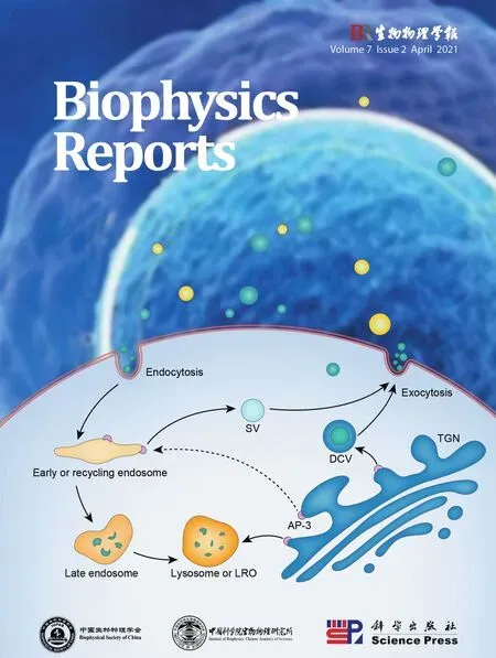
By recruiting cargoes and interacting with coated or accessory proteins and lipids, AP-3 participates in the transport of both membrane and molecules from the TGN and/or endosomes to lysosomes or lysosomerelated organelles (LROs) (Fig. 2). Inhibition of AP-3 function by RNAi leads to a misrouting of both LAMP1 and LAMP2 to the cell surface (Groux-Degroote et al.2008). This result has been further confirmed in AP-3-deficient cells, in which LAMP1, LAMP2 and melanosomal membrane protein tyrosinase are mislocalized (Dell'Angelica et al. 1999b). OCA2, the product of the gene oculocutaneous albinism type 2,resides in the endoplasmic reticulum, and it also localizes to melanosomes. OCA2 contains the dileucinebased signal, LL1, and is an AP-3 cargo according to in vitro binding experiments (Sitaram et al. 2009).Tyrosinase-related protein 1 (Tyrp1), also known as TRP1, is significantly decreased in AP-3-deficient primary melanocytes. This phenomenon is suppressed by treatment with a lysosome protease inhibitor,indicating that TRP1 is missorted to lysosomes for degradation when AP-3 is absent (Di Pietro et al. 2006).However, TRP1 does not directly interact with AP-3 but instead acts through an association with tyrosinase(Kobayashi and Hearing 2007). AP-3 physically interacts with biogenesis of lysosome-related organelles complex-1 (BLOC-1), and simultaneous deficiencies in both AP-3 and BLOC-1 affect the targeting of LAMP1, PI4KIIα and VAMP7 (Salazar et al.2006). The δ subunit of AP-3 interacts with the v-SNARE protein, VAMP7, and mislocalization of the δ subunit causes diminished lysosomal secretion as well as several neurological problems (Martinez-Arca et al.2003). Deletion of AP-3 in yeast is associated with mislocalization of the Vamp3p t-SNARE protein,vacuolar proteins and alkaline phosphatase (Cowles et al. 1997).

Fig. 2 Schematic representation of AP-3 subcellular localization and mediated trafficking. The AP-3 complex localizes to the TGN, early endosomes and recycling endosomes, and it is responsible for regulation of the trafficking of cargo proteins from the TGN and/or endosomes to lysosomes or LROs. The AP-3 complex is also responsible for the biogenesis of SVs in neurons and of DCVs in endocrine cells,and it is involved in the recycling of receptors in immune cells and the regulation of PCP proteins. The corresponding AP-3-mediated trafficking pathways are indicated by arrow lines
In addition, AP-3 is involved in the biogenesis of SVs from endosomes in neurons. In vitro experiments show that only the neuronal AP-3B generates synaptic vesicles from endosomes although it is not the predominant form of AP-3 in brain (Blumstein et al.2001). In mocha mice, an AP-3-deficient mouse model,zinc transporter 3 (ZnT3) and chloride channel (ClC-3)are reduced in SVs, suggesting that the trafficking of ZnT3 and CIC-3 to SVs is regulated by AP-3 (Salazar et al. 2004a, b). Hippocampal neurons lacking neuronal AP-3 have diminished asynchronous release, which is accomplished by SVs and impairs the precision of action potential timing (Evstratova et al. 2014). In midbrain dopamine neurons, loss of AP-3 reduces vesicular monoamine transporter 2 (VMAT2)localization to release sites and impairs the release of dopamine vesicles (Silm et al. 2019). AP-3 targets vesicular glutamate transporter (VGLUT) to SVs with low release probability, suggesting its involvement in VGLUT recycling (Evstratova et al. 2014; Li et al. 2017).Calcyon, a single transmembrane protein that binds to clathrin light chain in neurons, directly interacts with AP-3 μ3 subunit, and it regulates the trafficking of SVs and the targeting of AP-3 cargoes, including ZnT3 and PI4KIIα (Muthusamy et al. 2012).
AP-3 is also involved in the exocytosis of DCVs in endocrine cells. The neuroendocrine cells and endocrine cells from mocha mice have increased constitutive exocytosis of DCV cargoes, including soluble cargoes and membrane proteins, but they have a decreased response to stimulation, suggesting that AP-3 deficiency potentially leads to a disturbance of DCV biogenesis in the regulated exocytosis (Grabner et al. 2006). In PC12 cells, loss of AP-3 impairs DCV formation and substantially decreases synaptotagmin 1, indicating that AP-3 may concentrate the cargoes required for regulated exocytosis (Asensio et al. 2010).
AP-3 is also involved in the recycling of receptors in immune cells and the regulation of planar cell polarity(PCP) proteins. TLR2 stimulation and cytokine activation are involved in the inflammatory responses in innate immunity. AP-3 is required for the trafficking of TLR2 and its ligands to lysosomes for degradation(Petnicki-Ocwieja et al. 2015). The trafficking of human CD1b is related to the lipid antigen-presenting process from the sorting of endosomes to late endosomes; this process is mediated by AP-3 (Briken et al. 2002). PCP is established by the polarized and asymmetric localization of several membrane PCP proteins,including Vangl2 and Frizzled6. The subcellular localization of Vangl2 is abnormal in AP-3-depleted cells and mocha mice, causing auditory and vestibular dysfunction in mutant mice (Tower-Gilchrist et al. 2019).
Proteomics and proximity strategies have been used to identify the potential cargoes involved in AP-3-dependent trafficking. Purification of AP-3-dependent vesicles from PC12 cells using ZnT3, a known AP-3 cargo,identified enriched components, including AP-3 subunits, BLOC-1 and PI4KIIα (Salazar et al. 2004b).Combination of organelle density gradients and proteome analysis characterized the AP-3-dependent cargo protein profile in the Arabidopsis AP-3β mutant,indicating disturbed aquaporins and phosphorylation patterns (Pertl-Obermeyer et al. 2016). However, more research is needed to unravel the precise mechanism underlying the interaction between AP-3 and the newly identified cargoes.
PHYSIOLOGICAL FUNCTIONS AND ASSOCIATED DISEASES OF AP-3
Functional defects in AP complexes will cause incorrect localization of cargo proteins, subsequently affecting a wide range of cellular activities, including cell signal transduction, organelle dynamics and cellular homeostasis (Park and Guo 2014). The physiological roles of AP-3 have been identified through genetic studies and related inherited diseases. We summarized the related diseases in fruit fly, mouse, dog and human caused by the mutations in different subunits of AP-3(Table 1).

Table 1 Diseases and genetic disorders caused by AP-3 mutations
The large δ subunit of AP-3 is defective in the fruit fly garnet eye color mutant, which results in a reduced level of pigment granules, indicating that AP-3 is involved in the biogenesis and sorting pathway of pigment granules (Ooi et al. 1997). Mocha mice, which have δ subunit mutations, present with color defects,inner ear degeneration and neurologic defects, whichare associated with abnormalities in cargo sorting from the TGN to the LROs and in SV transport (Kanthetiet al.1998). In humans, mutation of the δ subunit causes destabilization of AP-3 in cells and leads to Hermansky-Pudlak syndrome-10 (HPS10), a novel type of HPS with severe neurologic involvement. Using retroviral reconstitution in the patients’ T cells may restore the formation of AP-3 and reverse degranulation defects(Ammannet al. 2016).
Mutation of the β3A subunit in dogs causes various phenotypes characterized by canine cyclic hematopoiesis (gray collie syndrome) and decreased coat pigmentation (Bensonet al. 2003). Genetic deficiencies of the β3A subunit inpearlmice present as color defects, abnormal LROs, melanosomes and platelet granules without any neurological defects (Fenget al. 1999). In humans, β3A mutation causes Hermansky-Pudlak syndrome-2 (HPS2), a rare autosomal recessive disorder characterized by oculocutaneous albinism and prolonged bleeding(Dell'Angelicaet al. 1999b). In these cases, the biogenesis and function of LROs, including melanosomes and platelet granules, are abnormal in AP-3-deficient cells. Additionally, AP-3 deficiency leads to increased expression of LAMP-1, LAMP-2 and CD63 on the cell surface of fibroblasts; these markers normally localize to late endosomes or lysosomes(Dell'Angelicaet al. 1999b). Although CD63 mislocalizes to the cell membrane, perforin and granzymes are correctly localized to the lytic granules in cytotoxic T lymphocytes (CTLs). However, these lytic granules in AP-3-deficient CTLs are unable to move along microtubes towards the docking domain of immunological synapses, thereby preventing their secretion, resulting in no CTL-mediated killing (Clarket al. 2003).
Knockout of neuronal β3B in mice results in several neurological and behavioral impairments (Seonget al.2005). The β3B mutation in humans causes severe developmental delay, poor visual contact with optic atrophy and postnatal microcephaly, called early-onset epileptic encephalopathy (EOEE) (Assoumet al. 2016).
In addition, μ3B subunit-deficient mice show spontaneous seizures without any deafness or balance problems (Nakatsuet al. 2004). To the best of our knowledge, the diseases caused by mutations in other subunits have not yet been reported and remain to be further determined.
CONCLUSION AND PERSPECTIVES
Over the last several decades, research on AP-3 has expanded to a great extent, and many experimental approaches have been employed to understand the function and molecular mechanism of AP-3 in cellular trafficking. The biological significance of AP-3 has been well identified, but the following aspects remain unclear: the trafficking steps involved from the TGN to lysosomes or other organelles; the fate of AP-3 during vesicle maturation; the associated coat proteins other than clathrin; the associated lipids and their functions in AP-3 trafficking; and the recycling pathway of AP-3.The use of combinations of powerful biochemical methods, genetic tools and imaging techniques,including electron microscopy and super-resolution microscopy, holds great promise for resolving these unknowns that remain in this field. Thus, a more thorough understanding of AP-3 will shed light on the cellular functions and biological significance of the AP-3 complex.
Abbreviations
ADP Adenosine diphosphate
AP Adaptor protein
ARF ADP-ribosylation factor
BLOC-1 Biogenesis of lysosome-related organelles complex-1
BMP Bis-monoacylglycerophosphate
ClC-3 Chloride channel 3
DAG Diacylglycerol
DCV Dense core vesicles
EOEE Early-onset epileptic encephalopathy
GEF Guanine nucleotide exchange factor
GTP Guanosine triphosphate
HOPS Homotypic fusion and vacuole protein sorting complex
HPS Hermansky-Pudlak syndrome
LAMP1 Lysosome-associated membrane glycoprotein 1
LAMP2 Lysosome-associated membrane glycoprotein 2
LRO Lysosome-related organelle
OCA2 Oculocutaneous albinism type 2
PCP Planar cell polarity
PE Phosphatidylethanolamine
PI(3,4)P2Phosphatidylinositol 3,4-bisphosphate
PI(3,4,5)P3Phosphatidylinositol 3,4,5-trisphosphate
PI(4)P Phosphatidylinositol 4-phosphate
PI(4,5)P2Phosphatidylinositol 4,5-bisphosphate
PI3P Phosphatidylinositol 3-phosphate
PI4KIIα Phosphatidylinositol 4-kinase II α
PS Phosphatidylserine
SNARE Soluble NSF Attachment Protein Receptor
SV Synaptic vesicle
TfR Transferrin receptor
TGN Trans-Golgi network
TLR2 Toll-like receptor 2
Tyrp1(TRP1) Tyrosinase-related protein 1
VAMP7 Vesicle-associated membrane protein 7
VGLUT Vesicular glutamate transporter
VMAT2 Vesicular monoamine transporter 2
VPS41 Vacuolar protein sorting-associated protein 41
ZnT3 Zinc transporter 3
Acknowledgements We apologize to the scientists in this field,whose publications were not cited due to space limitations. This work was supported by grants from the Ministry of Science and Technology of the People’s Republic of China(2018YFA0507101, 2016YFA0500203), the National Natural Science Foundation of China (31770900, 31730054), the Beijing Natural Science Foundation (5212016), and the Youth Innovation Promotion Association of the Chinese Academy of Sciences (2011087). All authors participated in the design and discussion of manuscript conception and outline, contributed to editing of the draft manuscript.
Compliance with Ethical Standards
Conflict of interest Zhuo Ma, Md. Nur Islam, Tao Xu and Eli Song declare that they have no conflict of interest.
Human and animal rights and informed consent This article does not contain any studies with human or animal subjects performed by any of the authors.
Open Access This article is licensed under a Creative Commons Attribution 4.0 International License, which permits use,sharing, adaptation, distribution and reproduction in any medium or format, as long as you give appropriate credit to the original author(s) and the source, provide a link to the Creative Commons licence, and indicate if changes were made. The images or other third party material in this article are included in the article’s Creative Commons licence, unless indicated otherwise in a credit line to the material. If material is not included in the article’s Creative Commons licence and your intended use is not permitted by statutory regulation or exceeds the permitted use, you will need to obtain permission directly from the copyright holder. To view a copy of this licence, visit http://creativecommons.org/licenses/by/4.0/.
登录APP查看全文
- Biophysics Reports的其它文章
- Optimizing weighting functions for cryo-electron microscopy
- Anti-oxidative effect of zinc in human umbilical cord mesenchymal stem cells
- Mammalian mitochondrial iron–sulfur cluster biogenesis and transfer and related human diseases
- Genetic variation and function: revealing potential factors associated with microbial phenotypes
- Live-cell fluorescence imaging of ciliary dynamics
- Potential of adipose derived stem cell preparations in neurological repair and regeneration