Assess on rabbit conjunctival lymphatic vessels distribution in different age groups
2022-03-31 01:03JingYiWu1DieHu2LongFangZhou2YingSu2QianWenBu2XiaoJingPan2AlexHuang
国际眼科杂志 2022年4期
Jing-Yi Wu1,2,3,4, Die Hu2,3,4, Long-Fang Zhou2,3,4, Ying Su2,3,4, Qian-Wen Bu2,3,4, Xiao-Jing Pan2,3,4, Alex Huang
1Weifang Medical University, Weifang 261021, Shandong Province, China 2Eye Institute of Shandong First Medical University; Qingdao Eye Hospital of Shandong First Medical University, Qingdao 266071, Shandong Province, China 3State Key Laboratory Cultivation Base, Shandong Provincial Key Laboratory of Ophthalmology, Qingdao 266071, Shandong Province, China 4School of Ophthalmology, Shandong First Medical University, Qingdao 266071, Shandong Province, China 5Shiley Eye Institute; Hamilton Glaucoma Center; Viterbi Family Department of Ophthalmology, San Diego 92101, CA, USA 6University of California, San Diego 92101, CA, USA
Abstract
INTRODUCTION
The lymphatic system is an integral part of the body’s circulatory system.It plays various essential roles: maintaining fluid homeostasis, transporting immune cells and fat and macromolecules from the digestive system[1-3].Total body fluid, cells, protein, lipids, and macromolecules extravasated from the interstitial spaces of the body are reabsorbed by lymphatic vessels and returned to the venous systemviathe thoracic duct[4-7].When the lymphatic physiology is disrupted, extracellular fluid can accumulate in the interstitial space of tissues, resulting in increased pressure and lymphedema,etc[6,8-10].
Natively, the subconjunctival space is a potential space.Subconjunctival injections are a simple method for drug delivery and treating ophthalmic diseases, but drugs are absorbed by subconjunctival lymphatic vessels to limit the effect[11-13].The subconjunctival space is also used in ocular hypertensive treatment for glaucoma.Under normal physiological conditions, aqueous humor exits the eyeviathe conventional and unconventional aqueous humor outflow pathways.These pathways can be disrupted in glaucoma, leading to poor outflow and ocular hypertension.One way to lower intraocular pressure(IOP)is to perform glaucoma surgeries(such as trabeculectomies)that drain the aqueous from the anterior chamber directly into the subconjunctival space.In this case, continued subconjunctival outflow is desired.In conclusion, it is imperative to understand the process of fluid elimination from the subconjunctival space[14-19].
Previous studies have confirmed the subconjunctival lymphatic system as the main drainage pathway for the subconjunctival space[11].Subconjunctival lymphatic functional studies and structural characterization have been performed in enucleated human and pig eyes and live mouse, monkey, and human eyes[11,20].In pigs, it was further found that the subconjunctival lymphatic vessels were more densely distributed on the nasal side than in other regions[21-22].This was confirmed in adult mouse eyes with a developmental study explaining.Murine subconjunctival lymphatics develop in the embryonic and post-natal period from a nasal root before wrapping around the limbus temporally.
There is a lack of specific research on the morphology and distribution of the subconjunctival lymphatic vessels in New Zealand white rabbits, limiting the application of research involving the subconjunctival lymphatic vessels in rabbits’ eyes.This is important because rabbits are a standard model for trabeculectomy research and developing subconjunctival minimally invasive glaucoma surgery(MIGS).Small studies have previously shown that subconjunctival trypan blue injection leads to a visible bleb with distinct lymphatic outflow pathways[11].This study further evaluates the subconjunctival outflow pathways of live rabbit eyesviatrypan blue injection, followed by evaluation of bleb-related outflow pathways based upon location and age.
MATERIALS AND METHODS
AnimalModelsFive healthy adult male New Zealand rabbits weighing 3-4 kg and five 3-4wk juvenile male New Zealand white rabbits weighing 1-2 kg were purchased by the Animal Management Center of Qingdao Eye Institute Affiliated to Shandong First Medical University.They were separately reared with a good diet and health, and the eyeball tissue structures of conjunctiva and cornea were healthy.All experimental animals were fed and used following the ARVO Statement issued.
AnimalTreatmentNew Zealand white rabbits were anesthetized by intravenous injection of 30 mg/kg 3% pentobarbital sodium at ear margin, and then 0.6%-1% trypan blue(Solarbio T8070; Beijing, China)0.02-0.1 mL was injected into conjunctiva with sterile 30G injection needle to form blebs with a diameter of 2-3 mm.One of the two eyes of each rabbit was randomly selected as the injection eye.Injection sites include nasal side 2∶00-4∶00 for the right eye and 8∶00-10∶00 for the left eye.At the superior between 11∶00 and 1∶00.Temporal side: 8∶00-10∶00 for the right eye and 2∶00-4∶00 for the left eye.From 5∶00-7∶00 in the inferior part, video recording was performed under the surgical video system(Zeiss).

RESULTS
DistributionofConjunctivalLymphaticVesselsinRabbitsTo study the morphology distribution of the conjunctival lymphatic vessels in rabbits, we injected trypan blue subconjunctival in four quadrants: nasal, temporal, superior, and inferior.The visible blue drainage pathways did not coincide with observable ocular surface blood vessels(Figure 1), supporting a non-blood vessel identity.Because the lymphatic vessels of rabbit conjunctiva are small in diameter and difficult to fill[8], we mainly studied the middle part(2-5 mm from the cornea)and peripheral part of the conjunctiva(near the fornix part).All injection site were 1-2 mm near the corneal limbus.

Figure 1 Distribution of lymphatic vessels in rabbit conjunctiva A: Distribution of lymphatic vessels in the conjunctiva in the nasal quadrant(supranasal quadrant); B: Distribution of lymphatic vessels in the conjunctiva in the superior quadrant; C: Distribution of lymphatic vessels in the conjunctiva in the temporal quadrant(supratemporal quadrant); D: Distribution of lymphatic vessels in the conjunctiva in the inferior quadrant.Take the left eye as an example to explain the distribution of lymphatic vessels in rabbit conjunctiva.The white arrow refers to the thick circular lymphatic vessels near the limbus part of the conjunctiva, and the black arrow refers to the single collecting lymphatic vessels in the peripheral part of the conjunctiva.
The conjunctiva of the nasal quadrant is shown in Figure 1A.The injection site was between 8∶00-10∶00 of the left eye and between 2∶00-4∶00 of the right eye.A thick annular lymphatic vessel was observed in the middle part of the conjunctiva, and there were many branches in this part.The lymphatic vessels converge into a single collecting lymphatic vessel at the peripheral part.The conjunctiva in the superior quadrant was visible in Figure 1B, and the injection site was 10∶00-2∶00 of the conjunctiva.The middle part of the conjunctiva was widely distributed with thick annular lymphatic vessels that had many branches, and all the annular lymphatic vessels were connected.The lymphatic vessels converge into a single collecting lymphatic vessel in the peripheral part.The conjunctiva of the temporal quadrant was visible in Figure 1C, and the injection site was between 2∶00-4∶00 on the left eye and between 8∶00-10∶00 on the right eye.The lymphatic vessels in the middle part of the conjunctiva were annular and had many branches, and they gathered into a single collecting lymphatic vessel at the peripheral part.The conjunctiva in the inferior quadrant could be seen in Figure 1D, and the injection site was between 4∶00-8∶00 of the eye.The distribution of lymphatic vessels in the inferior conjunctiva was similar to that in the superior part.The lymphatic vessels in the middle part of the conjunctiva were widely distributed, with thick annular lymphatic vessels having many branches connected by communication branches.The peripheral parts could be seen to converge into a single collecting lymphatic vessel.In conclusion, regardless of the quadrant, the conjunctival lymphatic vessels in the middle part were relatively thick and had many branches, which converged into a single collecting duct in the peripheral part.
MorphologyandDistributionofLymphaticVesselsinConjunctivaofJuvenileRabbitsTo compare the morphology and distribution of the conjunctival lymphatic vessels between adult rabbits and juvenile rabbits, trypan blue was injected into the conjunctiva of the juvenile rabbits and observed through the operation video system.It could be seen that the diameter of the conjunctival lymphatic vessels in the juvenile rabbits was smaller than that in the adult rabbits, and it was slower to fill(Figure 2).The morphology and distribution of their lymphatic vessels were consistent with those of adult rabbits; that is, the middle part of the lymphatic vessels was thick and communicated with each other(Figure 2), and a single collecting lymphatic vessels could be seen at the peripheral part(Figure 2).What is different from adult rabbits in distribution is that the conjunctival lymphatic vessels of juvenile rabbits have few branches.
TheDifferenceintheNumberofLymphaticVesselsinDifferentQuadrantsandTheirRelationshipwithAgeThe average number of lymphatic vessels in the nasal quadrant, the superior quadrant, the temporal quadrant, and the inferior quadrant of adult rabbit eyes was 2.4±0.20, 2.2±0.15, 2.6±0.10, and 2.6±0.10, respectively.The average number of lymphatic vessels in the nasal quadrant, the superior quadrant, the temporal quadrant, and the inferior quadrant of the juvenile rabbit eyes were 2.0±0.13, 1.6±0.10, 1.6±0.09, and 1.8±0.15, respectively.There was no significant difference in the average number of lymphatic vessels in adult and juvenile rabbits in each quadrant.ThenP=0.838 in each quadrant of the adult rabbit eyes(Figure 3), andP=0.750 in each quadrant of the juvenile rabbit eyes(Figure 3).Under the null assumption, there was no significant difference in the number of lymphatic vessels in each quadrant of rabbit eyes conjunctiva in each group.Subsequently, pairedt-test of the number of lymphatic vessels in the same quadrant between the adult rabbit eyes and the juvenile rabbit eyes revealed thatP=0.612 for the nasal lymphatic vessel,P=0.591 for the superior lymphatic vessel,P=0.219 for the temporal lymphatic vessel, andP=0.724 for the inferior lymphatic vessel(Figure 3).There was no difference in the number of lymphatic vessels in the same quadrant between the two age groups.
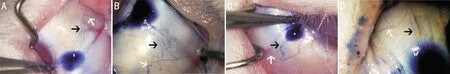
Figure 2 Distribution of lymphatic vessels in juvenile rabbit conjunctiva A: Distribution of lymphatic vessels in the conjunctiva in the nasal quadrant(supranasal quadrant in left eye); B: Distribution of lymphatic vessels in the conjunctiva in the superior quadrant(left eye shown); C: Distribution of lymphatic vessels in the conjunctiva in the temporal quadrant(supratemporal quadrant in left eye); D: Distribution of lymphatic vessels in the conjunctiva in the inferior quadrant(right eye shown).The juvenile lymphatic vessels after injection of trypan blue under the conjunctiva.The white arrow refers to the thick circular lymphatic vessels near the limbus part of the conjunctiva, and the black arrow refers to the single collecting lymphatic vessels in the peripheral part of the conjunctiva.

Figure 3 Comparison of the number of conjunctival lymphatic vessels in rabbits Compare the number of lymphatic vessels in different quadrants of adult rabbits; Comparison of the number of lymphatic vessels in different quadrants of juvenile rabbits; Comparison of the number of lymphatic vessels in the same quadrant between the adult rabbit and the juvenile rabbit.
DISCUSSION
Conjunctival lymphatic drainage plays a vital role in ocular drainage[23], and there are few studies on the morphology and distribution of conjunctival lymphatic vessels.In recent years, Yuetal[11]and Guoetal[21]have found in a series of studies that the bulbar conjunctiva is rich in lymphatic vessels by subconjunctival injection of trypan blue.
In addition, in a study, by injecting contrast into isolated porcine, bovine, human, and mouse eyeballs, they observed that the related outflow pathways(lymphatic vessels)had an extremely high similarity, which revealed that the morphology of conjunctival lymphatic vessels of different species had certain common characteristics[20].
In this study, the conjunctival lymphatic vessels were visualized by trypan-blue angiography.The number of lymphatic vessels was an innovative quantitative analysis to better study its morphology and distribution.Trypan blue, an anionic azo dye, is a standard tracerinvivoand used in anterior segment surgery[24-26].Yuetal[11]studied the morphology of lymphatic vessels in monkey and rabbit eyes by subconjunctival injection of trypan blue.They obtained good results that confirmed the subconjunctival injection of trypan blue is an experimental reliability method for studying the conjunctival lymphatic vessels.New Zealand white rabbit is a standard animal model in lymphatic vessel-related research.According to the experimental results, we have the following conclusions: 1)The conjunctival lymphatic vessels of rabbits are widely distributed on the conjunctiva and are similar in different quadrants; 2)Compared with the conjunctival lymphatic vessels of adult rabbits, the lymphatic vessels of juvenile rabbits have fewer branches; 3)There is no significant difference in the number of lymphatic vessels distributed between the quadrants of the conjunctiva of rabbits, and it is not related to age.
In terms of the pattern of lymphatic vessels distribution, it may be related to the direction of lymphatic drainage.In the early studies, it was generally believed that conjunctival lymph flowed out from the peripheral lymphoid ring of the cornea and drained in four directions: upper, lower, medial and lateral in different quadrants[19,27-28].Other studies have shown that the conjunctival lymphatic vessels present two large lymphatic trunks at a distance of 7-8 mm from the cornea, namely, the nasal trunk and the temporal trunk.The nasal trunk lymphatic vessels drain into the maxillary inferior lymph nodes, and the temporal trunk lymphatic vessels drain into the auricular junction lymph nodes[3,28-31].This shows many branches of lymphatic vessels near the limbus part of the conjunctiva.The lymphatic vessels in the peripheral limbus part of the conjunctiva are thick collecting channels and drain to the orbit.
It has been found that new lymphatic vessels originate from the nasal side and grow clockwise and counter clockwise in the first two weeks of life, eventually covering the limbus and the entire conjunctiva[11,22].A series of recent studies have further proved that due to the mode of lymphatic trunk entering the eyeball from the inner can thus region during development, more lymphatic branches were produced on the nasal side[22].The results of the above studies are related to the preferential drainage of the nasal side of the lymphatic vessels.Recent conjunctival lymphangiography studies have further revealed that the nasal lymph vessels were more abundant in distribution[22,30].Consistent with the above results, our study found that young rabbits had fewer lymphatic branches compared with adult rabbits, which verified in the profile that rabbit conjunctival lymphatic vessels produced more branches during development.However, the difference was that in our experiment, we found that the distribution numbers of lymphatic vessels in each quadrant of rabbit conjunctiva between the two age groups were not significantly different and were not related to age.
There are potential limitations in this study.First, the model has a small sample size and lack of reliable data, making the experimental results may have limited generalizability.Second, the lack of existing research on this topic may affect the study results.Nevertheless, in this experiment, the classical trypan blue angiography was used, and a trend was obtained, which illustrated the application of trypan blue angiography in rabbits of two ages and the similarities and differences of lymphatic vessels.Our results indicate that future research needs a more adequate and representative sample to further explore the differences between conjunctival lymphatic vessels in different ages.
In conclusion, the results of this study provided detailed information on the morphology and distribution of the conjunctival lymphatic vessels in New Zealand white rabbits by using trypan blue, which was the critical basis for further research on the structural and functional characteristics of the conjunctival lymphatic vessels in both standard and diseased conditions.This study confirmed the similarities and differences between rabbits and other mammals, which provided a new animal model and reference significance for the research related to improving aqueous humor drainage after glaucoma surgery and the research on ocular tumors,etc.Detailed knowledge of the distribution of conjunctival lymphatics has significant relevance to a better understanding of immunology, drug delivery, glaucoma filtration surgery in the conjunctiva.This also indicated that New Zealand white rabbits, as mammals with moderate size, easy operation, and low price, were a suitable animal model for studying bulbar conjunctival lymphatic vessels.