
A case of conjunctival intraepithelial neoplasia with spheroidal degeneration: a clinicopathological study
2022-08-10 01:39ShoichiroOtaSatoruKaseTakayukiTanakaSusumuIshida
Dear Editor,
I am Shoichiro Ota from Hokkaido University hospital,Japan. Conjunctival intraepithelial neoplasia (CIN) is a disease concept that includes dysplasia and carcinoma
.Pathologically, CIN is characterized by tumor cell proliferation located within the epithelium with preservation of the basement membrane
. In addition to histological diagnosis, anterior segment optical coherence tomography (ASOCT) plays an important role in clinical diagnosis, showing diffuse epithelial thickening with homogeneous high-intensity reflection in case of CIN
.
Spheroidal degeneration is a collection of golden spheres deposited in the keratoconjunctival tissue, first reported by Fraunfelder
in 1972. It is usually found in the pinguecula, and identified as cell-free amorphous deposits in histopathology
; however, the origin and OCT findings of spheroidal degeneration have yet been elucidated. The aim of this study is to report a case of CIN with spheroidal degeneration, and to analyze the correlation between ASOCT findings and histopathology. Written informed consent was obtained about the use of medical record for the case report from this patient. This case study adhered to tenets of Declaration of Helsinki. Institutional review board waived approval of this study as a clinical study application because this is a single case report.
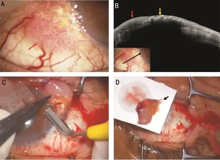
A 76-year-old man was aware of right conjunctival hyperemia and visited a nearby doctor. He has been observed for normal tension glaucoma. The patient was followed up for 2mo as a clinical diagnosis of an inflammatory lesion, but the symptom did not improve. Since the conjunctival tumor was then suspected, he was referred to our department. The best-corrected visual acuity at the first visit was 1.2 and 0.1 in his right eye (OD) and left eye(OS), respectively. The intraocular pressure is 12 mm Hg OD and 13 mm Hg OS. Slit-lamp examination showed a sessile elevated papillary mass lesion at the inferotemporal conjunctiva OD. In that area, fireworks-like blood vessels on the tumor surface together with dilated blood vessels flowing into the tumor were found. In addition, there were yellow granular lesions on the corneal limbus continuous with the papillary tumor (Figure 1A). The OCT findings of the tumor lesion displayed diffuse homogenous high-intensity reflection(Figure 1B, red arrow), as well as a heterogenous band-shaped hyper-intensive reflection corresponding to the yellow granular lesion (Figure 1B, yellow arrow). From clinical findings,CIN and squamous cell carcinoma were listed as differential diagnoses, and total tumor resection with 3 mm surgical margins and cryopexy were performed (Figure 1C). The resected tumor tissue was placed on a disinfected filter paper to clarify the location of the yellow granular lesion in the corneal limbus, and pathological sections were made accordingly,looking at the cross section of the granular lesion (Figure 1D,arrow). The patient was eventually diagnosed with CIN based on the pathological findings, where the surgical margins were free of tumor cells, and no additional treatment was performed.The patient was well without local recurrence or systemic metastasis 2mo after the surgery.
开关变换器的主电路与反馈控制电路构成了一个自动控制系统。常用的开关变换器控制类型有:电压型控制、电流型控制以及电压、电流结合型控制[12-14]。文中所采用的电压控制环路设计如图2所示。
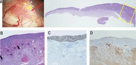
The pathological findings of this case demonstrated the atypical epithelium toward the conjunctival side (Figure 2A),while normal corneal epithelium was found in the corneal margin. Abnormal squamous epithelial cells were found in the central papillomatous tumor without sub-basement membrane invasion, which allowed us to diagnose CIN. Pathological findings at the yellow granular lesion depicted by slit-lamp examination showed basophilic, spherical uniform nonstructured configurations beneath the tumor epithelium, which is consistent with spheroid degeneration (Figure 2B, asterisks).A collection of degenerated elastic fibers consistent with solar elastosis was found around the spheroidal degeneration(Figure 2B, arrows). Immunostaining for Ki67, a proliferation marker, was positive for atypical epithelial cells, consistent with findings of CIN (Figure 2C). Spheroidal degeneration and solar elastosis were positive for IgG (Figure 2D). The margin of excision was free of tumor cells.
This study for the first time demonstrated the clear clinicopathological correlation in CIN containing spheroidal degeneration. Although the origin of spheroidal degeneration has not been elucidated yet, Johnson and Overall
suggested that fibrinoid degeneration is correlated with the degeneration,where IgG plasma protein deposition may also be contained.CIN commonly shows diffuse thickening of the epithelium and high-intensity reflections by ASOCT
; however, OCT findings of spheroidal degeneration have not been reported.In this study, ASOCT demonstrated heterogeneous granular hyper-intensity reflection in spheroidal degeneration.Immunohistochemistry revealed IgG accumulation in the degenerated spheroidal region, as reported in corneal spheroidal degeneration
. Solar elastosis is usually found in pinguecula tissues, where ASOCT showed a subepithelial diffuse mass beneath the thinner epithelium with mild reflection
. Almost half of conjunctival squamous cell neoplasia contains solar elastosis
, and the ASOCT findings of the CIN display diffuse homogenous high-intensity reflection
. However,the origin of spheroidal degeneration or its correlation with solar elastosis has not been determined. Taken together,heterogenous hyper-reflective lesions observed by OCT in this study feature conjunctival spheroidal degeneration,which might result from the microscopical properties and/or accumulation of high molecules such as IgG. The authors here propose that the ASOCT findings in spheroidal degeneration be “hyper-reflective foci”.
DISCUSSION
2.2 术后安静时VAS评分比较 各组VAS评分均随着术后时间增长呈下降趋势。术后2 h SB1组、SB2组镇痛效果起效均较S组快,SB1组和SB2组各时点VAS评分均显著低于S组,且SB1组和SB2组间VAS评分比较差异无统计学意义(P>0.05)。三组在组间、时点间比较差异均有统计学意义(P<0.01),但组间和时点间交互作用差异无统计学意义(P>0.05)。见表3。
There is no previous report of CIN with spheroidal degeneration like this case, that could be considered extremely rare. As described above, spheroidal degeneration is often found in pinguecula and is histologically seen as acellular deposits
. In this case, there are 2 possibilities of the pathologies underlying CIN containing spheroidal degeneration: CIN was originated from the epithelium of the pinguecula, or CIN arising around the pinguecula infiltrated the pinguecula. In this case, the main tumor is an elevated pinkish papillary tumor, and posed the fireworks-like neovascularization typical of CIN. In addition, the papillary tumor was continuous with the yellow granular lesion due to spheroidal degeneration on the corneal side, where the Ki67-positive tumor epithelium became flattened. Taken together,CIN together with spheroidal degeneration might come from bulbar conjunctival tumor cell invasion to the pinguecula.
Surgical treatment options for CIN are commonly local tumor excision including partial lamellar scleroconjunctivectomy technique or enucleation. In the former excision, limbusbased pentagonal or circular conjunctival incision with 3-4 mm outside the tumor margins revealed favorable outcomes with low recurrence rates
. However, in this case, although the question was whether the safety margin should include yellow lesions observed by slit-lamp examination, the safety margins of 3 mm were set including the yellow lesions in this case. Histopathology subsequently proved intraepithelial tumor invasion upon the spheroidal degeneration, while there was free of tumor cells in all the surgical margins. Therefore,in case of CIN combined with spheroidal degeneration, the preoperative safety margin should be carefully considered.In conclusion, this is the first case with CIN containing spheroidal degeneration. ASOCT is critical to identify the spheroidal degeneration, depicting heterogenous band-shaped hyper-reflective foci.
坝体稳定泄流与形成冲切条件泄流两者之间最大的区别是泄流渠的稳定问题,后者要对渠道形成溯源冲刷,而前者则要形成稳定的渠道,具体区别参见表3。
ACKNOWLEDGEMENTS
None;
None;
None;
None.
1 Ramberg I, Toft PB, Georgsen JB, Siersma VD, Funding M, Jensen DH, von Buchwald C, Heegaard S. Conjunctival intraepithelial neoplasia and carcinoma: distinct clinical and histological features in relation to human papilloma virus status.
2021;105(6):878-883.
2 Nahon-Estève S, Martel A, Maschi C, Baillif S, Lassalle S, Caujolle JP. Swept-source and spectral-domain OCT imaging of conjunctival tumors.
2021;128(6):947-950.
3 Nanji AA, Sayyad FE, Galor A, Dubovy S, Karp CL. High-resolution optical coherence tomography as an adjunctive tool in the diagnosis of corneal and conjunctival pathology.
2015;13(3):226-235.
4 Fraunfelder FT, Hanna C, Parker JM. Spheroid degeneration of the cornea and conjunctiva.
1972;74(5):821-828.
5 Bal S, Chang HY, Wolkow N. Pinguecula with spheroidal degeneration:clinicopathologic correlation.
2020;41(1):1.
6 Johnson GJ, Overall M. Histology of spheroidal degeneration of the cornea in Labrador.
1978;62(1):53-61.
7 Tulvatana W, Bhattarakosol P, Sansopha L, Sipiyarak W, Kowitdamrong E, Paisuntornsug T, Karnsawai S. Risk factors for conjunctival squamous cell neoplasia: a matched case-control study.
2003;87(4):396-398.
8 Deka AC, Dutta AM, Sarma PC, Baruah KC. Solar elastosis in conjunctival squamous cell neoplasm.
2014;51(3):245-246.
9 Shields CL, Shields JA. Tumors of the conjunctiva and cornea.
2004;49(1):3-24.
10 Mirzayev I, Gündüz AK, Ateş FSÖ, Özcan G, Işık MU. Factors affecting recurrence after surgical treatment in cases with ocular surface squamous neoplasia.
2019;12(9):1426-1431.
猜你喜欢
中国学校体育(2021年9期)2021-03-27
家庭百事通·健康一点通(2020年8期)2020-09-08
赢未来(2018年23期)2018-12-20
家庭百事通·健康一点通(2018年4期)2018-05-16
新课程·中旬(2017年7期)2017-08-13
中学生数理化·高二版(2016年5期)2016-05-14
中学生数理化·高二版(2016年5期)2016-05-14
莫愁·家教与成才(2016年4期)2016-04-13
科普童话·百科探秘(2015年7期)2015-07-25
发明与创新·中学生(2015年8期)2015-07-21
 International Journal of Ophthalmology2022年8期
- International Journal of Ophthalmology的其它文章
- Celastrol inhibits laser-induced choroidal neovascularization by decreasing VEGF induced proliferation and migration
- Vascular endothelial growth factor-165b protects the blood-retinal barrier from damage after acute high intraocular pressure in rats
- Expression profile analysis to identify potential gene changes induced by dexamethasone in the trabecular meshwork
- Atypical Adams-Oliver syndrome with typical ocular signs of familial exudative vitreoretinopathy
- Severe unilateral congenital ptosis with poor levator function: tarsoconjunctival mullerectomy plus levator resection vs frontalis sling procedure
- Methotrexate for chronic non-necrotizing anterior scleritis in Chinese patients