
Mechanism of stilbene glycosides on apoptosis of SH-SY5Y cells via regulating PI3K/AKT signaling pathway
2024-03-26 12:55KANGBiqianLIYueHEXiaoxuanXIAOZhenHURuiLUOChenliangQIAOMingyuWUGuiyouLIZhenzhongZHUXiaoyingHUANGZhongshi
KANG Bi-qian, LI Yue, HE Xiao-xuan, XIAO Zhen, HU Rui, LUO Chen-liang, QIAO Ming-yu, WU Gui-you, LI Zhen-zhong, ZHU Xiao-ying, HUANG Zhong-shi✉
1. School of Basic Medicine, Youjiang Medical University for Nationalities, Baise 533000, China
2. School of Pharmacy, Guangxi University of Chinese Medicine, Nanning 530200, China
3. School of Pharmacy, Youjiang Medical University for Nationalities, Baise 533000, China
4. School of Clinical Medicine, Youjiang Medical University for Nationalities, Baise 533000, China
Keywords:
ABSTRACT Objective: To investigate the effects of stilbene glycoside (TSG) on okadaic acid-induced apoptosis in human neuroblastoma cells (SH-SY5Y) via the PI3K/AKT pathway.Methods:The optimal concentration of OA was screened by CCK-8 assay, and SH-SY5Y cells were divided into control group, model group, TSG group, LY294002 group and LY294002+TSG group.The proliferation and apoptosis in each group were detected by CCK-8 and TUNEL assays; Western blotting method and real-time fluorescence quantitative polymerase chain reaction was used to detect the expression of PI3K, P-PI3K (Y607), AKT, P-AKT (Ser473),Bcl-2 and Bax proteins.The relative protein expression was represented by P-PI3K (Y607)/PI3K, P-AKT (Ser473) /AKT and Bcl-2/Bax gray ratio.Results: CCK-8 screened the optimal concentration of OA as 40 nmol/L.Compared with the control group, the model group increased relative cell viability, decreased apoptosis rate, the pathway and apoptotic proteins expression levels of P-PI3K (Y607) /PI3K, P-AKT (Ser473)/AKT and Bcl-2/Bax were decreased, and the mRNA expression levels of PI3K, AKT and Bcl-2 were decreased.Bax mRNA expression level increased (P< 0.05); Compared with model group, TSG group increased relative cell viability, decreased apoptosis rate, increased protein expression levels of P-PI3K (Y607)/PI3K, P-AKT (Ser473)/AKT, Bcl-2/Bax, and increased mRNA expression levels of PI3K, AKT, and Bcl-2.Bax mRNA expression decreased (P<0.05), LY294002 group decreased relative cell viability, increased apoptosis rate, P-PI3K (Y607) /PI3K protein expression levels were significantly decreased (P<0.05), P-AKT (Ser473) /AKT and Bcl-2/Bax protein expression levels were significantly decreased, but there was no statistical significance,PI3K, AKT and Bcl-2 mRNA expression levels were decreased, and Bax mRNA expression levels were increased (all P< 0.05); Compared with LY294002 group, LY294002+TSG group increased relative cell viability, decreased apoptosis rate, and the protein expression levels of P-PI3K (Y607) /PI3K, P-AKT (Ser473) /AKT and Bcl-2/Bax were increased.The mRNA expression levels of PI3K, AKT, Bcl-2 were increased, Bax was decreased (all P<0.05).Conclusion: Stilbene glycoside may alleviate okadaic acid-induced apoptosis in SHSY5Y cells by interfering with the PI3K/AKT signaling pathway, which in turn regulates the expression of apoptotic factors such as Bcl-2 and Bax.
1.Introduction
Neuronal loss is one of the causes of the pathogenesis of several degenerative diseases.Alzheimer’s disease (AD), a common neurodegenerative disease, is the fourth leading cause of death in the elderly population after heart disease, malignant tumors and stroke[1].The pathogenesis of AD is still unclear, but two common hallmark pathological changes have been demonstrated: β-amyloid plaque deposition and hyperphosphorylated Tau neurofibrillary tangles[2], both of which cause progressive neuronal loss.
It has been shown that the phosphatidylinositol 3-kinase (PI3K)/protein kinase B (AKT) signaling pathway plays a key role in the regulation of cell proliferation and differentiation and growth[3].This pathway has been found to be involved in the formation of two specific pathological structures in AD[4], and can regulate the expression of mitochondrial membrane permeability proteins Bcl-2 and Bax[5], so activation of the PI3K/AKT signaling pathway may delay the progression of AD pathogenesis by protecting neurons from Aβ-induced neurotoxicity and reducing neuronal loss.
Neurodegenerative diseases are usually associated with excessive apoptosis[6].the Bcl-2 family of proteins strictly regulates mitochondrial apoptosis.Anti-apoptotic proteins (e.g., Bcl-xl, Bcl-2,etc.) exert anti-apoptotic effects by stabilizing the permeability of the mitochondrial membrane and preventing the release of mitochondrial cytochrome C.Pro-apoptotic proteins (e.g., Bak,Bax, etc.) permeabilize the outer mitochondrial membrane and promote the release of cytochrome C and other apoptotic factors from mitochondria in order to trigger cytochrome and cysteoaspartic acid enzymes to activate cellular protease and apoptotic cells[7].The balance of anti-apoptotic proteins and pro-apoptotic proteins determines cell survival[8], and the ratio of the two can be altered through many signaling pathways to efficiently transmit cellular stress information.
The traditional Chinese medicine He Shouwu (Polygonum multiflorum Thunb) has the efficacy of preventing and treating neurological lesions and improving memory[9], and it is often used as a traditional Chinese medicine compound or specialized formula for the treatment of AD, whereas stilbene glycoside, as an active ingredient of He Shouwu, is the main basis of the medicinal effect of He Shouwu [9].In recent years, domestic and foreign scholars have found that stilbene glycoside (2, 3, 5, 4’ - Tetrahydroxy stilbene 2-Ο-β-D-glucoside, TSG) has a good neuroprotective effect, and the previous study of our group found that stilbene glycoside has a better anti-dementia effect, which can reduce the expression of Aβ and protect the neurotoxic damage of the metabolites of Aβ [10-12].On this basis, this experiment aims to explore the specific target and mechanism of action of TSG in regulating SH-SY5Y cell apoptosis by interfering with the PI3K/AKT signaling pathway.
2.Materials and Methods
2.1Materials
Human neuroblastoma cells SH-SY5Y were purchased from Wuhan Punosai Life Science and Technology Co.Stilbene glycoside(batch no.: CHB180810) was purchased from Chengdu Cloma Biotechnology Co.Ltd; DMEM-F12 basal medium, 0.25% trypsin,and double antibiotic were purchased from Gibco, USA; fetal bovine serum (batch no.: 1943609-65-1) was purchased from Sigma,USA; LY294002 (batch no.: GC15485), CCK-8 reagent (batch no.:GK10001) was purchased from Shanghai Hongye Biotechnology Co.Ltd; Okadaic acid (batch no.: AO1IS211161) was purchased from Shanghai Yuanye Biotechnology Co.Ltd; TUNEL apoptosis kit (No.C1806) was purchased from Biyuntian Biotechnology Co.Ltd; ECL Luminescent Substrate Detection Kit (batch no.:HR20230328M) The goat anti-rabbit immunoglobulin G (IgG)secondary antibody (batch no.SA00001-2) was purchased from Wuhan Three Eagles, the rabbit anti-AKT was purchased from abclonal, the rabbit anti-PI3K, P-PI3K, AKT, P-AKT, Bcl-2, and Bax were all purchased from Affinity.the cell culture incubator was purchased from Thermo, USA, the fluorescence inverted microscope was purchased from Leica, the fluorescence inverted microscope was purchased from Leica, and the fluorescence inverted microscope was purchased from Leica, and the fluorescence inverted microscope was purchased from Leica.The cell culture incubator was purchased from Thermo, the fluorescence inverted microscope was purchased from Leica, the fluorescence quantitative PCR instrument was purchased from Hangzhou BORI, and the gel electrophoresis instrument was purchased from Beijing Liuyi Technology.
2.2 Experimental Methods
2.2.1 Cell culture and passaging
Add 15% fetal bovine serum, 1% penicillin-streptomycin mixture into DMEM-F12 basal medium to form complete medium, place the cells containing complete medium in 37 ℃, 5% CO2constant temperature incubator for culture, observe the cell and medium status every 12~24 h, and change the liquid in time.Observe the cell growth to the bottom of the cell culture flask 80% ~ 90% and good growth status, 1 PBS washed three times with 1 mL of 0.25%trypsin repeatedly and uniformly blown to make the cell suspension,to be a single cell under the microscope, uniform form when the termination of the digestion, replaced with fresh complete culture medium, evenly distributed to the new 2-3 flasks into the incubator,so that the cells continue to grow.
2.2.2 Screening out the most suitable OA concentration
When the cell growth reached 80%~90% and the growth status was good, the original complete medium was discarded, and the digested cells were inoculated into 96-well plates with 5×10⁴cells per well,and when the cell adherence growth reached about 80%, complete medium with final OA concentrations of 0, 10 20, 40, 80, 160 nmol/L was added according to the experimental setups, and 5 replicate wells were set up for each group.In the outer ring of blank wells,100 μL of complete culture medium was added to each well, and the plate was placed in a constant temperature incubator to continue incubation for 24 h.Then 10 μL of CCK-8 reagent was added to each well, and the absorbance at 450 nm (OD value) was measured by an enzyme marker after incubation for 1 h in the incubator at 37 ℃, and the survival rate was calculated.Survival rate = (experimental group- blank group)/(control group - blank group) × 100%.
2.2.3 Cell grouping and treatment
Cells with logarithmic growth period and good growth status were taken and divided into control group, model group, TSG group,PI3K inhibitor LY294002 group and LY294002+TSG group.The control group was cultured in complete culture medium for 24 h.The model group and TSG group were cultured in complete medium containing OA (40 nmol/L) for 24 h.The TSG group was further cultured in 100 μmol/L TSG solution configured in complete medium for 24 h.(This concentration and the duration of the effect were determined based on the results of the preliminary pre-test of the group.) On the basis of the administration of the drug in the model group, LY294002 group and LY294002+TSG group were pretreated with complete medium containing LY294002 (20 μmol/L)for 6 h, and then LY294002+TSG group was added with complete medium containing TSG (100 μmol/L) to continue incubation for 24 h.In the model group, LY294002 and LY294002+TSG groups were pretreated with complete medium containing LY294002 (20 μmol/L)for 6 h.
2.2.4 Detection of cell survival in each group by CCK-8 method
Treat the cells in each group according to the method in 1.2.3,inoculate them in 96-well plates, set 5 replicate wells in each group,with a density of 5×10⁴/well, add 10 μL of CCK8 reagent to each well after 24 h of incubation, and then measure the absorbance(OD) at 450 nm in each well with an enzyme counter for 1 h after incubation in a 37 ℃ incubator, and calculate the survival rate.Survival rate = (experimental group - blank group) / (control group -blank group) × 100%
2.2.5 Detection of apoptosis in each group by TUNEL method
Vicki and Cindy replied that they had closed their eyes and run down the hill, holding hands, in the opposite direction from Edward. And what did you do? my mother asked me.
Cells in each group were treated according to the method in 1.2.3, after washing once with PBS, the cells were fixed with 4%paraformaldehyde for 30 min, then incubated with PBS containing 0.3% Triton X-100 for 5 min at room temperature, configured with TUNEL assay according to the instructions of the apoptosis detection kit, 50 μL of TUNEL assay solution was added into the samples, and the samples were incubated at 37 ℃ for 60 min protected from light.Subsequently, DAPI was added and incubated for 5 min, and the samples were sealed with anti-fluorescence quenching sealing solution and then observed under a random field of view in a fluorescence microscope.The TUNEL-positive cells(apoptotic cells) and DAPI-labeled cells (total number of cells) were counted by Imagy J software, and the apoptosis rate = TUNELpositive cells/total number of cells 100%.
2.2.6 Western Blot detection of P-PI3K/PI3K, P-AKT/AKT,Bcl-2/Bax protein expression
Treat the cells in each group according to the method in 1.2.3,collect the cells, wash them three times with pre-cooled PBS,add lysis solution (RIPA: protease inhibitor: protein phosphatase inhibitor = 100:1:1) and lysed on ice for 30 min, collect the lysate and centrifuge the cells at 12,000 r/min for 5 min, then remove the supernatant, and then quantify the proteins using the BCA method,and measure the absorbance value at 562 nm by an enzyme marker,then measure the protein expression of PI3K/PI3K, P-AKT/AKT,Bcl-2/Bax by Western Blot.The protein was quantified by the BCA method, and the absorbance value at 562 nm was measured by an enzyme counter, and a standard curve was made.The prepared protein samples were subjected to protein electrophoresis using 10% sodium dodecyl sulfate polyacrylamide gelelectrophoresis(SDS-PAGE) with 80 V for 30 min before electrophoresis, and 120 V after electrophoresis to the separation gel.The “sandwich membrane structure” was placed in the membrane transfer tank, and the membrane transfer buffer was added, and the membrane was placed in an ice bath with a constant flow rate of 300 mA for 1 h.The power supply was cut off after the membrane was transferred.The membrane was closed with 5% skimmed milk powder at room temperature for 1 h.Add PI3K (1:1,000), P-PI3K (1:500), AKT(1:1,000), P-AKT (1:500), Bcl-2, Bax (1:500) and other primary antibodies at 4 ℃ overnight, and then wash the membrane with TBST three times, each time for 10 min, and then incubate the membrane with secondary antibody (1:5,000) at room temperature for 1 h.The membrane was washed with TBST three times, each time for 10 min.The membrane was washed three times with TBST for 10 min each time, and finally, the washed membrane was put into ECL luminescent reagent for soaking and wetting, and then protected from light for developing.The results were analyzed by Image J.The gray values of protein bands were expressed as P-PI3K/PI3K, P-AKT/AKT, Bcl-2/Bax gray ratios, and normalized to each group.
2.2.7 Real-time fluorescence quantitative polymerase chain reaction (qRT-PCR) detection of PI3K, AKT, Bcl-2, Bax mRNA expression
Cells were collected, RNA was extracted, and the OD 260/280 values were determined by UV spectrophotometer, which ranged from 1.8 to 2.0 indicating that the samples were extracted with a high concentration of RNA, and could be subjected to subsequent reverse transcription experiments.According to the instructions of cDNA reverse transcription kit 20 μL reaction system was configured, and the reaction conditions were as follows: incubation at 25 ℃ for 10 min, incubation at 42 ℃ for 15 min, and heating at 85 ℃ for 5 s to reverse transcription of RNA to cDNA.qRT-PCR reaction system was configured, and the reaction conditions were as follows: predenaturation at 95 ℃ for 600 s, denaturation at 95 ℃ for 5 s, and annealing and extension at 60 ℃ for 20 s, and the reaction system was amplified for 40 cycles.cycles.The results of the relative expression of each target gene were calculated by using GAPDH as the internal reference gene, and the CT value of the target gene was calculated by the 2-△△CTmethod.The specific primer sequences are shown in Table 1.

Tab 1 Primer sequence and product length
2.3 Statistical methods
GraphPad Prism 8.0.2 software was used to analyze the experimental data, and the results were expressed as (±s), the comparison of means between the two groups was performed using the t-test of completely randomized design information, and the comparison of means between the groups was performed using the one-way analysis of variance (ANOVA), and the difference was statistically significant at P<0.05.
3.Results
3.1 Effects of different concentrations of OA on cell viability
The experiments showed that after SH-SY5Y cells were treated with 0, 10 20, 40, 80, 160 nmol/ L concentration gradient of OA for 24 h, the cell survival rate showed a gradual decreasing trend with the increase of OA concentration.Compared with the control group, the cell survival rates in the 40, 80, and 160 nmol/ L OA concentration groups were statistically significant (t=3.022, P<0.05;t=13.041, P<0.01; t=15.176, P<0.01).When the OA concentration was less than or equal to 40 nmol/ L, the cell survival rate was above 80%, in order to try to ensure the cell state in the subsequent experiments, this experiment decided to adopt 40 nmol/ L as the final OA dosing concentration.See Figure 1.

Fig 1 Effect of different concentrations of OA on cell viability(n=3,±s)
3.2 Effect of stilbenosides on cell proliferation
Compared with the control group, the cell viability of the model group was significantly decreased (P<0.01).Compared with the model group, the cell viability of the TSG group was effectively alleviated (P<0.05), and the cell viability of the LY294002 group was significantly decreased (P< 0.01).Compared with LY294002 group, the cell viability of LY294002+TSG group was significantly improved (P<0.05).See Figure 2.

Fig 2 Cell survival in each group
3.3 Detection of apoptosis rate of cells in each group by diphenylyl glycoside
According to the operation of TUNEL kit, the DAPI group indicated that the cells in each group were stained with blue fluorescence under the microscope field of view, while the apoptotic cells would be stained with green fluorescence, i.e., TUNEL-positive cells, and the apoptotic cells and non-apoptotic cells were in the same field of view after Merge to facilitate the observation.The results showed that compared with the control group, the number of TUNEL positively stained cells and apoptosis rate in the model group were significantly increased (P<0.01); compared with the model group,the number of TUNEL positively stained cells and apoptosis rate in the TSG group were significantly decreased (P<0.05), and the number of TUNEL positively stained cells and apoptosis rate in the LY294002 group were significantly increased (P<0.05); and the number of TUNEL positively stained cells and apoptosis rate in the LY294002 group were significantly increased (P<0.05).After adding TSG, the number of TUNEL positively stained cells and apoptosis rate were significantly decreased in LY294002 + TSG group compared with LY294002 group (P<0.05).See Figure 3 and Table 2.

Fig 3 Apoptosis rate in each group (TUNEL method, ×200)
Tab 2 Rate of apoptosis in TUNEL-positive cells in each group(n=3, ±s)

Tab 2 Rate of apoptosis in TUNEL-positive cells in each group(n=3, ±s)
Note: Compared to the control group, **P<0.01; Compared to the model group, #P<0.05; Compared to the LY294002 group, △P<0.05.
Group apoptosis(%)control group 6.33±1.54 model group 39.58±1.87**TSG group 11.13±6.40#LY294002 group 61.61±12.92#LY294002+TSG group 34.72±11.00△F 22.7 P<0.001
3.4 Expression of signaling pathway proteins in cells of each group by diphenylyl glycosides
Compared with the control group, the relative expression of P-PI3K (Y607)/ PI3K and P-AKT (Ser473)/ AKT proteins in the model group was significantly reduced (P<0.01); compared with the model group, the relative expression of P-PI3K (Y607)/ PI3K and P-AKT (Ser473)/ AKT proteins in the TSG group was significantly increased (P<0.01) The relative expression of P-PI3K (Y607)/ PI3K protein in LY294002 group was significantly decreased (P<0.01),and the relative expression of P-AKT (Ser473)/ AKT protein was seen to be more significantly decreased, with no statistically significant difference (P>0.05); compared with the LY294002 group, the relative expression of P-PI3K (Y607)/ PI3K protein in the LY294002 + TSG group was significantly increased (P<0.01), with no statistically significant difference (P>0.05); compared with the LY294002 group, the relative expression of P-PI3K ( Y607)/ PI3K,P-AKT (Ser473)/ AKT protein relative expression was significantly increased in the LY294002 + TSG group (P<0.05).See Figure 4.
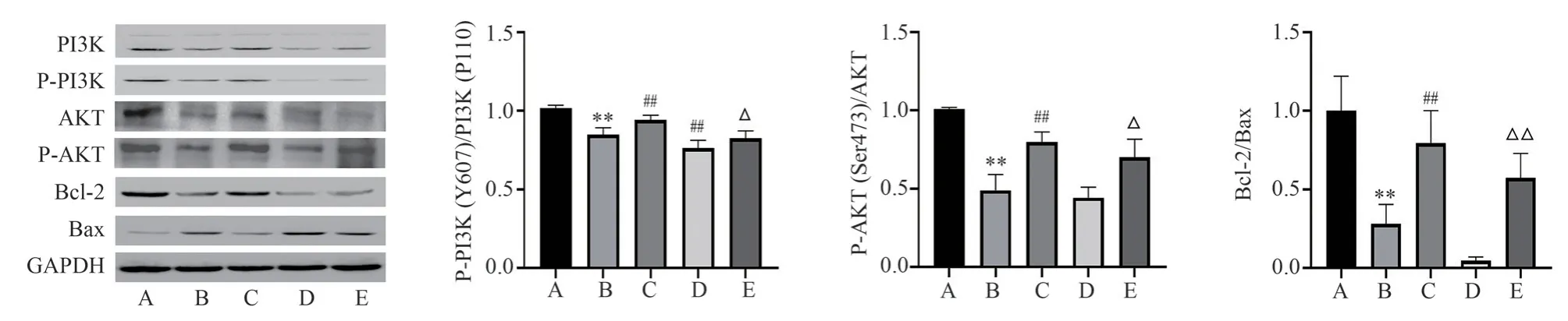
Fig 4 Comparison of protein expression in cells of each group
3.5 Expression of apoptotic proteins by diphenylyl glycosides in each group
Compared with the control group, the relative expression of Bcl-2/Bax protein in the model group was significantly decreased(P<0.01); compared with the model group, the relative expression of Bcl-2/Bax protein in the TSG group was significantly increased(P<0.01), and a more significant decrease in the relative expression of Bcl-2/Bax protein was seen in the LY294002 group, which did not have any statistical difference (P>0.05); compared with the Compared with LY294002 group, the relative expression of Bcl-2/Bax protein in LY294002 + TSG group increased significantly(P<0.01).See Figure 4.
3.6 Expression of PI3K and AKT mRNA in cells of each group by diphenylstilbene glycoside
Compared with the control group, the relative expression of PI3K and AKT mRNA in the model group was significantly reduced(P<0.01); compared with the model group, the relative expression of PI3K and AKT mRNA in the TSG group was significantly increased(P<0.05), and the relative expression of PI3K and AKT mRNA in the LY294002 group was significantly reduced (P<0.05); compared with the LY294002 group Compared with the LY294002 + TSG group,the relative expression of PI3K and AKT mRNA in the LY294002 +TSG group was significantly increased (P<0.05).See Table 3.
Tab 3Comparison of relative expression of PI3K, AKT and apoptosis factor mRNA in cells of each group(n=3, ±s)
Note: Compared to the control group, **P<0.01; Compared to the model group, ##P<0.01; Compared to the LY294002 group, △P<0.05, △△P<0.01.
Group PI3K AKT Bcl-2 Bax control group 1.00±0.00 1.00±0.00 1.00±0.00 1.00±0.00 model group 0.51±0.10** 0.46±0.08** 0.45±0.06** 8.86±2.75**TSG group 0.90±0.15## 0.78±0.10# 0.83±0.11## 3.53±0.74#LY294002 group 0.25±0.04# 0.21±0.04## 0.18±0.02## 13.90±1.86#LY294002+TSG group 0.53±0.07△ 0.37±0.11△ 0.43±0.07△ 7.84±0.99△△F 32.76 57.91 64.02 29.86 P<0.001 <0.001 <0.001 <0.001
3.7 Expression of apoptotic factor mRNA by diphenylyl glycosides in each group
Compared with the control group, the relative expression of Bcl-2 mRNA in the model group was significantly decreased, and the relative expression of Bax mRNA was significantly increased(both P<0.01); compared with the model group, the relative expression of Bcl-2 mRNA in the TSG group was significantly increased (P<0.01), and the relative expression of Bax mRNA was significantly decreased (P<0.05), and in the LY294002 group Bcl-2 mRNA relative expression was significantly decreased (P<0.01) and Bax mRNA relative expression was significantly increased (P<0.05)in the LY294002 group; compared with the LY294002 group, Bcl-2 mRNA relative expression was significantly increased (P<0.01) and Bax mRNA relative expression was significantly decreased in the LY294002 + TSG group (P<0.05 ).See Table 3.
4.Discussion
The PI3K/AKT signaling pathway has a wide range of functions,such as regulating cell survival, cell proliferation, growth,differentiation, motility, intracellular transport, and more complex processes, etc.PI3K is an important signaling molecule in the cell, which is divided into class I, II, and III, with class I being the most important and commonly used, and it can be activated by the signaling pathways of nerve growth factor, insulin, and so on, and the subtypes of class I are heterodimers composed of regulatory subunits ( P85) and catalytic subunit (P110)[13], and further divided into class IA (PI3K α, β, and ) and class IB (PI3Kγ)according to their mode of regulation.The regulatory subunit P85 binds to phosphorylated tyrosine residues on activated receptors through its SH2 structural domain, and then recruits the catalytic subunit P110 in order to form a fully active PI3K enzyme[13].When neurotrophic factors or extracellular growth factors bind to transmembrane tyrosine kinase receptors (RTKs), the RTKs then undergo autophosphorylation, which recruits PI3K to the site of RTKs phosphorylation[14].Activation of the PI3K/AKT pathway begins with the activation of PI3K by RTKs, and activated PI3K activates AKT, which is structurally conformationally altered to expose the phosphorylation sites at Thr308 and Ser473, leading to phosphorylation of the Thr308 residue by PDK1 and a second phosphorylation at Ser473.Activated AKT is involved in the regulation of apoptosis by regulating the activity of downstream target proteins such as Bcl-2 (B-cell lymphoma/leukemia-2);Bcl-2xL (B-cell lymphoma factor-2XL) through activation or inhibition[15, 16].This pathway has a close connection with AD pathogenesis, in which Wang et al[17] found that activation of the PI3K/AKT signaling pathway improved neuroinflammatory responses and cognitive deficits in AD mice, and Guo et al[18]demonstrated that activation of the PI3K/AKT signaling pathway reduced apoptosis in the brain of AD mice.In this experiment, PI3K P110 catalytic subunit and AKT Ser473 phosphorylation site were selected for protein assay, and found that the ratio of P-PI3K/PI3K and P-AKT/AKT protein expression was significantly decreased after adding PI3K inhibitor, and the ratio of P-PI3K/PI3K and P-AKT/AKT protein expression was significantly increased after TSG intervention, suggesting that TSG may reduce apoptosis by activating the PI3K/AKT signaling pathway.
Massive neuronal loss is more pronounced in the AD patient population than in the normal aging population.The mechanism of neuronal loss in AD has not been widely finalized, and the prevailing forms are apoptosis, necrosis, pyroptosis and other forms of cell death (e.g., iron death, copper death, etc.)[16].In this experiment,apoptosis, which has been most widely explored in the mechanism of neuronal loss in AD, was selected for study, and OA was used for the induction of human neuroblastoma cells SH-SY5Y, which is an easy-to-handle and proliferate cell line with a well-established differentiation method for generating stable neuronal cultures[19]and obtaining mature neuron-like features, which is commonly utilized in the in the study of the pathogenesis of neurodegenerative diseases.OA, as a protein phosphatase inhibitor, can inhibit phosphatase activity, and in vitro and in vivo experiments have shown that OA induces phosphorylation of Tau protein, which can be used to establish a model for the study of AD-like cells[20-21].In this experiment, compared with the model group, cell viability increased, apoptosis rate decreased significantly, and PI3K/AKT pathway protein and gene expression increased significantly in the TSG group, suggesting that TSG may reduce apoptosis to a certain extent by interfering with the PI3K/AKT pathway.LY294002, as an inhibitor of the PI3K/AKT pathway, promotes apoptosis and inhibits PI3K/AKT The protein and gene levels of the PI3K/AKT signaling pathway were inhibited.Compared with the LY294002 group, apoptosis was reduced in the LY294002 + TSG group, and the protein and gene expression of the PI3K/AKT signaling pathway showed an increase, which further proved that the effect of TSG on the reduction of apoptosis was manifested through the PI3K/AKT signaling pathway.
In summary, TSG may inhibit apoptosis to some extent by interfering with the PI3K/AKT signaling pathway.In the future,we can continue to explore the effects of TSG on the upstream and downstream signaling molecules of PI3K/AKT to fully elucidate its specific mechanism of action.
Authors’ contribution
Huang Zhongshi : experimental design and article review; Zhu Xiaoying and Li Zhenzhong: provided technical guidance for Western blot experiments; Li Yue and He Xiaoxuan : responsible for cell culture and drug intervention; Xiao Zhen, Hu Rui, Luo Chenliang, Qiao Mingyu and Wu Guiyou were responsible for the indicator detection and data analysis; Kang Biqian: wrote the paper.
All authors declare no conflict of interest.
 Journal of Hainan Medical College2024年1期
- Journal of Hainan Medical College的其它文章
- Protective effect of camellia oil on H2O2-induced oxidative stress injury in H9C2 cardiomyocytes of rats
- Mechanism of Yanghe Pingchaun granules on airway remodeling in asthmatic rats based on IL-6/JAK2/STAT3 signaling axis
- Ghrelin regulates insulin resistance by targeting insulin-like growth factor-1 receptor via miR-455-5p in hepatic cells
- Pharmacodynamic study and mechanism of action of Linggui Zhugan Decoction in the intervention of Nonalcoholic fatty liver disease
- Bioinformatics and network pharmacology identify the therapeutic role and potential targets of diosgenin in Alzheimer disease and COVID-19
- Meta analysis of the efficacy of western medicine combined with Qiliqiangxin capsule versus western medicine alone in the treatment of chronic heart failure