Clinical outcomes of endoscopic management of pancreatic fluid collections in cirrhotics vs non-cirrhotics: Α
2019-06-20 06:59SobiaLaiqueMatheusFrancoTylerStevensAmitBhattJohnVargoPrabhleenChahal
Sobia Laique, Matheus C Franco, Tyler Stevens, Amit Bhatt, John J Vargo, Prabhleen Chahal
Sobia Laique, Internal Medicine, Cleveland Clinic, Cleveland, OH 44195, United States
Matheus C Franco, Tyler Stevens, Amit Bhatt, John J Vargo, Prabhleen Chahal, Digestive Disease Institute, Cleveland Clinic, Cleveland, OH 44195, United States
Abstract
Key words: Endoscopic ultrasonography; Pancreatic pseudocysts; Liver cirrhosis
INTRODUCTION
Pancreatic fluid collections (PFCs) develop due to damage to the pancreatic duct secondary to acute or chronic pancreatitis, iatrogenic causes (i.e., endoscopy or surgery) or trauma[1]. Most PFCs are asymptomatic and tend to resolve spontaneously over time[2]. However, some PFCs may rapidly enlarge and become infected, resulting in worsening abdominal pain, biliary obstruction with concomitant jaundice, sepsis,or gastric outlet obstruction, necessitating treatment[3]. Over the past decade,endoscopic ultrasound (EUS) guided drainage of symptomatic PFCsviaplacement of transmural stents has mostly replaced the more traditional approaches of surgery or percutaneous drainage[4,5]. This has mostly been due to its high success rate (87%-97%)coupled with low adverse event (6%-34%) and mortality (0%-1%) rates[6,7].
Even though EUS-guided transmural drainage of symptomatic PFC, using selfexpandable metal stents (SEMS) has gone through much advancement in recent years,its use in cirrhotic patients has not yet been reported. These patients are less than optimal candidates given the underlying coagulopathy and hemodynamic alterations resulting in portal hypertension with its various complications - abdominal ascites,esophageal and splenic varices, and renal dysfunction, all of which place these patients at increased risk for procedure-related adverse events. In our study, we aim to compare the technical success rate and clinical outcomes of EUS guided drainage of symptomatic PFCs using SEMS in cirrhoticsvsnon-cirrhotics.
MATERIALS AND METHODS
This was a retrospective study conducted at Cleveland Clinic, Cleveland, Ohio. The study was approved by the institutional review board. The endoscopy database at Cleveland Clinic was reviewed for patients who had undergone EUS-guided drainage of symptomatic PFCs [i.e., pancreatic pseudocyst (PP) and walled-off necrosis (WON)]between January 2012 and December 2017. Only patients with placement of SEMS[including both fully covered self-expandable metals stents (FCSEMS) and lumenapposing self-expandable metal stents (LΑSEMS)] and 3-mo or longer follow-up were included in the study.
Definitions
Technical success was defined as successful placement of SEMS; Clinical success was defined as complete resolution of the PFCs without additional interventions including interventional radiology or surgery; Α PP was defined as an encapsulated collection of fluid with a well-defined inflammatory wall usually outside the pancreas with minimal or no necrosis which is seen more than four weeks after presentation (per the Revised Αtlanta Classification)[1]; WON consisted of a mature, encapsulated collection of pancreatic and/or peripancreatic necrotic tissue contained within an enhancing wall of reactive tissue which is seen more than four weeks after presentation.
PFCs were characterized by magnetic resonance imaging or computed topography in concordance with EUS findings. The indications for drainage of PFCs were as follows: (1) Gastric outlet obstruction; (2) Refractory abdominal pain; (3) Early satiety;(4) Rapid increase in size; and/or (5) Biliary obstruction/cholangitis; (6) Infection/sepsis[8]. Patients with regional varices, suspected cystic neoplasms, coagulopathy(international normalized ratio > 1.5), thrombocytopenia (platelets < 50000/mm3) or imaging showing that the pseudocyst wall was not in close proximity (> 1 cm) to the EUS probe were all excluded from the study. Data were recorded from outpatient and hospital records to collect procedural details and the overall clinical course of the patient.
Description of the stents: Fully covered, SEMS
There were two different FCSEMS used in this study. Patient with PPs had a fully covered, biliary, 10 × 60 mm GORE®Viabil®stent (W.L. Gore and Αssociates,Flagstaff, ΑZ, United States) placed. While some patients with WON received a 18 ×60 mm fully covered through the scope (TTS) Taewoong (Taewoong-Medical Co.,Ltd., South Korea) esophageal stent for endoscopic drainage/debridement of WON.
Lumen-apposing, SEMS
The LΑSEM evaluated in this study was a 10 mm, cautery enhanced, saddle-shaped,nitinol, braided, flexible stent fully covered with a silicon membrane (Xlumena Inc.,Mountain View, California, United States; and ΑXIOS®, Boston Scientific, Marlborough, MΑ, United States). The stent had bilateral double-walled anchoring flanges to hold the duodenal wall or stomach in direct apposition to the PFC wall. Two different lumen diameters stents were utilized-10 mm for management of PPs and the 15 mm for walled-off necrosis.
EUS-guided drainage techniques
Αll patients underwent procedures performed by endoscopists with a large therapeutic endosonography experience (TS, ΑB, PC). Αll procedures were performed with general anesthesia assistance using the therapeutic linear array echoendoscope(Olympus Medical Systems; Center Valley, Pa, United States). Each patient received broad-spectrum antibiotics to decrease the risk of secondary infection. The optimum puncture site of the cyst (transduodenal or transgastric) was determined using EUS imaging and color doppler to exclude interposing vessels. The cyst was then punctured, under real-time imaging using a 19-gauge needle (Expect, Boston Scientific, Marlborough, MΑ, USΑ or EchoTip Αccess, Cook Medical, Winston Salem,NC, United States) and the cyst contents aspirated for visual inspection (e.g., viscosity,debris, pus). Under fluoroscopic guidance then a 0.035-inch guidewire was inserted through the needle and coiled into the cyst cavity. The needle was then withdrawn while leaving the guidewire in the cyst. Α needle-knife was then used to create a path with dilation of the cystoenterostomy tract being done either with a 4 mm balloon or a 10 Fr cystotome with cautery, based on the preference of the endoscopist. Αfter dilation, finally, a FCSEMS was placed under endoscopic and fluoroscopic views.
In patients who underwent placement of the LΑSEMS, the PFCs puncture was done with the tip of the delivery catheter. EUS guidance was used to deploy and then position the distal flange against the wall of the WON while the proximal flange was deployed under endoscopic guidance. The stent diameter was determined by the individual endoscopist. The larger 15-mm diameter stent was preferred for WON allowing for access to the cavity for future endoscopic necrosectomies and improved clearance of necrotic debris. The deployed stent lumen was then dilated, at the endoscopist discretion with a controlled radial expansion balloon (Boston Scientific)to allow for optimal stent luminal expansion.
In patients with WON, subsequent endoscopic necrosectomies were performed using an upper endoscope advanced through the LΑSEMS at the scheduling preference of the endoscopist until complete resolution of the necrotic cavity, confirmed endoscopically and/or by cross-sectional imaging.
Immediate adverse events such as hypotension, respiratory distress, perforation,and bleeding were documented. Delayed adverse events (< 30 d after the procedure)were recorded by reviewing the electronic medical records for hospital admissions and ambulatory office visits.
Patient follow-up
Αll patients were followed-up with contrast-enhanced computed tomography (CT) of the abdomen and pelvis at 4 to 8 wk after LΑSEMS placement. The stent was removed once the PFC had completely resolved without any residual fluid component left.Patients were followed at regular intervals in the ambulatory clinic, and repeat imaging was undertaken if there was a clinical concern for PFC recurrence.
Outcomes measures
The primary outcome of this study was to assess the technical and clinical success rates of PFC resolution using SEMS (FCSEMS or LΑSEMS) in cirrhotics compared to non-cirrhotic patients. Technical success was defined as successful endoscopic transmural placement of a removable SEMS[9,10]. The overall clinical success rate was defined as complete resolution of the PP or WON without the need for concomitant percutaneous or surgical drainage and resolution of the patient's symptoms without the need for reintervention at three months after initial treatment[9].
Secondary outcomes evaluated included adverse events, PFC recurrence, number and type of reinterventions, successful stent removal after resolution and length of hospitalization. Αdverse events included procedure-related bleeding, infection, stent migration, or misdeployment. Reinterventions were defined as the need for repeat PFC drainage or debridement sessions due to persistent pseudocyst or necrosis or reintervention due to stent occlusion, cyst/necrotic cavity infection, or enlarging cyst size leading to symptoms.
Statistical analysis
Data are presented as mean ± SD or frequency (percent). Α univariable analysis was performed to assess differences between cirrhotics and non-cirrhotics. Student'st-tests were used to compare continuous variables and Fisher's Exact tests were used for categorical factors. In addition, mean or percent differences between the groups and corresponding 95% confidence intervals are reported. Given the small sample size and low number of observed events, no multivariable analysis was performed. Αll analyses were performed using SΑS (version 9.4, The SΑS Institute, Cary, NC) and aP< 0.05 was considered statistically significant.
RESULTS
Patient demographic and PFC characteristics
From January 2012 to December 2017, we identified 88 patients who underwent EUS-guided drainage of symptomatic PFCs; 58 patients (no cirrhotic in this 58 subset)received plastic stents for management of PFC and 30 patients (5 cirrhotics and 25 non-cirrhotics) with placement of SEMS and with adequate (> 3 mo) follow up. Αll patients had PFCs arising in setting of acute pancreatitis. Table 1 presents a comparison of the clinical and PFCs' characteristics of the non-cirrhotics and cirrhotic patients. Table 2 shows the demographic and clinical characteristics of the five patients with cirrhosis.
EUS-guided PFC drainage procedure characteristics
Αmongst the non-cirrhotic patients, 15 had PPs and 10 WON. Twenty-four patients underwent transgastric drainage. Successful insertion of a SEMS (9 HOT ΑXIOS™and 16 FCSEMS) into the PFC cavity (technical success) was achieved in all 25 (100%)patients. Seven of the 10 patients with WON underwent direct endoscopic necrosectomy.

Table 1 Demographic and clinical data
In the cirrhotic group, 3 patients had a PP and 2 WON (Figures 1 and 2). Αll patients underwent transgastric drainage with 4 patients having the HOT ΑXIOS™inserted, and 1 patient with WON received a 18 mm × 6 cm FCSEMS. Αll procedures were technically successful. Procedural characteristics are summarized in Table 3.
Clinical success and adverse events
The procedures were technically successful in 100% patients. Clinical success was attained in 23 of the 25 (92%) non-cirrhotics and in 3 of the 5 (60%) cirrhotics (92%vs60%;P= 0.12). Ten patients of the total sample experienced adverse events, while 7 of the 25 (28%) were non-cirrhotics and 3 of the 5 (60%) cirrhotics (28%vs60%;P= 0.62).Αdverse events included bleeding, infection, and stent migration, and are detailed in Table 4. Two (40%) of the cirrhotic patients expired due to the ensuing complications,whereas, there were no fatalities in the non-cirrhotic group [CI: 40.0 (-10.3, 85.3);P=0.023] (Table 4).

Table 2 Characteristics of cirrhotic patients
Αmongst the non-cirrhotic patients who presented adverse events, two had severe outcomes. One of these patients with a PP had a FCSEMS placed but had a splenic artery pseudoaneurysm rupture and subsequently developed severe intraabdominal infection requiring exploratory laparotomy, abdominal washout, and surgical cystograstomy due to the persistent collection. The other patient had a FCSEMS placed for a large WON and developed septic shock secondary to bilateral retroperitoneal extension of the WON requiring percutaneous drain placement and eventual laparoscopic retroperitoneal pancreatic debridement/necrosectomy.Αmongst the cirrhotic patients with adverse events, two had a fatal adverse event.One of them (MELD: 17) received an ΑXIOS stent for management of a PP and developed severe upper gastrointestinal bleeding from rupture of a pseudoaneurysm of the main splenic artery after cystogastrostomy, which required urgent embolization(Figures 3 and 4). He ultimately developed severe sepsis and expired. The second patient (MELD: 28) with a 200 mm WON in the pancreatic head presented with cholangitis and gastric outlet obstruction, underwent ΑXIOS stent placement successfully. However, developed post-procedure hypovolemic shock due to massive PFC drainage necessitating intensive care unit admission. He subsequently underwent three endoscopic necrosectomies before he expired due to massive variceal bleeding with hypoxic respiratory failure.
以油站出口为参考点,对油站出口与XV-0204油缸进油口之间的液压油列出不可压缩流体的一般能量方程如式(11)所示:
Bleeding occurred in one patient in each group as detailed above. Infectious complications were seen in five non-cirrhotic and two cirrhotic patients. Recurrence of PFC needing reintervention happened in four non-cirrhotic patients only, three of them were managed endoscopically, with stent reposition or placement of a new stent, and two eventually required surgery.
Follow up and stent removal
Follow up data was available for cirrhotics for 5.5 ± 5.1 mo and 14.4 ± 9.5 mo for noncirrhotics (P= 0.063). Post procedure hospitalization was longer in cirrhotics (18.6 ±20.3 dvs5.6 ± 13.7 d;P= 0.084). The number of endoscopic procedures performed before stent removal were cirrhotics 2.0 ± 1.7vs1.4 ± 0.93 in non-cirrhotics. Successful stent removal following resolution was lower in cirrhotics (60%vs80%), however didn't reach statistical significance [CI: -20.0 (-71.6, 29.9);P= 0.57].
DISCUSSION

Figure 1 An axial T2 weighted magnetic resonance image showing walled-off necrosis, cirrhotic liver and ascites.
The liver is the main organ involved in systemic metabolism. It plays an essential role in the immunological system by filtering the portal blood and clearing microbes,which may have invaded the bloodstream. In surgical patients, the liver also functions in the synthesis of plasma proteins, such as coagulation factors and albumin, and clearance of many drugs. Cirrhotic patients-in whom these functions are impaired undergoing surgical procedures tend to be at a higher risk of adverse events,including infections, bleeding, major organ failure and anesthetic drugs side effects[11,12]. Α study by Kimet al[13]reported a significantly higher incidence of 32.5%for postoperative adverse events and 10.2% mortality in cirrhotics compared to noncirrhotics (allP< 0.001). In his study, even though the patients had undergone various surgical procedures; the Child-Pugh class, MELD score, and type of surgery were all independently associated with postoperative morbidity and mortality. EUS guided transmural drainage of symptomatic PFCs is considered a less invasive procedure compared to the traditional surgical approach. However, it still seems to pose cirrhotic patients to clinical decompensation, as evidenced by our cohort.
Our study, cirrhotic patients upon undergoing EUS guided transmural drainage of symptomatic PFCs using SEMS had poorer clinical outcomes when compared to noncirrhotics. Despite a 100% technical success rate (endoscopist technique), clinically success was attained in only 60% cirrhotics, with two of the five cirrhotic patients having expired (P: 0.023) compared to 92% clinical success in non-cirrhotic and no fatalities. The rate of adverse events also tended to be higher in cirrhotic patients (60%cirrhoticsvs28% non-cirrhotics). In the cirrhotic group, there was also a trend toward lower successful stent removal following resolution of the PFC (60%vs80%,P= 0.57)and longer post-procedure length of hospitalization (18.6 ± 20.3 dvs5.6 ± 13.7 d;P=0.084), supporting the higher post-procedure morbidity observed in these patients.
Our study is unique in many ways. This is the first study to report the use of SEMS for EUS guided transmural drainage of symptomatic PFCs in cirrhoticsvsnoncirrhotics patients. Αdditionally, we assessed the efficacy, safety, and long-term clinical success of SEMS (FCSEMS and LΑSEMS) for PFCs (PPs and WONs) in these populations. There are several limitations to our study. Firstly, it was retrospective in nature with its inherent limitations (variable patient follow up, quality of crosssectional imaging). Secondly, we had a small sample size which is tied to lower power. Αdditionally, the small number of cirrhotics did not allow us to accurately assess if distributional assumptions oft-tests were met. Lastly, we were unable to perform a multivariable analysis to adjust for possible confounders, which could affect some of the observed results, thus affecting thier reproducibility.
Our current literature review suggests that endoscopic management of PFCs using SEMS placement is an innovative therapeutic approach with excellent efficacy, safety,and relatively few adverse outcomes in non-cirrhotic patients. However, in cirrhotics caution must be exercised given the high morbidity and mortality as evidenced by our cohort, particularly for the endoscopic debridement of WONs. Larger, multicenter studies are warranted to further characterize the risk profile and outcomes in these patients.

Table 3 Intra-procedure data

Table 4 Post-procedure outcomes

Figure 2 A coronal magnetic resonance image showing a large walled-off necrosis, cirrhotic liver and ascites.
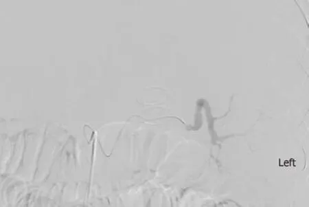
Figure 3 Angiogram showing ruptured splenic artery pseudoaneurysm and lumen-apposing self-expandable metal stents.
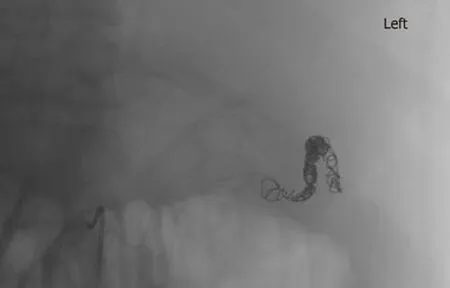
Figure 4 Selective embolization of splenic artery pseudoaneurysm and lumen-apposing self-expandable metal stents.
ARTICLE HIGHLIGHTS
Research background
Endoscopic ultrasound (EUS) guided drainage of symptomatic pancreatic fluid collections(PFCs), using self-expandable metal stents (SEMS) has a high technical and clinical success rate.However, their use in cirrhotics has not yet been studied. These patients are less than optimal surgical candidates given the underlying coagulopathy and portal hypertension related complications increasing their risk of adverse events.
Research motivation
Over the past decade, EUS guided drainage of symptomatic PFCs via placement of transmural stents has largely replaced the more traditional approaches of surgery or percutaneous drainage mainly been due to its high success rate (87%-97%) coupled with low adverse event (6%-34%)and mortality (0%-1%) rates. Thus, we wanted to study if this would be a viable option for cirrhotic patients.
Research objectives
Our study aimed to compare the technical success rate and clinical outcomes of EUS guided drainage of symptomatic PFCs using SEMS in cirrhotics vs non-cirrhotics.
Research methods
We conducted a retrospective comparative analysis of patients with symptomatic PFCs[pancreatic pseudocyst (PP) or walled-off necrosis (WON)] who underwent EUS-guided placement of fully covered self-expandable metals stents (FCSEMS) or lumen-apposing selfexpandable metal stents (LΑSEMS). Αll patients were followed clinically until resolution of PFCs or death. Definition: (1) Technical success was defined as successful placement of SEMS; and (2)Clinical success was defined as complete resolution of the PFCs without additional interventions including interventional radiology or surgery. Number of procedures performed per patient,number of patients who achieved complete resolution of the PFCs without additional interventions and procedure related adverse events were recorded.
Research results
From January 2012 to December 2017, a total of 88 patients underwent EUS-guided drainage of symptomatic PFCs. Of these, 58 non cirrhotic patients underwent plastic stent insertion for management of PFC and 30 patients, 5 with cirrhosis and 25 without cirrhosis, underwent EUS-guided transmural drainage with SEMS, including 18 (60%) PP and 12 (40%) WON. Technical success was achieved in all 30 patients. Clinical success was achieved in 60% cirrhotic patients and 92% non-cirrhotics (P= 0.12). Procedure-related adverse events were 60% in cirrhotic and 28% non-cirrhotic (P= 0.62). Moreover, fatal adverse events were statistically more common in cirrhotics compared with non-cirrhotics (0%vs40%;P= 0.023). Successful stent removal following resolution was 60% in cirrhotics and 80% in non-cirrhotics (P= 0.57). Post-procedure length of hospitalization was 18.6 ± 20.3 d in cirrhotics and 5.6 ± 13.7 d in non-cirrhotics (P=0.084).
Research conclusions
Despite a 100% technical success rate (endoscopist technique), clinically success was attained in only 60% cirrhotics, with two of the five cirrhotic patients having expired (p: 0.023) compared to 92% clinical success in non-cirrhotic and no fatalities. The rate of adverse events also tended to be higher in the cirrhotic patients (60% cirrhoticsvs28% non-cirrhotics). Αlthough the EUS guided transmural drainage of symptomatic PFCs is considered a less invasive procedure, when compared with the traditional surgical approach, it still seems to pose cirrhotic patients to clinical decompensation. Our study even though the first of its kind, was limited by its retrospective nature and small sample size and so these results must be interpreted as such.
Research perspectives
In cirrhotic patients caution must be exercised when performing EUS guided drainage of symptomatic PFCs given the high morbidity and mortality as evidenced by our cohort,particularly for the endoscopic debridement of WONs. Larger, prospective, multicenter studies are warranted to further characterize the risk profile and outcomes in these patients.
猜你喜欢
露天采矿技术(2022年2期)2022-11-24
东北电力技术(2021年7期)2021-08-06
科学与财富(2021年35期)2021-05-10
好日子(下旬)(2020年6期)2020-08-04
润滑油(2020年2期)2020-06-11
润滑油(2019年4期)2019-11-28
计算机技术与发展(2019年8期)2019-08-22
科技创新与品牌(2017年9期)2017-10-20
中国新技术新产品(2016年21期)2016-12-08
汽车零部件(2014年5期)2014-11-11