
尿酸对乳鼠心肌成纤维细胞胶原合成及炎性分泌影响
2019-09-10 07:22王妍琦肖帅孙海鹏王春鹏王其新
青岛大学学报(医学版) 2019年3期
王妍琦 肖帅 孙海鹏 王春鹏 王其新
[摘要]目的探讨尿酸对原代培养的乳鼠心肌成纤维细胞(CFs)胶原合成及炎性分泌的影响,以及micro-RNA-155(miR-155)在其中的作用。方法分离培养乳鼠CFs,并用不同浓度(0、200、400、600、800 μmol/L)的尿酸处理。采用MTT法检测CFs的增殖情况,羟脯氨酸试剂盒检测胶原含量,酶联免疫吸附试验(ELISA)法检测炎性因子白细胞介素-6(IL-6)、白细胞介素-1β(IL-1β)和肿瘤坏死因子-α(TNF-α)表达水平。将miR-155 inhibitor转染入CFs,选择前述实验结果较为稳定的600 μmol/L尿酸刺激CFs 24 h,采用荧光定量PCR检测miR-155、IL-6、IL-1β、TNF-α、Ⅰ型胶原(Col-Ⅰ)、Ⅲ型胶原(Col-Ⅲ)的表达水平。结果与0 μmol/L尿酸组相比较,400、600、800 μmol/L尿酸组CFs的增殖和胶原含量增加(F=18.46、11.82,P<0.05),200、400、600、800 μmol/L尿酸组CFs IL-6、IL-1β、TNF-α的表达增加(F=29.32~69.76,P<0.05)。miR-155 inhibitor可明显抑制尿酸诱导的IL-6、IL-1β、TNF-α、Col-Ⅰ、Col-Ⅲ表达增加(F=72.74~275.32,P<0.05)。结论尿酸可以通过miR-155促进CFs的胶原合成及炎性分泌。
[关键词]尿酸;心肌;成纤维细胞;微RNAs;胶原;炎症
[中图分类号]R542.33[文献标志码]A[文章编号]2096-5532(2019)03-0290-05
[ABSTRACT]ObjectiveTo investigate the effects of uric acid on collagen synthesis and inflammatory secretion in primary cultured neonatal rat cardiac fibroblasts (CFs) and the role of microRNA-155 (miR-155) in the process. MethodsThe CFs of neonatal rats were isolated and treated with different concentrations of uric acid (0, 200,400,600, and 800 μmol/L). The proliferation of CFs was evaluated by MTT assay, the collagen content was determined by hydroxyproline assay kit, and the expression of inflammatory factors interleukin-6 (IL-6), interleukin-1β (IL-1β), and tumor necrosis factor-α (TNF-α) was determined by enzyme-linked immunosorbent assay. The CFs were then transfected with miR-155 inhibitor and stimulated with 600 μmol/L uric acid, which yielded stable results in the above test, for 24 h. The expression of miR-155, IL-6, IL-1β, TNF-α, collagen type Ⅰ (Col-Ⅰ), and collagen type Ⅲ (Col-Ⅲ) was determined by quantitative real-time PCR. ResultsCompared with the group treated with 0 μmol/L uric acid, the groups treated with 400, 600, and 800 μmol/L uric acid had significantly increased proliferation and collagen content of CFs (F=18.46 and 11.82,P<0.05), and the groups treated with 200, 400, 600, and 800 μmol/L uric acid had significantly increased expression of IL-6, IL-1β, and TNF-α in CFs (F=29.32-69.76,P<0.05). However, the increased expression of IL-6, IL-1β, TNF-α, Col-Ⅰ, and Col-Ⅲ induced by uric acid was significantly inhibited by miR-155 inhibitor (F=72.74-275.32,P<0.05). ConclusionUric acid acts through miR-155 to promote collagen synthesis and inflammatory secretion of CFs.
[KEY WORDS]uric acid; myocardium; fibroblasts; microRNAs; collagen; inflammation
心力衰竭(簡称心衰)是众多心血管疾病的最终归宿,心脏重塑和炎症是心衰发展过程中的关键病理标志。心肌纤维化是心脏重塑的关键原因,也是多种心脏疾病发展至一定阶段的常见病理改变[1]。心肌成纤维细胞(CFs)是心脏中数量最多的细胞,其主要功能是调节细胞外基质(ECM)平衡。当心脏受损时(缺血、低氧、物理化学刺激等),CFs增殖活化,分泌炎性细胞因子,合成胶原增多,这是心肌纤维化的主要病理途径[2]。大量流行病学研究表明,高尿酸血症是多种心血管疾病的独立危险因素[3-7],血尿酸水平升高可以增加病人心衰及不良预后的风险,且与心室重构有着密切关系[8-9]。近年来,尿酸对心血管疾病影响的基础研究主要集中在心肌细胞的损伤上[10-11],而关于尿酸对心肌纤维化和CFs影响的研究报道相对较少。MicroRNA-155(miR-155)已被证明可以在多种心血管疾病中发挥作用[12-15]。最近的研究表明,miR-155能够增加心肌重构过程中蛋白的过度沉积[12,16],但是miR-155在CFs炎症反应中的作用研究相对较少。本研究应用尿酸刺激CFs,观察其对细胞炎性分泌及胶原合成的影响,并探讨miR-155在其中的作用。
1材料与方法
1.1材料
1.1.1动物出生2~3 d的Wistar大鼠,雌雄不限,购自青岛大任富城畜牧有限公司。
1.1.2试剂尿酸(Sigma公司),DMEM培养液、2.5 g/L胰酶(含0.2 g/L EDTA)和胎牛血清(Hyclone公司),MTT试剂盒(Solarbio公司),羟脯氨酸试剂盒(南京建成科技有限公司),Lipofectamin 2000(Invitrogen公司),大鼠白细胞介素-6(IL-6)、白细胞介素-1β(IL-1β)和肿瘤坏死因子-α(TNF-α)酶联免疫吸附试验(ELISA)检测试剂盒(欣博盛公司),miR-155 inhibitor及其阴性对照、miRNA PCR试剂盒(广州锐博生物科技有限公司),总RNA试剂盒(美国Omega公司),逆转录试剂盒及荧光定量PCR试剂盒(天根生物公司),PCR引物由生工生物工程(上海)股份有限公司合成。
1.2CFs的分离培养
取10~15只出生 2~3 d的Wistar大鼠,无菌条件下取出心脏,放入预冷D-Hanks液中洗净淤血,剪取心室,将组织剪成大小约1 mm3的碎块,加入0.8 g/L胰蛋白酶2 mL,置于 37 ℃水浴中消化10 min,吹打1 min,吸除上清,再加入0.8 g/L胰蛋白酶2 mL消化2~3 min,吸取上清至5 mL胎牛血清中终止消化,如此反复10~15次至组织完全消化。用 200 目筛网过滤细胞悬液,收集滤液至离心管中以l 500 r/min离心10 min,将所得细胞用含体积分数0.10胎牛血清的 DMEM 培养液重懸,充分吹打后将细胞转入培养瓶中,置于37 ℃含体积分数0.05 CO2的培养箱中差速贴壁培养1 h,弃未贴壁细胞,加入含体积分数0.10胎牛血清的 DMEM 培养液继续培养,取2~4代细胞进行后续实验。
1.3MTT法检测CFs增殖情况
取培养状态良好的CFs,接种于96孔板,每孔100 μL(5 000个细胞),培养24 h,分别用不同浓度(0、200、400、600、800 μmol/L)的尿酸处理24 h,加入MTT溶液每孔10 μL,孵育4 h,弃上清,再加入DMSO每孔100 μL,置摇床上低速震荡10 min,用酶标仪检测490 nm波长处吸光度(A)值。每组设4个复孔。
1.4羟脯氨酸法检测CFs胶原含量
取培养状态良好的CFs,接种于6孔板培养24 h,分别给予不同浓度的尿酸(0、200、400、600、800 μmol/L)处理24 h,取细胞培养液,按照试剂盒说明书操作,用羟脯氨酸法检测胶原蛋白含量。
1.5ELISA 法检测CFs炎性因子表达
取培养状态良好的CFs,接种于6孔板培养24 h,分别给予不同浓度的尿酸(0、200、400、600、800 μmol/L)处理24 h,用ELISA 法检测细胞培养上清液中IL-6、IL-1β和TNF-α的表达水平。
1.6细胞转染
取培养状态良好的CFs,接种于6孔板培养24 h。按照说明书分别将miR-155 inhibitor及其阴性对照与Lipofectamin 2000温和混匀,室温孵育20 min,后将混合液加入到各孔中,置于培养箱中培养 6 h。选择前述实验结果较为稳定的600 μmol/L尿酸进行刺激。实验设尿酸组、尿酸+阴性对照组以及尿酸+inhibitor组。采用荧光定量PCR方法检测各组CFs中miR-155的表达情况。
1.7荧光定量PCR检测炎性因子及胶原mRNA的表达
实验设正常对照组(A组)、尿酸组(B组)和尿酸+inhibitor组(C组)。采用荧光定量PCR方法检测各组CFs中IL-6、IL-1β、TNF-α、Ⅰ型胶原(Col-Ⅰ)和Ⅲ型胶原(Col-Ⅲ) mRNA表达水平。
1.8统计学分析
采用SPSS 22.0软件进行统计学处理,所得数据均符合正态分布以±s表示,多组比较采用单因素方差分析,以P<0.05 为差异有统计学意义。
2结果
2.1乳鼠CFs的形态
倒置显微镜下观察,培养3~5 d时CFs呈融合状态,细胞大且薄,呈长梭形或多角形,略透明,排列紧密,交叉重叠生长,无自发搏动。
2.2不同浓度尿酸对CFs增殖的影响
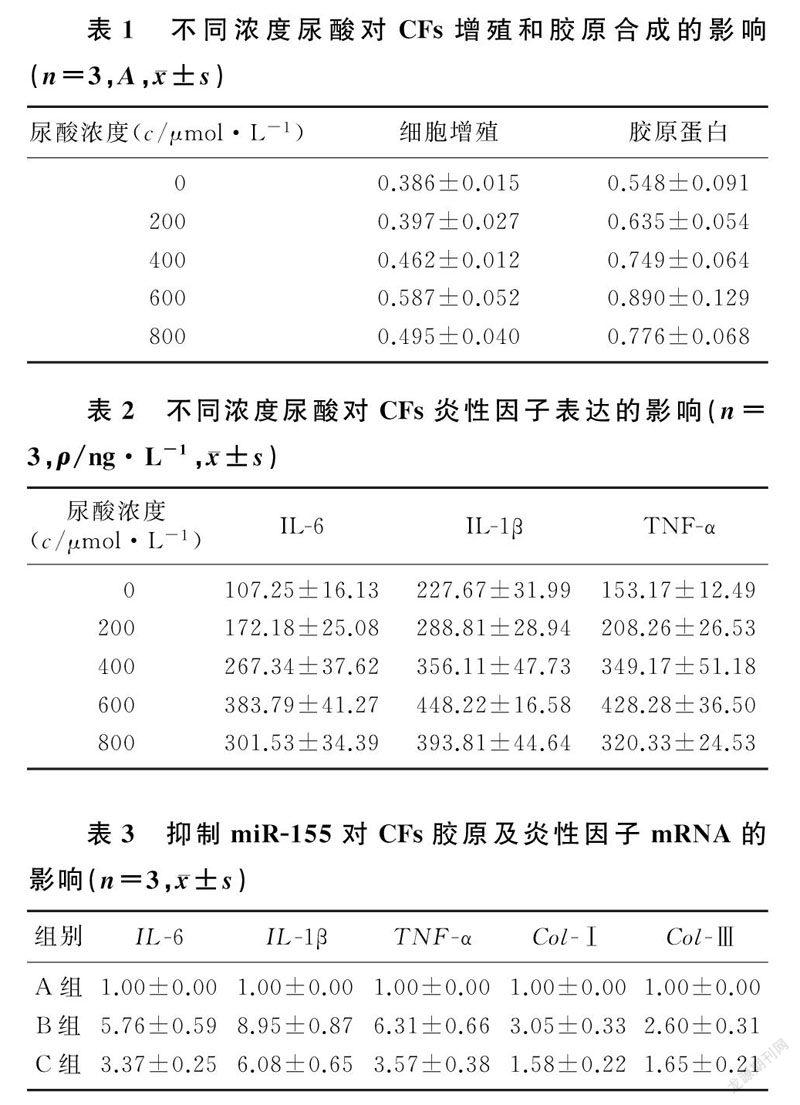
尿酸作用24 h后,400、600、800 μmol/L尿酸组MTT法所测的A值较0 μmol/L尿酸组明显增加(F=18.46,P<0.05),而200 μmol/L尿酸组与0 μmol/L尿酸组相比较差异无显著性(P>0.05)。见表1。
2.3不同浓度尿酸对CFs胶原含量的影响
尿酸作用24 h后,400、600、800 μmol/L尿酸组CFs胶原蛋白含量较0 μmol/L尿酸组明显增加(F=11.82,P<0.05),而200 μmol/L尿酸组与0 μmol/L尿酸组相比较差异无显著性(P>0.05)。见表1。
2.4不同浓度尿酸对CFs炎性因子表达的影响
不同浓度尿酸作用24 h后,与0 μmol/L尿酸组相比,200、400、600、800 μmol/L尿酸组CFs炎性因子IL-6、IL-1β、TNF-α的表达均显著增加(F=29.32~69.76,P<0.05)。见表2。
2.5转染CFs后各组miR-155的表达
以600 μmol/L尿酸处理细胞24 h后,尿酸组CFs miR-155的相对表达量为1.00±0.00,尿酸+阴性对照组为1.07±0.10,尿酸+inhibitor组为0.31±0.08,尿酸+inhibitor组miR-155的相对表达量明显低于尿酸组及尿酸+阴性对照组(F=172.65,P<0.05)。
2.6MiR-155对尿酸诱导的CFs中炎性因子及胶原mRNA表达的影响
荧光定量PCR检测显示,miR-155 inhibitor可以明显抑制600 μmol/L尿酸诱导的IL-6、IL-1β、TNF-α、Col-Ⅰ、Col-Ⅲ的表达增加(F=72.74~275.32,P<0.05)。见表3。
3讨论
高尿酸血症是一种常见的代谢性疾病,与高血压、血脂异常、糖代谢受损等密切相关[5],是心血管疾病的一个重要的独立危险因素[4-7]。有临床研究显示,在普通人群中,血尿酸水平与左心室肥厚存在独立相关性[17];在老年心衰病人中,血尿酸水平与心室重构明显相关且独立于肾功能之外[7,18]。动物实验研究显示,血尿酸水平升高可以诱导小鼠心室重塑、炎症、心肌纤维化以及胶原相关酶的分泌异常[19-21]。在细胞水平,尿酸可以诱导CFs增殖、内皮素-1(ET-1)表达增加和活性氧(ROS)生成增多等氧化应激反应[22],但是尿酸对CFs胶原合成及炎性因子分泌的影响尚未见报道。
心肌纤维化是心脏受损后,炎症介导心肌细胞坏死、凋亡,ECM异常增多和过度沉积的病理过程。心脏ECM主要由胶原、蛋白多糖、糖蛋白、生长因子和蛋白酶等构成,其中的胶原主要是Col-Ⅰ和表1不同浓度尿酸对CFs增殖和胶原合成的影响(n=3,A,±s)尿酸浓度(c/μmol·L-1)细胞增殖胶原蛋白Col-Ⅲ[23]。生理状态时,CFs通过合成胶原、分泌胶原水解酶,合成新胶原蛋白替代老化的胶原蛋白,从而稳定心脏ECM的成分和结构[24]。心脏损伤后,受到自分泌和旁分泌的调控,CFs活化并迅速增殖,向损伤区域迁移,分泌ECM(主要是胶原),从而促进伤口愈合和瘢痕形成[25-26],当心脏持续接受外界刺激时,则会造成胶原的过度沉积,形成心肌纤维化[27]。本实验结果显示,CFs的增殖能力以及合成Col-Ⅰ和Col-Ⅲ的量在一定范围内随尿酸浓度的增加而增加,表明较高浓度的尿酸可以诱导CFs过度合成胶原,导致心肌纤维化的发生。
心脏炎症可以触发CFs的表型转化,增加心肌胶原沉积,是心脏重构的一个病理关键[28]。心脏损伤后,CFs可以产生促炎性细胞因子、趋化因子(如IL-1β、IL-6、TNF-α等),募集炎性细胞(中性粒细胞、巨噬细胞、单核细胞等)到心脏组织中,CFs与炎性细胞相互作用,引发心脏炎症的恶性循环[29-30]。高尿酸血症病人血清炎症指标升高,说明高尿酸血症可引起机体炎症反应[31]。最近有研究结果表明,CFs中NLRP3炎症小体的激活在心脏炎症反应中起关键作用[32-33]。而尿酸可以通过激活NLRP3炎症小体,释放促炎性细胞因子到细胞外,介导炎症反应[34]。因此,我们推测尿酸可以诱导CFs的炎症反应。本实验结果显示,一定浓度尿酸可刺激CFs分泌炎性细胞因子(IL-6、IL-1β、TNF-α),可见尿酸可以在一定程度上促进心脏炎症的发生发展。
研究已证明,某些microRNAs在某些特定的疾病中过度表达,它们通过单独或者联合作用调节疾病的发生发展。miR-155在各种炎症性心脏病,包括心肌肥大、心肌炎、动脉粥样硬化和心衰中起重要作用[35]。最近研究发现,miR-155参与心脏重塑的病理过程,miR-155敲除可以改善血管紧张素Ⅱ(AngⅡ)诱导的心脏重塑,减轻胶原沉积,而miR-155的过度表达则可促进CFs的表达转化[12]。本研究通过抑制miR-155表达来探讨miR-155在尿酸诱导的心肌纤维化中的作用,结果表明miR-155在尿酸诱导的心肌纤维化中起促进作用,它参与了CFs的胶原合成及炎性分泌。
综上所述,尿酸可以通过miR-155促进CFs的胶原合成及炎性分泌,抑制miR-155则可减轻上述反应,提示靶向miR-155可能作为心肌纤维化的潜在治疗方法。
[参考文献]
[1]施洋,李澜,邢晓雪,等. 心肌纤维化与慢性充血性心力衰竭研究進展[J]. 中国临床药理学杂志, 2016,32(1):87-90.
[2]LINDNER D, ZIETSCH C, TANK J, et al. Cardiac fibroblasts support cardiac inflammation in heart failure[J]. Basic Research in Cardiology, 2014,109(5):428.
[3]GUDIO G . Hyperuricemia as a risk factor for cardiovascular disease: clinical review[J]. Medwave, 2016,16(10):e6606.
[4]BORGHI C, VERARDI F M, PAREO I, et al. Hyperuricemia and cardiovascular disease risk[J]. Expert Review of Cardiovascular Therapy, 2014,12(10):1219-1225.
[5]YOSHIMURA A, ADACHI H, HIRAI Y, et al. Serum uric acid is associated with the left ventricular mass index in males of a general population[J]. International Heart Journal, 2014,55(1):65-70.
[6]PALMER T M, NORDESTGAARD B G, MARIANNE B, et al. Association of plasma uric acid with ischaemic heart disease and blood pressure: mendelian randomisation analysis of two large cohorts[J]. British Medical Journal, 2013,347:f4262.
[7]FILIPPATOS G S, AHMED M I, GLADDEN J D, et al. Hyperuricaemia, chronic kidney disease, and outcomes in heart failure: potential mechanistic insights from epidemiological data[J]. European Heart Journal, 2011,32(6):712-720.
[8]KAUFMAN M, GUGLIN M. Uric acid in heart failure: a biomarker or therapeutic target[J]? Heart Failure Reviews, 2013,18(2):177-186.
[9]RADOVANOVIC S, SAVIC-RADOJEVIC A, PEKMEZO-VIC T, et al. Uric acid and gamma-glutamyl transferase activity are associated with left ventricular remodeling indices in patients with chronic heart failure[J]. Revista Espaola de Cardiología, 2014,67(8):632-642.
[10]LI Zhi, SHEN Yang, CHEN Yingqun, et al. High uric acid inhibits cardiomyocyte viability through the ERK/P38 pathway via oxidative stress[J]. Cellular Physiology and Bioche-mistry, 2018,45(3):1156-1164.
[11]YAN Meiling, CHEN Kankai, HE Li, et al. Uric acid induces cardiomyocyte apoptosis via activation of calpain-1 and endoplasmic reticulum stress[J]. Cellular Physiology and Bioche-mistry, 2018,45(5):2122-2135.
[12]WEI Yuzhen, YAN Xiaofei, YAN Lianhua, et al. Inhibition of microRNA-155 ameliorates cardiac fibrosis in the process of angiotensin Ⅱ-induced cardiac remodeling[J]. Molecular Medicine Reports, 2017,16(5):7287-7296.
[13]NAZARI-JAHANTIGH M, WEI Y Y, NOELS H, et al. MicroRNA-155 promotes atherosclerosis by repressing Bcl6 in macrophages[J]. The Journal of Clinical Investigation, 2012,122(11):4190-4202.
[14]WANG Hui, BEI Yihua, HUANG Peipei, et al. Inhibition of miR-155 protects against LPS-induced cardiac dysfunction and apoptosis in mice[J]. Molecular Therapy, Nucleic Acids, 2016,5(10):e374.
[15]BALA S, CSAK T, SAHA B, et al. The pro-inflammatory effects of miR-155 promote liver fibrosis and alcohol-induced steatohepatitis[J]. Journal of Hepatology, 2016,64(6):1378-1387.
[16]HE Wangwei, HUANG He, XIE Qiang, et al. MiR-155 knockout in fibroblasts improves cardiac remodeling by targeting tumor protein p53-Inducible nuclear protein 1[J]. Journal of Cardiovascular Pharmacology and Therapeutics, 2016,21(4):423-435.
[17]DOEHNER W, JANKOWSKA E A, SPRINGER J, et al. Uric acid and xanthine oxidase in heart failure-Emerging data and therapeutic implications[J]. International Journal of Car-diology, 2016,213:15-19.
[18]張佩迪,刘培钊,杨艳敏. 血尿酸水平与老年慢性心力衰竭心室重构的关系[J]. 中国老年学, 2017,37(15):3714-3716.
[19]JIA G H, HABIBI J, BOSTICK B P, et al. Uric acid promotes left ventricular diastolic dysfunction in mice fed a wes-tern diet[J]. Hypertension, 2015,65(3):531-539.
[20]SHIMIZU T, YOSHIHISA A, KANNO Y, et al. Relationship of hyperuricemia with mortality in heart failure patients with preserved ejection fraction[J]. American Journal of Phy-siology-Heart and Circulatory Physiology, 2015,309(7):H1123-H1129.
[21]CHEN C C, HSU Y J, LEE T M. Impact of elevated uric acid on ventricular remodeling in infarcted rats with experimentalD, hyperuricemia[J]. American Journal of Physiology-Heart and Circulatory Physiology, 2011,301(3):H1107-H1117.
[22]CHENG T H, LIN J W, CHAO H H, et al. Uric acid activates extracellular signal-regulated kinases and thereafter endothelin-1 expression in rat cardiac fibroblasts[J]. Internatio-nal Journal of Cardiology, 2010,139(1):42-49.
[23]RIENKS M, PAPAGEORGIOU A P, FRANGOGIANNIS N G, et al. Myocardial extracellular matrix an ever-changing and diverse entity[J]. Circulation Research, 2014,114(5):872-888.
[24]NGU J M, TENG G Q, MEIJNDERT H C, et al. Human cardiac fibroblast extracellular matrix remodeling: dual effects of tissue inhibitor of metalloproteinase-2[J]. Cardiovascular Pathology, 2014,23(6):335-343.
[25]DEB A, UBIL E. Cardiac fibroblast in development and wound healing[J]. Journal of Molecular and Cellular Cardiology, 2014,70(9):47-55.
[26]HUANG Ying, QI Yuan, DU Jianqing, et al. MicroRNA-34a regulates cardiac fibrosis after myocardial infarction by targeting Smad4[J]. Expert Opinion on Therapeutic Targets, 2014,18(12):1355-1365.
[27]MOORE-MORRIS T, GUIMARAES-CAMBOA N, YUT-ZEY K E, et al. Cardiac fibroblasts: from development to heart failure[J]. Journal of Molecular Medicine, 2015,93(8):823-830.
[28]CHEN W, FRANGOGIANNIS N G. Fibroblasts in post-infarction inflammation and cardiac repair[J]. Biochimica et Biophysica Acta-Molecular Cell Research, 2013,1833(4,SI):945-953.[29]TANG Xilan, LIU Jianxun, DONG Wei, et al. Protective effect of kaempferol on LPS plus ATP-induced inflammatory response in cardiac fibroblasts[J]. Inflammation, 2015,38(1):94-101.
[30]WU L, ONG S, TALOR M V, et al. Cardiac fibroblasts mediate IL-17A-driven inflammatory dilated cardiomyopathy[J]. Journal of Experimental Medicine, 2014,211(7):1449-1464.
[31]TURAK O, OZCAN F, TOK D, et al. Serum uric acid, inflammation, and nondipping circadian pattern in essential hypertension[J]. Journal of Clinical Hypertension, 2013,15(1):7-13.
[32]ZHANG W B, XU X M, KAO R, et al. Cardiac fibroblasts contribute to myocardial dysfunction in mice with sepsis: the role of NLRP3 inflammasome activation[J]. PLoS One, 2014,9(9):e107639.
[33]SANDANGER , RANHEIM T, VINGE L E, et al. The NLRP3 inflammasome is up-regulated in cardiac fibroblasts and mediates myocardial ischaemia-reperfusion injury[J]. Cardiovascular Research, 2013,99(1):164-174.
[34]GALLEGO-DELGADO J, TY M, ORENGO J M, et al. A surprising role for uric acid: the inflammatory malaria response[J]. Current Rheumatology Reports, 2014,16(2):1-6.
[35]ZHANG Dong, CUI Yongchun, LI Bin, et al. miR-155 regulates high glucose-induced cardiac fibrosis via the TGF-beta signaling pathway[J]. Molecular Biosystems, 2017,13(1):215-224.
(本文編辑 马伟平)
猜你喜欢
中国典型病例大全(2022年10期)2022-05-10
茶道(2022年3期)2022-04-27
家庭科学·新健康(2022年2期)2022-03-07
保健与生活(2021年11期)2021-06-10
家庭医药(2019年8期)2019-08-27
老年世界(2018年2期)2018-05-21
中国现代医生(2018年5期)2018-03-29
智富时代(2017年9期)2017-11-04
智富时代(2017年9期)2017-11-04
中国美容医学(2017年3期)2017-06-26
