不同矢状骨面型后牙微种植体植入安全区研究
2017-11-13 00:53王巧静黄振贤潘颖丹肖立伟
实用口腔医学杂志 2017年5期
王巧静 黄振贤 潘颖丹 肖立伟
不同矢状骨面型后牙微种植体植入安全区研究
王巧静 黄振贤 潘颖丹 肖立伟
目的CBCT探究不同矢状骨面型后牙区植入微种植体的安全区域特征。方法纳入骨性I、II、III类患者各20 名的CBCT数据,运用InVivo 5.0软件重建,选择上下颌第一前磨牙远中至第二磨牙近中区域,分别测量距牙槽嵴顶2、 4、 6、 8、 10 mm处的近远中向宽度,采用SPSS 19.0对测量结果进行统计学分析。结果上颌距牙槽嵴高度4~8 mm,下颌距牙槽嵴高度超过4 mm根尖间隔区域是安全适宜的微种植体植入部位。 上颌第一前磨牙和第二前磨牙根间骨量在骨性Ⅰ、Ⅱ类组>骨性Ⅲ类组(P<0.05),其它测量部位各组间无统计学差异。 下颌后牙区各根间骨量多数表现为骨性Ⅲ类组>骨性Ⅰ、Ⅱ类组(P<0.05)。结论上颌距牙槽嵴高度4~8 mm的第二前磨牙和第一磨牙间,下颌距牙槽嵴高度超过4 mm的第一和第二磨牙间的根尖间隔区域是相对安全的植入部位,不同骨面型存在一定差异。
CBCT; 矢状骨面型; 微种植体; 安全区域

1 材料与方法
1.1 研究对象
1.2 研究方法
使用Kavo 3D exam CBCT(KavoSybron,USA),所有研究对象在相同参数下(扫描范围高13 cm、直径16 cm,球管电压120 kV,管电流5 mA,扫描时间16.9 s, 分辨率0.25立体像素)进行扫描。扫描时受检者面部中线与地面垂直,眶耳平面与地平面平行,扫描范围自眶上缘至颏部,在牙尖交错位下进行扫描。
1.3 研究内容
将所得CBCT数据以DICOM 3.0标准文件格式导入In Vivo Dental 5(Anatomage,USA)软件,进行三维重建,重建层厚为0.4 mm。重建出颌骨水平面、矢状面(图 1A~B)。调整水平面使矢状观察轴通过相邻两牙根中心。调整矢状面,使水平观察轴与要测量的两牙根间的牙槽嵴顶平行,在矢状面上分别测量距离牙槽嵴顶5 个不同高度水平(2、 4、 6、 8、 10 mm)的近远中向骨量(图 1C)。
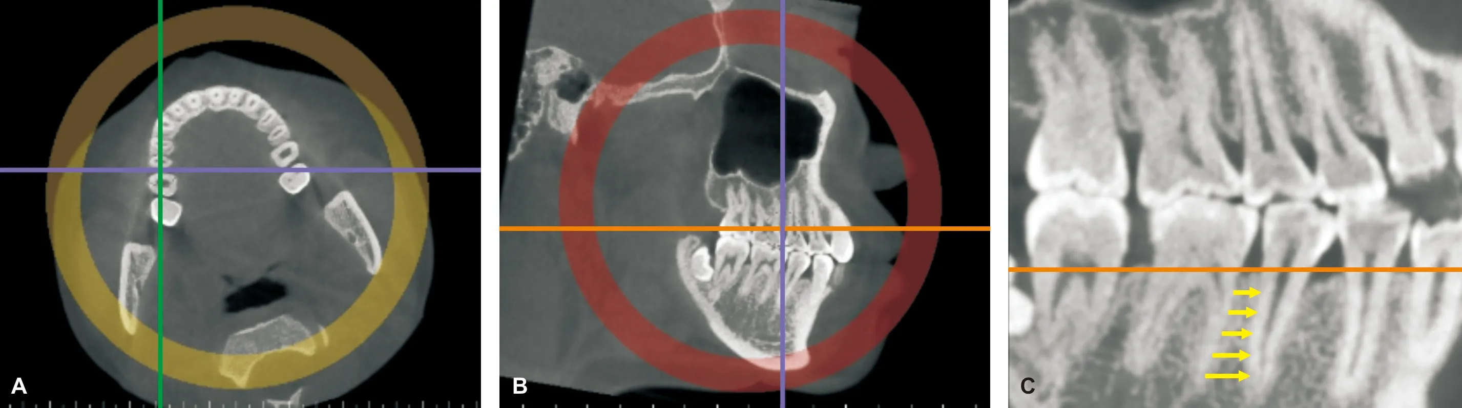
图 1 重建颌骨水平面(A)、矢状面(B,C)
1.4 统计学分析
用SPSS 19.0软件对实验数据进行统计学处理,若满足正态性和方差齐性,用单因素方差分析(One-factor analysis of variance, ANOVA), LSD检验进行两两比较。若不满足正态性和方差齐性,则用多个独立样本比较的Kruskal-WallisH检验。
2 结 果
对2 次的实验测量结果进行重复性检验,检验结果为0.358,无明显统计学差异(P>0.05)。
测量结果显示上颌后牙区相邻两牙根间近远中向最大距离均位于第二前磨牙与第一磨牙间,随着距牙槽嵴顶高度增加根间骨量逐渐增加,第一磨牙与第二磨牙间最窄,根间骨量自距牙槽嵴顶2~6 mm处逐渐减少,后逐渐增加(P<0.05)。上颌第一前磨牙和第二前磨牙间不同矢状骨面型组存在差异(P<0.05),在距不同牙槽嵴顶高度时均表现为骨性Ⅰ、Ⅱ类组>骨性Ⅲ类组。其余部位各组间无统计学差异(表 1)。
下颌后牙区相邻两牙根间近远中距离以第一前磨牙与第二前磨牙间及第一磨牙与第二磨牙间较大,第二前磨牙与第一磨牙间最小(P<0.05),根间骨量均随着距牙槽嵴顶高度增加逐渐增加。不同矢状骨面型组间在距不同牙槽嵴顶高度时总体表现为骨性Ⅲ类组>骨性Ⅰ、Ⅱ类组(P<0.05)(表 2)。
3 讨 论
微螺钉种植体的稳定性主要受种植体周围骨组织情况、植入方式、植入角度、种植体自身设计等因素影响。微种植体与周围牙体组织接触是影响其稳定性的主要因素之一。目前多数建议微种植体与相邻牙根表面应至少保留1~3 mm的安全距离[4,8-9]。本研究结果显示,距牙槽嵴顶4~10 mm,上下颌后牙根间近远中向距离均大于3 mm。由于上颌窦的存在,上颌后牙区距牙槽嵴顶超过8 mm时,微种植体的植入将受限[4]。因此上颌后牙区距牙槽嵴顶4~8 mm,下颌距牙槽嵴顶超过4 mm可认为是相对安全的植入区域。
本研究统计结果证实,上颌后牙区根尖间隔最大近远中向骨量均处在上颌第二前磨牙与第一磨牙之间为相对安全区域。而下颌第一前磨牙与第二前磨牙间、第一磨牙与第二磨牙间的近远中向骨量均较充裕,但对拔牙患者,临床上常选择拔除前磨牙,植入该区的种植体在内收前牙过程中有与牙根接触的风险,故倾向选择下颌第一磨牙与第二磨牙之间区域。

Tab 1 The comparation of the interradicular spaces of maxillary posterior regions(n=60, mm, ±s)
注: 高度: 牙槽嵴顶向根方的距离; a、b、c分别表示上颌第一前磨牙与第二前磨牙、上颌第二前磨牙与第一磨牙、上颌第一磨牙与第二磨牙间水平距离
综上所述,上颌种植体植入相对安全区域在第二前磨牙和第一磨牙间,距牙槽嵴顶4~8 mm,下颌在第一磨牙和第二磨牙间,距牙槽嵴顶大于4 mm。可以根据患者的实际情况和矫治计划选择合适的植入位点,防止种植体损伤相邻牙体组织结构,提高稳定性。
[1] Chang HP,Tseng YC. Miniscrew implant applications in contemporary orthodontics[J]. Kaohsiung J Med Sci, 2014, 30(3): 111-115.
[2] Schätzle M, Männchen R, Zwahlen M, et al. Survival and failure rates of orthodontic temporary anchorage devices: A systematic review[J]. Clin Oral Implants Res, 2009, 20(12): 1351-1359.
Tab 2 The comparation of the interradicular spaces of mandiblular posterior regions(n=60, mm, ±s)
注: 高度,牙槽嵴顶向根方的距离; d、e、f分别表示下颌第一前磨牙与第二前磨牙、下颌第二前磨牙与第一磨牙、下颌第一磨牙与第二磨牙间水平距离
[3] Crismani AG, Bertl MH, Celar AG, et al. Miniscrews in orthodontic treatment: Review and analysis of published clinical trials[J]. Am J Orthod Dentofacial Orthop, 2010, 137(1): 108-113.
[4] Poggio PM, Incorvati C, Velo S, et al. “Safe zones”: A guide for miniscrew positioning in the maxillary and mandibular arch[J]. Angle Orthod, 2006, 76(2): 191-197.
[5] Chaimanee P,Suzuki B,Suzuki EY.“Safe zones” for miniscrew implant placement in different dentoskeletal patterns[J]. Angle Orthod 2011, 81(3): 397-403.
[6] Kuroda S, Yamada K, Deguchi T, et al. Root proximity is a major factor for screw failure in orthodontic anchorage[J]. Am J Orthod Dentofacial Orthop, 2007, 131(4 Suppl): S68-S73.
[7] 王巧静, 黄振贤, 肖立伟. 正畸负载下微种植支抗移位的研究进展[J]. 国际口腔医学杂志, 2015, 42(5): 572-574.
[8] Schnelle MA, Beck FM, Jaynes RM, et al. A radiographic evaluation of the availability of bone for placement of miniscrews[J]. Angle Orthod, 2004, 74(6): 832-837.
[9] Liou EJ, Pai BC, Lin JC. Do miniscrews remain stationary under orthodontic forces?[J]. Am J Orthod Dentofacial Orthop, 2004, 126(1): 42-47.
[11]Yoon SS, Chung CH. Comparison of craniofacial growth of untreated Class I and Class II girls from ages 9 to 18 years: A longitudinal study[J]. Am J Orthod Dentofacial Orthop, 2015, 147(2): 190-196.
Thesafezonesofposteriorminiscrewimplantplacementindifferentsagittalskeletalfeatures
WANGQiaojing1,HUANGZhenxian2,PANYingdan1,XIAOLiwei1.
1. 410011Changsha,DepartmentofOrthodontics,StomatologyCenter,TheSecondXiangyaHospital,CentralSouthUniversity,China; 2.DepartmentofStomatology,ZhongshanHospital,XiamenUniversity
Objective: To measure the mesio-distal interradicular space of posterior teeth at different height by CBCT.Methods60 subjects with skeletal Class I, II and III patterns were included(n=20). From the distal of first premolar to the mesial of second molar,the mesiodistal width at the height of 2, 4, 6, 8 and 10 mm from the alveolar crest were measured and analysed.ResultsThe interradicular distance was larger than 3 mm within 4-10 mm height in maxilla and in mandible. The maxillary mesiodistal width values measured between the first premolar and second premolar in skeletal ClassⅠ and Class II pattern was greater than that in Class III(P<0.05) and there was no significant difference in other zones. In the mandible, the values of skeletal Class III pattern were greater than those of skeletal Class I and Class II pattern(P<0.05).ConclusionThe suitable interradicular zone is within 4-8 mm to the alveolar crest between the second premolar and first molar for miniscrew implant placement in maxilla,and over 4 mm between the first molar and second molar in mandible. The difference of interradicular spaces in sagittal skeletal features is existed.
CBCT;Sagittalskeletalfeatures;Miniscrewimplant;Safezones
410011 长沙, 中南大学湘雅二医院口腔医学中心[王巧静(现在厦门新开元医院口腔科) 潘颖丹 肖立伟]; 厦门大学附属中山医院口腔科(黄振贤)
肖立伟 E-mail: xlw69@163.com
R783.5
A
10.3969/j.issn.1001-3733.2017.05.019
(收稿: 2017-01-18 修回: 2017-04-08)
猜你喜欢
临床骨科杂志(2022年4期)2022-11-24
——该叫矢状脊还是矢状隆起?
化石(2022年2期)2022-06-23
医学研究杂志(2021年6期)2021-07-09
实用医药杂志(2020年8期)2020-08-26
现代临床医学(2019年6期)2019-12-07
安徽医药(2019年6期)2019-05-24
实用口腔医学杂志(2018年1期)2018-04-02
实用口腔医学杂志(2017年6期)2017-09-19
销售与市场·渠道版(2015年9期)2015-11-10
销售与市场(营销版)(2015年9期)2015-07-07