
3.0T磁共振血管壁成像技术对缺血性脑卒中颅内动脉斑块的分析研究
2021-03-22 22:13:57曹杰杨志宏曹家驹吴云钟美花
中国医学创新
2021年31期
曹杰 杨志宏 曹家驹 吴云 钟美花
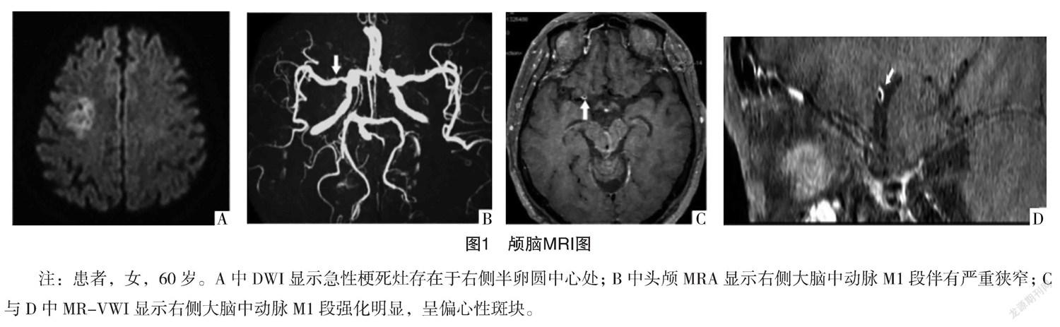
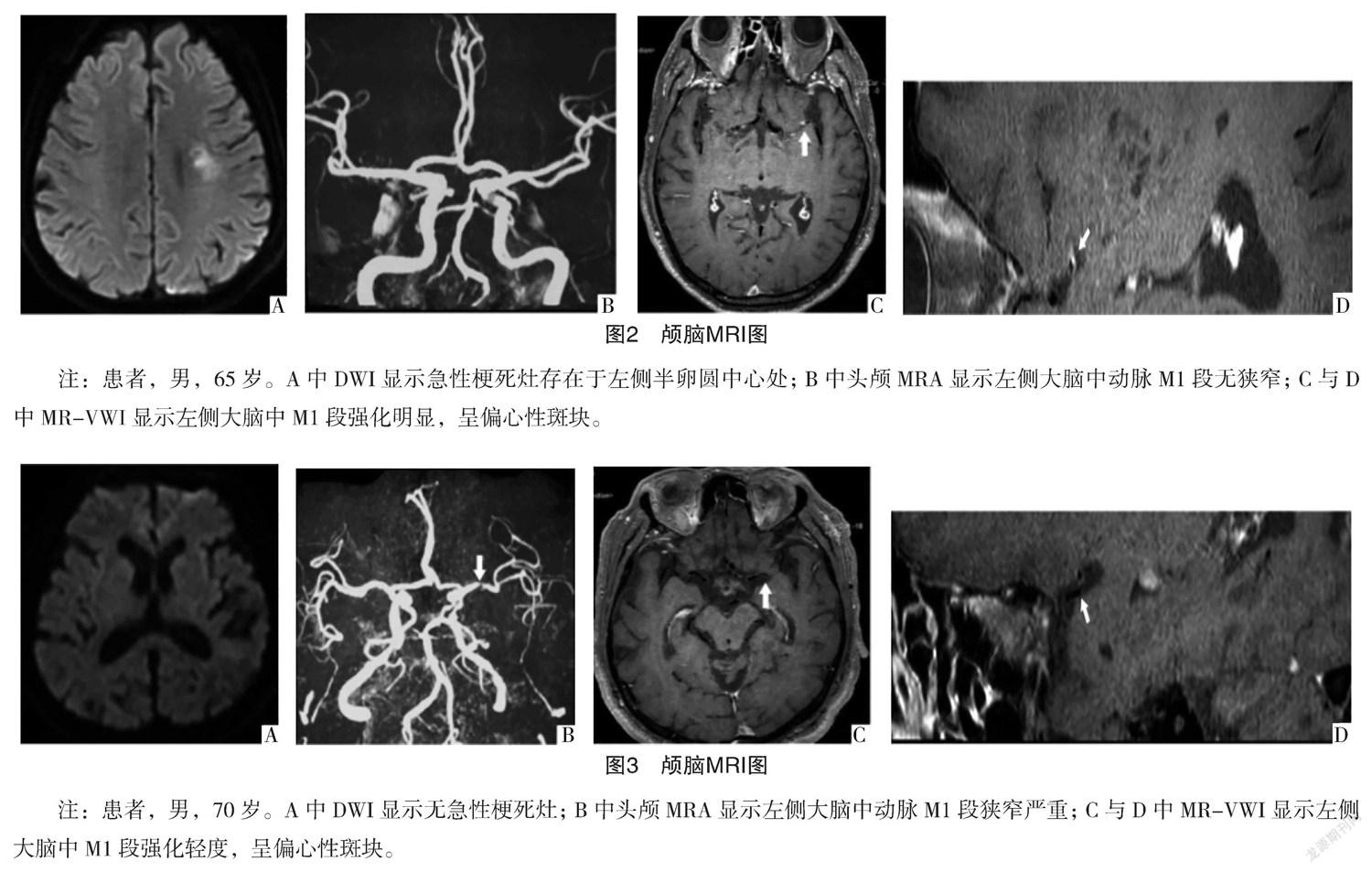
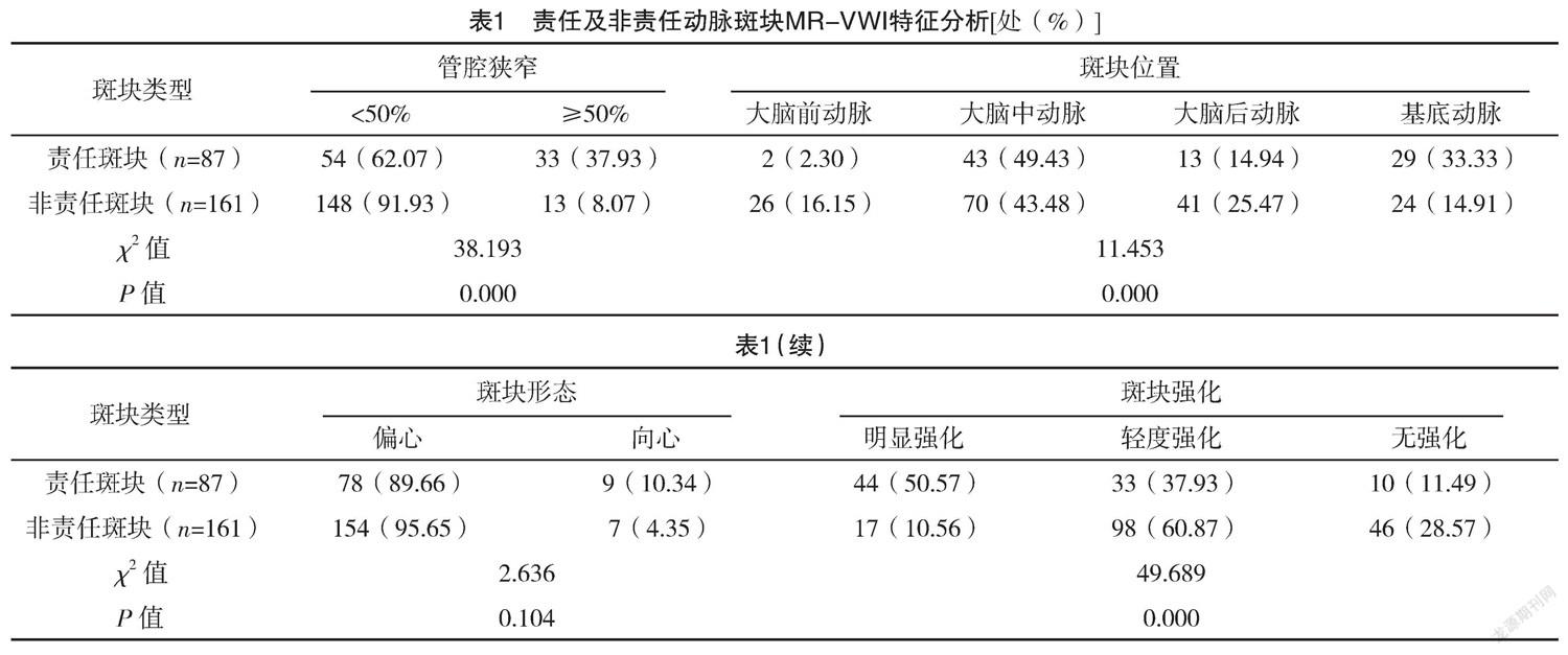
【摘要】 目的:研究3.0T磁共振血管壁成像技术(MR-VWI)在缺血性脑卒中(CIS)颅内动脉斑块的诊断价值。方法:回顾性分析2018年1月-2020年6月本院收治的62例CIS患者临床资料,均行颅内动脉MR-VWI检查,按斑块与缺血区责任动脉关系划分为责任动脉斑块组、非责任动脉斑块组。分析责任及非责任动脉斑块MR-VWI特征。结果:62例患者中共存在248处斑块,其中责任动脉斑块与非责任动脉斑块分别为87处(35.08%)、161处(64.92%)。责任斑块与非责任动脉斑块的管腔狭窄程度、斑块位置及强化情况比较,差异均有统计学意义(P<0.05)。责任斑块与非责任动脉斑块的斑块形态比较,差异无统计学意义(P>0.05)。责任斑块组管腔狭窄以<50%为主,斑块位置多见大脑中动脉与基底动脉,斑块强化成明显强化。结论:3.0T MR-VWI用于CIS效果较好,可明确颅内动脉斑块强化程度,评估疾病危险因素,值得推广及应用。
【关键词】 缺血性脑卒中 颅内动脉斑块 3.0T磁共振血管壁成像技术
3.0T MR Vessel Wall Imaging of Intracranial Arterial Plaque in Cerebral Ischemic Stroke/CAO Jie, YANG Zhihong, CAO Jiaju, WU Yun, ZHONG Meihua. //Medical Innovation of China, 2021, 18(31): 0-023
[Abstract] Objective: To study the diagnostic value of 3.0T magnetic resonance vascular wall imaging (MR-VWI) in the diagnosis of intracranial artery plaque in ischemic stroke (CIS). Method: The clinical data of 62 patients with CIS admitted to our hospital from January 2018 to June 2020 were retrospectively analyzed, all patients underwent MR-VWI examination of intracranial artery, they were divided into responsible artery plaque group and non-responsible artery plaque group according to the relationship between plaque and responsible artery in ischemic area. The MR-VWI characteristics of responsible and non-responsible arterial plaques were analyzed. Result: A total of 248 plaques were found in 62 patients, including 87 (35.08%) responsible artery plaques and 161 (64.92%) non-responsible artery plaques. There were significant differences in lumen stenosis degree, plaque location and enhancement between responsible plaque and non-responsible artery plaque (P<0.05). There was no significant difference in plaque morphology between responsible plaque and non-responsible plaque (P>0.05). In the responsible plaque group, lumen stenosis was less than 50%, and the middle cerebral artery and basilar artery were the most common plaque locations. Conclusion: 3.0T MR-VWI has a good effect on CIS, which can determine the degree of intracranial arterial plaque enhancement and evaluate the risk factors of disease, which is worthy of promotion and application.
[Key words] Cerebral ischemic stroke Intracranial arterial plaque 3.0T MR vessel wall imaging
First-authors address: Jiujiang First Peoples Hospital, Jiujiang 332000, China
doi:10.3969/j.issn.1674-4985.2021.31.005
缺血性腦卒中(cerebral ischemic stroke,CIS)是具有高发生率、高致残率及高致死率的脑血管疾病,因颅内动脉粥样硬化病变所致。近年来,随着我国老龄化局势加剧,该病也呈逐年增长趋势,对患者身心健康造成严重危害[1]。相关研究发现,CIS的发生、发展离不开动脉粥样硬化斑块内炎性范围,可用于评估斑块稳定性[2]。近年来随着医疗技术的更新与发展,3.0T磁共振血管壁成像技术(MR vessel wall imaging,MR-VWI)逐渐被用于诊断脑血管疾病,相比起常规MR扫描能够明确动脉粥样硬化程度及斑块情况[3]。因此,本研究对3.0T MR-VWI用于CIS颅内动脉斑块展开相应的分析,现报道如下。
1 资料与方法
1.1 一般资料 选取2018年1月-2020年6月本院收治的62例CIS患者,其中男37例,女25例;年龄30~74岁,平均(55.18±2.55)岁。纳入标准:(1)经临床影像学检查结合症状表现符合《中国CIS诊治指南》诊断标准[4];(2)年龄≥18岁。……
登录APP查看全文
