
原儿茶醛对大鼠脑缺血再灌注损伤后神经血管单元稳态破坏的保护作用
2021-12-10 17:09:00冯晋徐娅玲孟庆婷颜汉文何芳雁
中国药房
2021年15期
冯晋 徐娅玲 孟庆婷 颜汉文 何芳雁


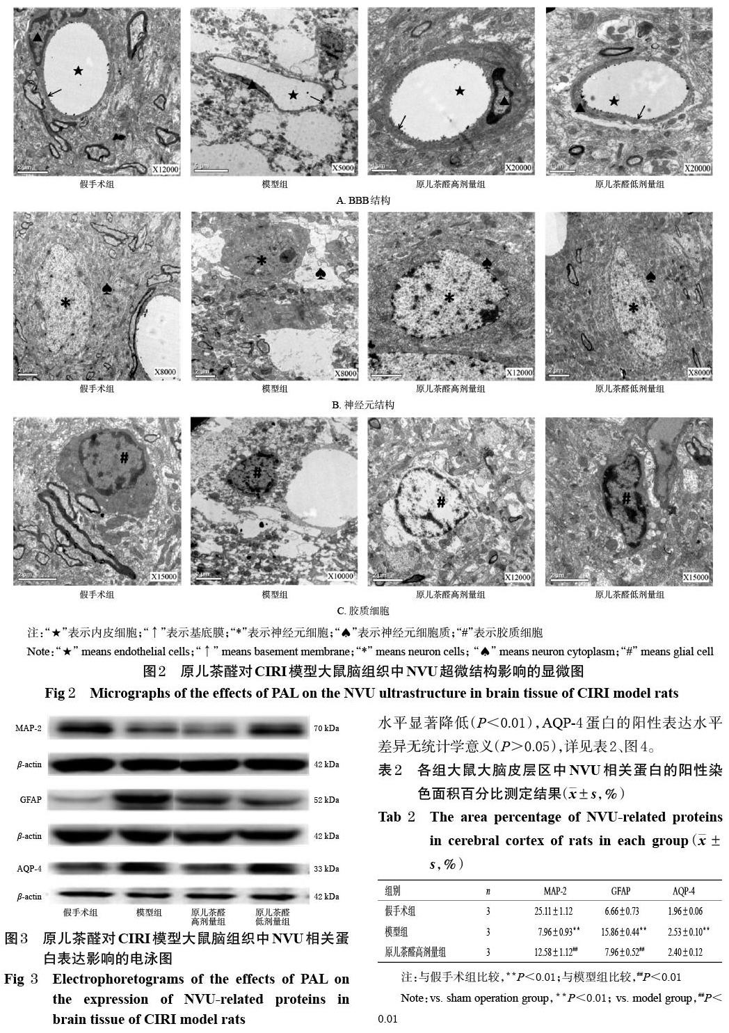
摘 要 目的:研究原兒茶醛对大鼠脑缺血再灌注损伤(CIRI)后神经血管单元(NVU)稳态破坏的保护作用。方法:将SD大鼠随机分为假手术组、模型组和原儿茶醛高、低剂量组(10、20 mg/kg),每组11只。给药组大鼠灌胃相应药物,假手术组和模型组大鼠灌胃等体积水,灌胃体积均为10 mL/kg,每日1次,连续5 d。末次给药后,采用线栓法复制大鼠CIRI模型,采用透射电镜观察大鼠脑组织中NVU超微结构的变化;采用Western blot法检测大鼠脑组织中NVU相关蛋白[神经元标志蛋白(MAP-2)、胶质纤维酸性蛋白(GFAP)、水通道蛋白(AQP-4)]的表达水平;采用免疫荧光染色法检测大鼠大脑皮层中上述蛋白的阳性表达水平。结果:与假手术组比较,模型组大鼠血脑屏障(BBB)结构被严重破坏,血管腔变窄,内皮细胞外侧严重水肿,基底膜厚薄不一;神经元核固缩,周围组织存在大面积水肿;胶质细胞结构严重破坏,胞体皱缩、细胞器损失;脑组织(或大脑皮层)中MAP-2蛋白表达水平(或阳性表达水平)均显著降低(P<0.05或P<0.01),GFAP、AQP-4蛋白表达水平(或阳性表达水平)均显著升高(P<0.01)。经原儿茶醛干预后,大鼠BBB损伤减轻,血管腔和基底膜形态未完全被破坏;神经元损伤减轻,神经元核固缩减少,染色质均匀、异染色质减少;胶质细胞结构破坏减轻,脑组织中GFAP、AQP-4(低剂量组除外)蛋白表达水平以及大脑皮层中MAP-2、GFAP蛋白阳性表达水平均显著逆转(P<0.05或P<0.01)。结论:原儿茶醛可保护CIRI模型大鼠的NVU稳态免受破坏;其作用机制可能与上调大鼠大脑皮层中MAP-2蛋白表达,下调脑组织中GFAP、AQP-4蛋白表达有关。
关键词 原儿茶醛;脑缺血再灌注损伤;神经血管单元;大脑皮层;大鼠
ABSTRACT OBJECTIVE: To observe the protective effect of protocatechuic aldehyde (PAL) on neurovascular unit (NVU) homeostasis damage in rats after cerebral ischemia-reperfusion injury (CIRI). METHODS: SD rats were randomly divided into sham operation group, model group, PAL high-dose and low-dose groups (10, 20 mg/kg), with 11 rats in each group. Administration groups were given relevant medicine intragastrically. Sham operation group and model group were given the same volume of normal saline intragastrically, 10 mL/kg once a day, for 5 days. After last administration, CIRI model was induced by suture method; the ultrastructural changes of NVU were observed by transmission electron microscope. Western blot assay was used to detect the expression of NUV related proteins (MAP-2, GFAP, AQP-4) in cerebral tissue. Immunofluorescence staining was used to observe the positive expression of above proteins in cerebral cortex. RESULTS: Compared with sham operation group, blood-brain barrier (BBB) structure of model group was destroyed severely, the vascular lumen became narrower, lateral edema of endothelial cells was severe, and the thickness of basement membrane varied; the nuclei of neurons were pyknosis and there was a large area of edema in the surrounding tissues; the structure of glial cells was seriously damaged, the cell body was shrunk and organelles were lost; protein expression (or positive expression) of MAP-2 in brain tissue (or cerebral cortex) were significantly decreased (P<0.05 or P<0.01), while protein expression (or positive expression) of GFAP and AQP-4 were increased significantly (P<0.01). After PAL intervention, the rats had less BBB damage, and the morphology of vascular lumen and basement membrane were not completely destroyed; the damage of neurons was alleviated, the pyknosis of neurons was decreased, the chromatin was homogeneous and the heterochromatin was decreased; the damage of glial cell structure was alleviated; protein expression of GFAP and AQP-4 (except for low-dose group) in cerebral tissue and positive expression of MAP-2 and GFAP protein in cerebral cortex were reversed significantly (P<0.05 or P<0.01). CONCLUSIONS: PAL can protect the stability of NVU from damage in CIRI model rats; the mechanism may be related to up-regulating the expression of MAP-2 protein in cerebral cortex and down-regulating the expression of GFAP and AQP-4 protein in brain tissue.
KEYWORDS Protocatechuic aldehyde; Cerebral ischemia-reperfusion injury; Neurovascular unit; Cerebral cortex; Rats
《中国脑卒中防治报告(2018)》显示,脑卒中居我国死亡原因之首[1]。其中,缺血性脑卒中(ischemic stroke,IS)是脑卒中发病率最高的病种,其是由于脑血流的突然中断导致脑细胞死亡和神经缺陷的一类疾病[2]。在IS的病理发展过程中,血栓脱落、血栓自溶以及缺血区侧支循环的建立与开放等均会引起缺血区的血流重建,从而造成脑缺血再灌注损伤(CIRI),进而导致IS恶化[3]。IS的发生不仅影响神经元,还影响位于“胶质细胞-神经细胞-血管小生态环境”中的所有脑细胞、细胞外基质和机体免疫系统。……
登录APP查看全文
