人喉鳞癌Hep-2细胞血红素加氧酶1表达及其对卡铂作用的影响
2015-02-19 07:17吕欣宋冬梅王宝山河北医科大学第一医院石家庄050032河北医科大学
山东医药 2015年36期
吕欣,宋冬梅,王宝山,2(河北医科大学第一医院,石家庄05003;2河北医科大学)
人喉鳞癌Hep-2细胞血红素加氧酶1表达及其对卡铂作用的影响
吕欣1,宋冬梅1,王宝山1,2
(1河北医科大学第一医院,石家庄050031;2河北医科大学)
摘要:目的探讨人喉鳞癌Hep-2细胞血红素加氧酶1(HO-1)表达对卡铂作用的影响及其可能的作用机制。方法将对数生长期Hep-2细胞分为六组。空白对照组不干预,ZnPP(HO-1抑制剂锌原卟啉)组加入ZnPP 10 μmol/L; Hemin(HO-1诱导剂氯化血红素)组加入Hemin 10 μmol/L;卡铂组加入卡铂10 μg/mL; Hemin联合卡铂组先加入Hemin 10 μmol/L,作用1 h后再加入卡铂10 μg/mL; ZnPP联合卡铂组先加入ZnPP 10 μmol/L,作用1 h后再加入卡铂10 μg/mL。每组设5个复孔,加药后培养24 h。采用MTT法观察各组细胞存活率; RT-PCR、Western blotting法检测各组细胞内HO-1mRNA和蛋白表达情况;dCFH-DA探针检测各组细胞内活性氧(ROS);流式细胞术检测各组细胞凋亡率。结果①细胞存活率:与空白对照组比较,卡铂组和ZnPP组明显降低,Hemin组明显升高(P均<0.05);与卡铂组比较,ZnPP联合卡铂组明显降低,Hemin联合卡铂组明显升高(P均<0.05)。②细胞HO-1mRNA和蛋白相对表达量:与空白对照组比较,卡铂组和Hemin组明显升高,ZnPP组明显下降(P均<0.05);与卡铂组比较,ZnPP联合卡铂组明显降低,Hemin联合卡铂组明显升高(P均<0.05)。③细胞凋亡率:与空白对照组比较,卡铂组、ZnPP组明显升高(P均<0.05);与卡铂组比较,ZnPP联合卡铂组明显升高,Hemin联合卡铂组明显降低(P均<0.05);④细胞内ROS活性:与空白对照组比较,卡铂组、ZnPP组显著增高(P均<0.05);与卡铂组比较,ZnPP联合卡铂组明显升高,Hemin联合卡铂组明显降低(P均<0.05)。结论HO-1在Hep-2细胞内的高表达可影响卡铂对Hep-2细胞的促凋亡作用,应用ZnPP后HO-1表达下调,同时提高了人喉鳞癌Hep-2细胞在卡铂作用下的凋亡率,这一过程可能与增强细胞内ROS水平相关。
关键词:喉鳞癌;血红素加氧酶1; Hep-2细胞;卡铂;细胞凋亡
HO-1 expression in human laryngeal squamous-cell carcinoma Hep-2 cells and its influence on Carboplatin’s effects
LYU Xin1,SONGdong-mei,WANG Bao-shan
(1 The First Hospital of Hebeimedical University,Shijiazhuang 050031,China)
Abstract:Objective To investigate the influence of heme oxygenase-1(HO-1)expression in human laryngeal squamous-cell carcinoma Hep-2 cells on the Carboplatin’s effects and its possiblemechanism.Methods Hep-2 cells in the logarithmic phase weredivided into six groups.The blank control group was not treated,ZnPP group was treated with 10 μmol/L HO-1 inhibitor,Hemin group was treated with 10 μmol/L HO-1 inducer,and Carboplatin group was treated with 10 μg/mL,Hemin combined with Carboplatin group was treated with 10 μmol/L Hemin alone for 1 h and then 10 μg/mL Carboplatin,ZnPP combined with Carboplatin group was treated with 10 μmol/L ZnPP alone for 1 h and then 10 μg/mL Carboplatin.These six groups all had five complex holes and were cultured for 24 h afterdosing.The survival rates were evaluated bymTT assay.The expression of HO-1mRNA and protein wasdetected by RT-PCR and Western blotting.Intracellular ROS activity was examined bydCFH-DA probe.The apoptosis rate was performed by flow cytometry.Results①the cell survival rate: compared with the control group,the survival rates of Hep-2 cells in the ZnPP and Carboplatin groups were significantlydecreased,but the survival rate in the Hemin group was increased(all P<0.05); compared with the Carboplatin group,the survival rate of the Znpp combined with Carboplatin group wasdecreased and the He-
min combined with Carboplatin group was increased(all P<0.05).②HO-1 inmRNA and protein levels: compared with the control group,the expression of HO-1mRNA and protein was increased in the Hemin and Carboplatin groups and wasdecreased in the ZnPP group(all P<0.05); compared with the Carboplatin group,the Znpp combined with Carboplatindecreased,but HO-1 expression was increased in the Hemin combined with Carboplatin group(all P<0.05).③the apoptosis rate: compared with the control group,the apoptosis rates in the Carboplatin and ZnPP groups were increased(all P <0.05); compared with the Carboplatin group,Znpp combined with Carboplatin increased the apoptosis rate,but Hemin combined with Carboplatindecreased the apoptosis rate(all P<0.05).④the ROS activity: compared with the control group,the the ROS activities in the Carboplatin and ZnPP groups were increased(all P<0.05); compared with the Carboplatin group,Znpp combined with Carboplatin increased the ROS activity,but Hemin combined with Carboplatindecreased the ROS activity(all P<0.05).Conclusions HO-1 overexpressionmay affect the pro-apoptotic effects of Carboplatin on Hep-2 cells,and the application of ZnPPdown-regulates the HO-1 expression and increases the apoptosis rate of Hep-2 cells with treatment of Carboplatin.In this process,the elevation of intracellular ROS activitymay be involved.
Key words:laryngeal squamous-cell carcinoma; heme oxygenase-1; Hep-2 cells; Carboplatin; apoptosis
血红素氧合酶1(HO-1)属于微粒体催化酶类,是降解血红素重要的限速酶之一,其可将血红素分解为一氧化碳、Fe2 +、胆绿素等[1~3]。HO-1是一种细胞保护性酶,具有抗炎、抗氧化及抑制细胞凋亡等重要生物学作用;但近年来研究发现,HO-1的这一细胞保护作用及抗凋亡特性可能是促进肿瘤细胞生长、转移及产生治疗抵抗的重要因素[4~6],但具体机制不清。目前,HO-1在头颈部肿瘤尤其是喉鳞癌中的作用鲜见报道。2014年11月~2015年5月,我们观察了HO-1诱导剂氯化高铁血红素(Hemin)和HO-1抑制剂锌原卟啉(ZnPP)对人喉鳞癌Hep-2细胞内HO-1表达的影响及HO-1表达对卡铂作用的影响,分析HO-1与喉鳞癌化疗抵抗的关系,为喉鳞癌的治疗寻找新的靶点。
1 材料与方法
1.1材料人喉鳞癌Hep-2细胞株(以下称Hep-2细胞)本实验室冻存。DMEM/F12购自Gibco公司;胎牛血清购自杭州四季青公司; Hemin购自美国Sigma公司(Lot NO.51280); ZnPP购自美国Santa Cruz Biotechnology公司(SC-200329);卡铂购自山东齐鲁制药有限公司(国药准字号H10920028);二甲基亚砜(DMSO)购自美国Sigma公司(Lot NO.D-5879);四甲基噻唑基四唑(MTT)购自美国Sigma公司(Lot NO.M2128);碘化丙啶(PI)购自美国Sigma公司(Lot NO.P4170); HO-1及内参GAPDH引物由上海生工合成;兔抗人HO-1单克隆抗体购自美国abcam公司(ab13243);内参β-actin购自北京博奥森试剂公司(bs-0061R);活性氧检测试剂盒购自江苏碧云天生物技术研究所(S0033); RT-PCR检测试剂盒购自立陶宛Fermentas公司(K1611,K1081)。
1.2细胞培养、分组及干预将Hep-2细胞置于含10%胎牛血清、100 U/mL青霉素、100 U/mL链霉素的DMEM/F12培养液中,于37℃、5% CO2的孵箱内培养。以0.25%胰酶消化传代,2~3d传代1次,实验细胞均处于对数生长期。将Hep-2细胞胰酶消化后,以每孔5×10³个细胞接种于96孔板,待细胞贴壁12 h后分为六组。空白对照组不干预,ZnPP组加入ZnPP 10 μmol/L; Hemin组加入Hemin 10 μmol/L;卡铂组加入卡铂10 μg/mL; Hemin联合卡铂组先加入Hemin 10 μmol/L,作用1 h后再加入卡铂10 μg/mL; ZnPP联合卡铂组先加入ZnPP 10 μmol/L,作用1 h后再加入卡铂10 μg/mL。每组设5个复孔,加药后均培养24 h。
1.3 Hep-2细胞存活率检测采用MTT法。每孔加入5mg/mLmTT 20 μL,继续培养4 h后,彻底去上清后加DMSO 200 μL/孔,振荡30min至结晶完全溶解,酶标仪测定570 nm处的光密度值(OD 值),实验重复5次。细胞存活率(%)=(OD试验孔/OD对照孔)×100%。
1.4 Hep-2细胞HO-1mRNA及HO-1蛋白表达检测采用RT-PCR法检测各组Hep-2细胞HO-1mRNA表达:各组加药培养24 h后TRIzol法提取总RNA,取等量RNA(500 ng)逆转录为cDNA。HO-1上游引物5'-CTTTGGTCAATGACACCGTG-3',下游引物5'-TGTTTGTGGTAAGCCATCCA-3',产物长度168 bp;β-actin上游引物5'-GTGGGGCGCCCCAGGCACCA-3',下游引物5'-CTCCTTAATGTCACGCACGATTT-3',产物长度540 bp。基因扩增反应条件: 95℃预变性5min; 95℃、30 s,57℃、30 s,72℃、30 s,β-actin 26个循环,HO-1 29个循环,72℃延伸10min,4℃、2 h。产物在1%的琼脂糖凝胶上电泳,利用Lab Work5.0系统进行图像分析。采用Western blotting法检测HO-1蛋白表达:各组加药24 h后收集细胞,加入4℃预冷的RIPA细胞裂解液300
μL冰上裂解30min,期间细胞刮刀刮3次,4℃、12 000 r/min离心20min,取上清液5 μL,BCA法蛋白定量,取总蛋白量一致后进行SDS-PAGE电泳,电转至PVDF膜上,然后置于5%脱脂奶粉TBST溶液封闭2 h,加入1∶2 000兔抗人HO-1一抗,同时用兔抗人β-actin(1∶500)作为内参,4℃过夜。辣根过氧化物酶(HRP)标记的羊抗兔二抗(1∶5 000)室温孵育2 h后,TBST溶液洗涤15min/2次,ECL发光显色,结果扫描,HO-1蛋白条带灰度值测定。
1.5Hep-2细胞内活性氧(ROS)检测采用DCFH-DA探针检测。加药培养24 h后,胰酶消化、终止、收集细胞,收集细胞后装载探针,按照1∶1 000用无血清培养基稀释DCFH-DA,使终浓度为10 μmol/L,细胞收集后悬浮于稀释好的DCFHDA中,细胞密度为1×106/mL,37℃细胞培养箱内孵育20min,无血清培养基洗3次,488 nm激发波长,525 nm发射波长,检测各组荧光强度,结果以平均荧光强度表示细胞内ROS的相对活性。
1.6 Hep-2细胞凋亡率观察加药后继续培养24 h,胰酶消化正常培养基终止消化后收集细胞,4℃预冷PBS洗涤细胞,1 500 r/min离心5min,重复2次,90%酒精固定-20℃保存12 h后,4℃预冷PBS洗涤细胞,1 500 r/min离心5min,重复2次,洗去酒精,制成单细胞悬液300 μL后加入PI使终浓度为10 μg/mL,室温避光孵育20min,流式细胞仪计算细胞凋亡率。数据由ModFit2.0软件系统分析。
1.7统计学方法采用SPSS13.0统计软件。计量资料以珋x±s表示,均数比较采用t检验,多组数据采用方差分析。P<0.05为差异有统计学意义。
2 结果
2.1细胞存活率细胞存活率空白对照组为96.8%±1.09%,卡铂组为75.3%±0.21%,ZnPP组为84.7%±0.04%,Hemin组为101.1%± 0.13%,ZnPP联合卡铂组为54.9%±0.31%,Hemin联合卡铂组为81.1%±0.19%; ZnPP组及卡铂组明显低于空白对照组; Hemin组明显高于空白对照组; ZnPP联合卡铂组明显低于卡铂组,Hemin联合卡铂组明显高于卡铂组; P均<0.05
2.2HO-1mRNA及HO-1mRNA蛋白表达与空白对照组比较,卡铂组和Hemin组HO-1mRNA和HO-1蛋白明显升高,ZnPP组明显下降(P均<0.05);与卡铂组比较,ZnPP联合卡铂组HO-1mRNA和HO-1蛋白明显降低,Hemin联合卡铂组明显升高(P均<0.05)。见表1、图1、图2。
表1 各组HO-1mRNA及蛋白相对表达(相对表达量,)
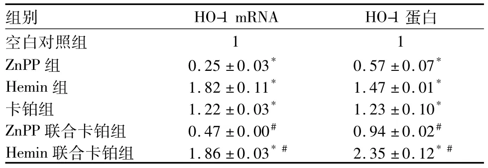
表1 各组HO-1mRNA及蛋白相对表达(相对表达量,)
注:与空白对照组比较,*P<0.05;与卡铂组比较,#P<0.05。
组别 HO-1mRNA HO-1蛋白空白对照组11 ZnPP组 0.25±0.03* 0.57±0.07*Hemin组 1.82±0.11* 1.47±0.01*卡铂组 1.22±0.03* 1.23±0.10*ZnPP联合卡铂组 0.47±0.00# 0.94±0.02#Hemin联合卡铂组 1.86±0.03* # 2.35±0.12*#
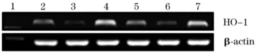
图1 各组HO-图1mRNA表达

图2 各组HO-图1蛋白表达
2.3ROS相对活性空白对照组、ZnPP组、Hemin组、卡铂组、ZnPP联合卡铂组、Hemin联合卡铂组平均荧光强度分别为1、1.79±0.12、0.59±0.09、2.30 ±0.11、3.91±0.04、1.21±0.02;与空白对照组比较,卡铂组、ZnPP组ROS活性显著增高(P均<0.05);与卡铂组比较,ZnPP联合卡铂组ROS活性明显升高,Hemin联合卡铂组明显降低(P均<0.05)。
2.4细胞凋亡率空白对照组、ZnPP组、Hemin组、卡铂组、ZnPP联合卡铂组、Hemin联合卡铂组细胞凋亡率分别为5.15%±1.27%、11.62%±1.02%、5.01%±0.94%、20.16%±1.02%、50.67%± 2.31%、13.62%±1.29%; Hemin组与空白对照组比较差异无统计学意义(P>0.05);卡铂组、ZnPP组明显高于空白对照组(P均<0.05); ZnPP联合卡铂组明显高于卡铂组,Hemin联合卡铂组明显低于卡铂组(P均<0.05)。
3 讨论
喉鳞癌早期症状隐匿、生长速度快,且对放化疗的敏感度低。HO-1与肿瘤发生、发展及化疗抵抗之间的关系目前虽不甚清楚,但近年来正逐渐引起临床重视[7,8]。研究表明,HO-1在一部分肿瘤组织中呈强阳性表达,增加或降低HO-1的表达可影响肿瘤细胞的增殖与凋亡,目前在肿瘤治疗过程中合理调控HO-1的表达正逐渐成为研究的一大热点[9,10]。
Tan等[11]研究证实,在乳腺癌细胞中HO-1的高表达可促进乳腺癌细胞的增殖,促进其侵袭及转
移,其潜在机制可能与HO-1促进细胞核增殖抗原及细胞周期调控蛋白Cyclin B1等表达有关。本研究结果表明,Hemin作用于Hep-2细胞后,细胞内HO-1mRNA和蛋白水平表达上调,而应用ZnPP后细胞内HO-1mRNA和蛋白水平表达下调,细胞活性降低。提示降低HO-1表达能够抑制Hep-2细胞的生长活性。单独应用化疗药物卡铂后,细胞内HO-1表达增高,这一结果与卡铂可促进细胞内ROS活性增强有关,ROS反应水平的增高可激活下游氧化应激反应原件ARE,进而激活Nrf2诱导HO-1表达,进而减弱卡铂对于Hep-2细胞的杀伤作用。而联合应用ZnPP抑制细胞内HO-1表达后,细胞存活率下降、凋亡率显著增加。由此提示Hemin可诱导HO-1表达,对人喉鳞癌Hep-2细胞抵抗氧化应激损伤及化疗药物的杀伤存在部分保护作用,而联合应用ZnPP阻断细胞内HO-1表达后,能够增强卡铂对Hep-2细胞的杀伤作用。该机制与HO-1在白血病、肺癌、胰腺癌等实体肿瘤细胞中的报道一致[12~17]。
目前研究证实,肿瘤细胞在放化疗过程中产生的自我保护及抵抗可能与HO-1的抗氧化应激反应有关。在肿瘤细胞中抗氧化酶系统如超氧化物歧化酶、过氧化氢酶等均表现为下调趋势;此时,HO-1及其代谢产物成为主要的抗氧化应激反应体系[18~20]。本实验结果显示,ZnPP下调人喉鳞癌Hep-2细胞内HO-1表达后,细胞存活率降低、凋亡率升高,细胞内ROS活性增加;联合应用卡铂化疗后细胞凋亡率显著升高,细胞内ROS活性进一步增加。而当卡铂联合应用Hemin后,细胞内ROS活性及细胞凋亡率下降,由此表明HO-1可能通过调节ROS活性影响卡铂对Hep-2细胞的杀伤作用。同时也有研究报道,HO-1还可通过调节Akt途径和P21蛋白等方式影响肿瘤细胞生物学行为,但具体机制不详,还有待于进一步研究证实。
综上所述,体外抑制HO-1表达能够降低Hep-2细胞活性,增强卡铂对Hep-2细胞的杀伤作用,其具体机制可能与增强细胞内ROS活性诱导细胞凋亡有关。HO-1可能在喉鳞癌发生、发展及治疗过程中发挥重要作用,合理调控肿瘤细胞中HO-1表达有望成为喉鳞癌临床治疗的新突破。
参考文献:
[1]Chen J,Emara N,Solomides C,et al.Resistance to platinumbased chemotherapy in lung cancer cell lines[J].Cancer Chemother Pharmacol,2010,66(6): 1103-1111.
[2]Zhang K,Yang H,Wang Y,et al.Expression and significance of nrf2 and its target genes in pulmonary adenocarcinoma a549 cells resistant to Cisplatin[J].Zhongguo Fei Ai Za Zhi,2009,12(11): 1150-1154.
[3]Wang LH,Li Y,Yang SN,et al.Gambogic acid synergistically potentiatescisplatin-induced apoptosis in non-small-cell lung cancer through suppressingNF-κB andmAPK/HO-1 signalling[J].Br J Cancer,2014,110(2): 341-352.
[4]Furfaro AL,Piras S,Passalacquam,et al.HO-1 up-regulation: a key point in high-risk neuroblastoma resistance to bortezomib[J].Biochim Biophys Acta,2014,1842(4): 613-622.
[5]Jais A,Einwallner E,Sharif O,et al.Heme oxygenase-1drivesmetaflammation and insulin resistance inmouse andman[J].Cell,2014,158(1): 25-40.
[6]Wagener FA,Dankers AC,van Summeren F,et al.Heme Oxygenase-1 and breast cancer resistance protein protect against heme-induced toxicity[J].Curr Pharmdes,2013,19(15): 2698-2707.
[7]Wegiel B,Gallod,Csizmadia E,et al.Carbonmonoxide expeditesmetabolic exhaustion to inhibit tumor growth[J].Cancer Res,2013,73(23): 7009-7021.
[8]Yun BR,LeemJ,Kim JH,et al.Enhancement of parthenolideinduced apoptosis by a PKC-alpha inhibition through heme oxygenase-1 blockage in cholangiocarcinoma cells[J].Expmolmed,2010,42(11): 787-797.
[9]Rushworth SA,Bowles KM,Raninga P,et al.NF-kappaB-inhibited acutemyeloid leukemia cells are rescued from apoptosis by heme oxygenase-1 induction[J].Cancer Res,2010,70(7):2973-2983.
[10]Salis O,Bedir A,Ozdemir T,et al.The relationship between anticancer effect ofmetformin and the transcriptional regulation of certain genes(CHOP,CAV-1,HO-1,SGK-1 and Par-4)onmCF-7 cell line [J].Eur Revmed Pharmacol Sci,2014,18(11):1602-1609.
[11]Tan Q,Wang H,Hu Y,et al.Src/STAT3-dependent heme oxygenase-1 inductionmediates chemoresistance of breast cancer cells todoxorubicin by promoting autophagy[J].Cancer Sci,2015,106(8): 1023-1032.
[12]Chen N,Wu L,Yuan H,et al.ROS/Autophagy/Nrf2 Pathwaymediated Low-Dose Radiation Induced Radio-Resistance in Human Lung Adenocarcinoma A549 Cell[J].Int J Biol Sci,2015,11(7): 833-844.
[13]La P,Fernando AP,Wang Z,et al.Zinc protoporphyrin regulates cyclind1 expression independent of heme oxygenase inhibition [J].J Biol Chem,2009,284(52): 36302-36311.
[14]Alaoui-JamalimA,Bismar TA,Gupta A,et al.A novel experimental heme oxygenase-1-targeted therapy for hormone-refractory prostate cancer[J].Cancer Res,2009,69(20): 8017-8024.
[15]Nowisd,Bugajskim,Winiarskam,et al.Zinc protoporphyrin IX,a heme oxygenase-1 inhibitor,demonstrates potent antitumor effects but is unable to potentiate antitumor effects of chemotherapeutics inmice[J].BMC Cancer,2008,8: 197.
[16]Mayerhoferm,Gleixner KV,Mayerhofer J.Targeting of heat shock protein 32(Hsp32)/heme oxygenase-1(HO-1)in leukemic cells in chronicmyeloid leukemia: a novel approach to overcome resistance against imatinib[J].Blood,2008,111(4): 2200-2210.
[17]HjortsMD,AndersenmH.The expression,function and targeting of haem oxygenase-1 in cancer[J].Curr Cancerdrug Targets,2014,14(4): 337-347.
[18]DomT,Kim HG,Khanal T,et al.Metformin inhibits heme oxygenase-1 expression in cancer cells through inactivation of Raf-ERK-Nrf2 signaling and AMPK-independent pathways[J].Toxicol Appl Pharmacol,2013,271(2): 229-238.
[19]Zhong Y,Zhang F,Sun Z,et al.Drug resistance associates with activation of Nrf2 inmCF-7/DOX cells,and wogonin reverses it bydown-regulating Nrf2-mediated cellulardefense response[J].Mol Carcinog,2013,52(10): 824-834.
[20]KimdH,Song NY,Kim EH,et al.15-deoxy-Δ12,14-prostaglandin induces p53 expression through Nrf2-mediated upregulation of heme oxygenase-1 in human breast cancer cells[J].Free Radic Res,2014,48(9): 1018-1027.
收稿日期:( 2015-06-30)
通信作者简介:王宝山(1963-),男,教授,博士研究生导师,研究方向为耳鼻咽喉头颈外科学。E-mail: wbsent@126.com
作者简介:第一吕欣(1983-),男,助理研究员,研究方向为耳鼻咽喉头颈外科学。E-mail: lvxin1983doctor@163.com
基金项目:河北省自然科学基金资助项目(H2013206264)。
文章编号:1002-266X(2015)36-0007-04
文献标志码:A
中图分类号:R739.65
doi:10.3969/j.issn.1002-266X.2015.36.003
猜你喜欢
云南医药(2021年3期)2021-07-21
四川文理学院学报(2020年5期)2020-02-12
实用药物与临床(2018年7期)2018-08-02
中日友好医院学报(2018年4期)2018-01-17
温州医科大学学报(2017年11期)2018-01-05
医学研究杂志(2015年9期)2015-07-01
医学研究杂志(2015年11期)2015-06-10
中国实用医药(2015年16期)2015-06-01
肝胆胰外科杂志(2015年2期)2015-02-27
癌变·畸变·突变(2015年3期)2015-02-27