不同剂量下腹腔注射人脐带间充质干细胞对治疗2型糖尿病大鼠的有效性研究
2019-09-28 13:39李翰宇连晓芬孙天慧杨旸卢东晖张帆
中国实用医药 2019年24期
李翰宇 连晓芬 孙天慧 杨旸 卢东晖 张帆


【摘要】 目的 評估腹腔注射不同剂量人脐带间充质干细胞(hUC-MSCs)对2型糖尿病(T2DM)大鼠治疗的有效性。方法 选取18只5~6周龄雄性SD大鼠, 随机选择3只分入组1, 予以正常饮食喂养;剩余15只予以高脂饮食喂养。8周后, 予15只大鼠按50 mg/kg剂量腹腔注射链脲佐菌素(STZ)1次, 注射STZ 1周后测空腹血糖, 达到组1的3倍则认为造模成功。将造模成功的9只大鼠随机分为组2、组3和组4, 每组3只。组3和组4立即腹腔注射1次hUC-MSCs, 注射细胞量为2×10⁶/kg, 组2进行腹腔注射等量磷酸盐缓冲液(PBS);1周后, 组4再次予以注射同等剂量hUC-MSCs, 测hUC-MSCs治疗2周后大鼠的空腹血糖、空腹胰岛素, 并行腹腔注射葡萄糖耐受试验(IPGTT), 比较分析四组大鼠血糖水平、胰岛素水平变化情况。治疗4周后, 留取四组大鼠胰腺组织行苏木精 — 伊红染色法(HE)染色和免疫组化, 并进行分析。结果 hUC-MSCs移植2周后, 四组的空腹血糖和HOMA-β水平组间两两比较差异均具有统计学意义(P<0.05);组2的空腹胰岛素(21.68±3.87)μIU/ml低于组1的(29.33±4.39)μIU/ml和组4的(28.95±2.41)μIU/ml, 差异均具有统计学意义(P<0.05);组1的HOMA-IR低于组2和组3, 组2的HOMA-IR高于组3和组4, 组3的HOMA-IR高于组4, 差异均具有统计学意义(P<0.05);组1的HOMA-IS高于组2和组3, 组2的HOMA-IS低于组3和组4, 组3的HOMA-IS低于组4, 差异均具有统计学意义(P<0.05)。hUC-MSCs移植后, 组3和组4的血糖水平均逐渐下降, 组4下降更明显, 但与组1比较差异具有统计学意义(P<0.01);IPGTT中, 组3和组4血糖高峰均在30 min出现, 与组1曲线特征相符合, 且组4更接近组1水平。hUC-MSCs注射后4周后取大鼠胰腺组织显微镜下观察, 组1大鼠胰腺组织形态完整, 胰岛内细胞排列整齐, 细胞界限清楚, 胰岛密度正常;组2大鼠胰岛形态不规则, 细胞排列紊乱, 胰岛数目减少;而组3和组4较组2有不同程度改善, 组4组织形态较组3胰腺组织形态完整, 细胞排列整齐。使用胰岛抗体免疫组化染色后可见, 组2大鼠胰岛细胞非常少, 组4组织形态较组3胰岛内胰岛细胞数目增多, 但较组1大鼠胰岛细胞少。结论 经腹腔途径注射hUC-MSCs可以有效的治疗T2DM大鼠, 且高剂量可以更好的修复胰腺组织, 改善β细胞功能, 增加β细胞数目, 改善胰岛素抵抗。
【关键词】 2型糖尿病;人脐带间充质干细胞;腹腔注射; 剂量;有效性
DOI:10.14163/j.cnki.11-5547/r.2019.24.108
Study on effectiveness of intraperitoneal injection of human umbilical cord mesenchymal stem cells at different doses in type 2 diabetes mellitus rats LI Han-yu, LIAN Xiao-fen, SUN Tian-hui, et al. Anhui Medical University, Hefei 230032, China
【Abstract】 Objective To evaluate the effectiveness of intraperitoneal injection of human umbilical cord-derived mesenchymal stem cells (hUC-MSCs) at different doses in type 2 diabetes mellitus (T2DM) rats. Methods A total of 18 male SD rats aged 5-6 weeks were selected. 3 rats were taken as group 1 and fed with normal diet, and the remaining 15 rats were fed with high-fat diet. 8 weeks later, 15 rats were intraperitoneally injected with streptozotocin (STZ) at a dose of 50 mg/kg once Fasting blood glucose was measured 1 week later, and 3 times of group 1 was considered to be successful. 9 rats with successful modeling were randomly divided into group 2, group 3 and group 4, with 3 rats in each group. Group 3 and group 4 were intraperitoneally injected with hUC-MSCs once immediately by 2×10⁶/kg. Group 2 was intraperitoneally injected with the same dose of phosphate buffered saline (PBS). 1 week later, group 4 was injected with the same dose of hUC-MSCs again. Fasting blood glucose and insulin were measured after 2 weeks of treatment. The changes of blood glucose and insulin levels were compared and analyzed by intraperitoneal glucose tolerance test (IPGTT). After 4 weeks of treatment, the pancreatic tissues of rats in four groups were taken for hematoxylin-eosin (HE) staining and immunohistochemistry. Results 2 weeks after hUC-MSCs transplantation, there was statistically significant differences in fasting blood glucose and HOMA-β levels among the four groups (P<0.05). Group 2 had lower fasting insulin as (21.68±3.87) μIU/ml than (29.33±4.39) μIU/ml in group 1 and (28.95±2.41) μIU/ml in group 4, and their difference was statistically significant (P<0.05). HOMA-IR in group 1 was lower than those in group 2 and group 3, HOMA-IR in group 2 was higher than those in group 3 and group 4, HOMA-IR in group 3 was higher than that in group 4. Their difference was statistically significant (P<0.05). HOMA-IS in group 1 was higher than those in group 2 and group 3, HOMA-IS in group 2 was lower than those in group 3 and group 4, and HOMA-IS in group 3 was lower than that in group 4. Their difference was statistically significant (P<0.05). After hUC-MSCs transplantation, the blood glucose levels of group 3 and group 4 gradually decreased, and that of group 4 decreased more significantly, but the difference was statistically significant compared with group 1 (P<0.01). In IPGTT, the blood glucose peaks of group 3 and group 4 appeared at 30 min, which was consistent with the curve characteristics of group 1, and group 4 was closer to the level of group 1. 4 weeks after hUC-MSCs injection, the pancreatic tissue of the rats was observed under microscope. The pancreatic tissue of group 1 was intact, the cells in the islets were arranged neatly, the cell boundaries were clear, and the islet density was normal. The islets of group 2 were irregular in shape, the cells were arranged disorderly, and the number of islets was decreased. Group 3 and group 4 were improved to different extents compared with group 2, and the morphology of group 4 was more complete than that of group 3, and the cells were arranged neatly. After immunohistochemical staining with islet antibody, there were very few islet cells in group 2, and the number of islet cells in group 4 was higher than that in group 3, but less than that in group 1. Conclusion Intraperitoneal injection of hUC-MSCs is effective in the treatment of T2DM rats. High dose of injection can better repair pancreatic tissue, improve the function of beta cells, increase the number of beta cells, and improve insulin resistance.
【Key words】 Type 2 diabetes mellitus; Human umbilical cord-derived mesenchymal stem cells; Intraperitoneal injection; Dose; Effectiveness
糖尿病是一类伴有不同程度胰岛素分泌缺乏或胰岛功能障碍的代谢性疾病, 2017年全球患病率为8.8%, 中国患病率为10.9%[1]。面对这如此庞大的患患者群, 现有的治疗手段却无法根治。根治糖尿病的关键就在于修复胰腺β细胞的功能。干细胞具有强大的分化潜能, 为修复胰腺提供了可能性。本研究拟用腹腔注射人脐带间充质干细胞(human Umbilical Cord-derived Mesenchymal Stem Cells, hUC-MSCs)途径, 探索hUC-MSCs对修复2型糖尿病(type 2 diabetes mellitus, T2DM)大鼠胰腺功能的有效性, 为临床干细胞治疗T2DM患者提供基础科研依据, 为T2DM患者寻求新的有效的临床治疗方法。现报告如下。
1x 材料与方法
1. 1 材料及主要试剂 选取雄性SD大鼠(5~6周龄)18只, 平均体重180~200 g, 购于广东省医学实验动物中心, 许可证号:SCXK(粤)2013-0002。人脐带来源间充质干细胞(P5-P6代细胞)由深圳市北科生物科技有限公司提供。
1. 2 研究方法
1. 2. 1 动物模型建立 在购入的18只5~6周龄的雄性SD大鼠中随机取15只作为造模组, 剩余3只作为对照组。①造
模组:高脂饲料饲喂8周, 8周后, 按50 mg/kg的剂量予以腹腔内注射链脲佐菌素(streptozotocin, STZ) 1次, 注射STZ 1周
后, 测大鼠体重、空腹血糖和空腹胰岛素水平。②对照组:正常饲料饲养8周, 8周后, 予以腹腔内注射等剂量磷酸盐缓冲液(phosphate buffered saline, PBS) 1次, 注射PBS 1周后, 测大鼠体重、空腹血糖和空腹胰岛素。造模成功检验方法:STZ注射1周后, 测所有大鼠体重、空腹血糖和空腹胰岛素, 取鼠尾血样前, 大鼠禁食过夜至少6 h, 造模组大鼠空腹血糖水平超过对照组3倍以上认为造模成功。造模结果:造模组15只SD大鼠, 经检验后, 成功9只, 失败6只, 造模成功率为60%, 无死亡。
1. 2. 2 干细胞治疗方法 实验分为4组, 将3只对照组SD大鼠分配至组1, 将9只造模成功的T2DM大鼠随机分配至组2、组3和组4, 每组3只。造模检验成功后, 立即予以组3和组4的T2DM大鼠经腹腔注射hUC-MSCs, 剂量为2×10⁶/kg;组2予以等剂量的PBS腹腔注射, 组1不予以任何处理;1周后, 再次予以组4大鼠腹腔注射剂量为2×10⁶/kg 的hUC-MSCs, 组1、组2、组3不予以任何处理。组2、组3和组4予以高脂饲料喂养, 组1予以正常饲料喂养。
1. 3 观察指标
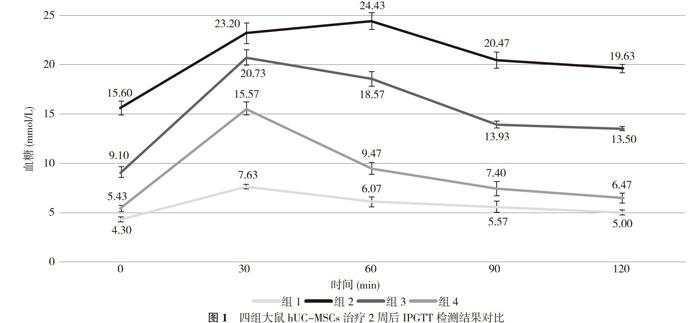
1. 3. 1 有效性指标 hUC-MSCs治疗2周后行腹腔注射葡萄糖耐受试验(intraperitoneal glucose tolerance test, IPGTT)。进行IPGTT之前, 大鼠禁食过夜至少6 h, 腹膜内注射葡萄糖(2.0 g/kg)。注射后0、30、60、90和120 min, 取鼠尾血样, 测定血糖和空腹胰岛素水平。
1. 3. 2 形态學和免疫组化 hUC-MSCs注射4周后, 过量麻醉法处死动物, 手术切除T2DM大鼠胰腺组织。4%多聚甲醛固定48 h, 常规石蜡包埋石蜡切片, 行HE染色, 并用胰岛素抗体进行胰岛免疫组化检测, 光镜下观察胰岛结构与α细胞、β细胞的分布情况。
1. 4 统计学方法 采用SPSS24.0统计学软件对数据进行处理。计量资料以均数±标准差( x-±s)表示, 采用t检验, 组间比较采用单因素方差分析+两两比较LSD法。P<0.05表示差异有统计学意义。
2 结果
2. 1 大鼠hUC-MSCs移植2周后血糖、胰岛素及β细胞功能比较 hUC-MSCs移植2周后, 四组的空腹血糖和HOMA-β水平组间两两比较差异均具有统计学意义(P<0.05);组2的空腹胰岛素(21.68±3.87)μIU/ml低于组1的(29.33±4.39)μIU/ml和组4的(28.95±2.41)μIU/ml, 差异均具有统计学意义(P<0.05);组1的HOMA-IR低于组2和组3, 组2的HOMA-IR高于组3和组4, 组3的HOMA-IR高于组4, 差异均具有统计学意义(P<0.05);组1的HOMA-IS高于组2和组3, 组2的HOMA-IS低于组3和组4, 组3的HOMA-IS低于组4, 差异均具有统计学意义(P<0.05)。hUC-MSCs移植后, 组3和组4的血糖水平均逐渐下降, 组4下降更明显, 但与组1比较差异具有统计学意义(P<0.01);IPGTT中, 组3和组4血糖高峰均在30 min出现, 与组1曲线特征相符合, 且组4更接近组1水平。见图1。
2. 2 四组大鼠hUC-MSCs移植4周后胰腺组织HE染色及胰岛抗体免疫组化染色分析 hUC-MSCs注射后4周后取大鼠胰腺组织显微镜下观察, 组1大鼠胰腺组织形态完整, 胰岛内细胞排列整齐, 细胞界限清楚, 胰岛密度正常;组2大鼠胰岛形态不规则, 细胞排列紊乱, 胰岛数目减少;而组3和组4较组2有不同程度改善, 组4组织形态较组3胰腺组织形态完整, 细胞排列整齐。见图2。使用胰岛抗体免疫组化染色后可见, 组2大鼠胰岛细胞非常少, 组4组织形态较组3胰岛内胰岛细胞数目增多, 但较组1大鼠胰岛细胞少。见图3。
3 讨论
目前虽然已有关于hUC-MSCs有效性的相关研究[2-4], 但是移植剂量没有统一的标准。为探寻β细胞修复程度与移植剂量的关系, 本实验设置了高低剂量组。关于输注途径, 对于动物实验而言, 动脉输注、胰腺包膜下注射和肾包膜下注射均需要手术, 创伤大, 会增加感染及死亡风险, 静脉输注相比腹腔注射途径治疗效果更优, 但操作难度相对较大, 虽然有实验[5, 6]显示腹腔注射途径效果不明显, 但是不排除与选择的干细胞种类、输注剂量和输注的模式有关, 故本实验拟采用腹腔注射途径, 去验证该途径的有效性。
hUC-MSCs治疗2周后, 接受干细胞治疗的大鼠血糖均较未接受干细胞治疗的T2DM大鼠下降明显, 且高剂量组的大鼠血糖下降更明显, 该结果提示干细胞治疗可以降血糖, 这与其他实验结果相符合[2-4]。目前, 干细胞治疗糖尿病的确切机制仍不明确, 从已有的研究中, 大家普遍认为主要有三大机制:①改善胰岛β细胞功能[2, 7-9];②改善外周组织胰岛素抵抗[10];③抗炎和免疫调节作用[11, 12]。在本实验中, 治疗后大鼠HOMA-β及空腹胰岛素水平明显升高, 且对于HOMA-β而言, 高剂量的效果更佳, 结合IPGTT、细胞学和免疫组化结果, 治疗后大鼠的胰腺形态得到了改善, 且胰岛细胞数目增多, 提示干细胞治疗不仅可以改善β细胞功能, 而且可以增加β细胞的数量, 这与其他实验相符合[7-9]。
但β细胞数目的增加是由于胰岛细胞的相互转化[13], 还是自身的再生[7-9], 亦或是凋亡的减少[2], 本实验尚不能说明。干细胞治疗后, HOMA-IS和HOMA-IR改善明显, 结合IPGTT结果, 治疗后的大鼠血糖曲线特征已符合正常大鼠的曲线特征, 且高剂量组更贴近正常组, 提示外周组织对胰岛素的反应性升高, 干细胞改善了外周胰岛素抵抗, 这与其他实验[2]相符合。可能与胰岛素靶组织中胰岛素受体底物-1, 也可能与改善体内代谢应激和全身慢性反应有关[14]。
综上所述, 经腹腔途径注射hUC-MSCs可以有效的治疗T2DM大鼠, 该途径有效;且高剂量的干细胞治疗, 可以更好的修复胰腺组织, 改善β细胞功能, 增加β细胞数目, 改善胰岛素抵抗。
参考文献
[1] Cho N H, Shaw J E, Karuranga S, et al. IDF Diabetes Atlas: Global estimates of diabetes prevalence for 2017 and projections for 2045. Diabetes Research & Clinical Practice, 2018(138):271.
[2] Xie Z, Hao H, Tong C, et al. Human umbilical cord-derived mesenchymal stem cells elicit macrophages into an anti-inflammatory phenotype to alleviate insulin resistance in type 2 diabetic rats. Stem Cells, 2016, 34(3):627-639.
[3] 阮光萍, 刘菊芬, 李自安, 等. 人脐带间充质干细胞对猕猴糖尿病治疗效果的研究. 中华细胞与干细胞杂志(电子版), 2017, 7(3):131.
[4] 董松, 王鸿, 雷蕾, 等. 人脐带间充质干细胞经动脉介入治疗2型糖尿病Beagle犬的作用. 中国医药导报, 2017, 14(22):16-20.
[5] 丁海霞, 王富军, 黄晓伟, 等. 不同途径移植脐带血干细胞对兔2型糖尿病的疗效观察. 中华细胞与干细胞杂志(电子版), 2016, 6(6):334-338.
[6] 徐谷根, 肖毅, 尹卓娜, 等. 不同部位脐血干细胞移植治疗糖尿病大鼠的效果研究. 中华临床医师杂志(电子版), 2015, 9(13):
134-136.
[7] Bell GI, Meschino MT, Hughes-Large JM, et al. Combinatorial human progenitor cell transplantation optimizes islet regeneration through secretion of paracrine factors. Stem Cells & Development, 2012, 21(11):1863.
[8] Cheng H, Zhang YC, Wolfe S, et al. Combinatorial treatment of bone marrow stem cells and stromal cell-derived factor 1 improves glycemia and insulin production in diabetic mice. Molecular & Cellular Endocrinology, 2011, 345(1-2):88-96.
[9] Karnieli O, Izhar-Prato Y, Bulvik S, et al. Generation of insulin-producing cells from human bone marrow mesenchymal stem cells by genetic manipulation. Stem Cells, 2010, 25(11):2837-2844.
[10] Si Y, Zhao Y, Hao H, et al. Infusion of Mesenchymal Stem Cells Ameliorates Hyperglycemia in Type 2 Diabetic Rats. Diabetes, 2012, 61(6):1616.
[11] Liu X, Zheng P, Wang X, et al. A preliminary evaluation of efficacy and safety of Whartons jelly mesenchymal stem cell transplantation in patients with type 2 diabetes mellitus. Stem Cell Research & Therapy, 2014, 5(2):57.
[12] Zhao Y, Jiang Z, Zhao T, et al. Targeting insulin resistance in type 2 diabetes via immune modulation of cord blood-derived multipotent stem cells (CB-SCs) in stem cell educator therapy: phase Ⅰ/Ⅱclinical trial. Bmc Medicine, 2013, 11(1):160.
[13] 申晶. 骨髓源間充质干细胞输注促进STZ诱导的糖尿病大鼠胰腺内α细胞向β细胞的转变:糖尿病治疗的新模式. 中国人民解放军医学院, 2013.
[14] Liu X, Zheng P, Wang X, et al. A preliminary evaluation of efficacy and safety of Whartons jelly mesenchymal stem cell transplantation in patients with type 2 diabetes mellitus. Stem Cell Research & Therapy, 2014, 5(2):57.
猜你喜欢
健康之家(2021年6期)2021-09-08
旗帜文摘(2020年5期)2020-06-09
人人健康(2020年4期)2020-05-25
科学大观园(2020年7期)2020-04-08
健康必读·下旬刊(2020年2期)2020-03-19
恋爱婚姻家庭·养生版(2016年11期)2016-11-03
延边医学(2015年27期)2015-10-21
中国民族民间医药·下半月(2011年10期)2011-12-27
中国现代医生(2009年13期)2009-06-03
中国实用医药(2009年8期)2009-05-06