43个萌出前牙冠内吸收发病情况调查及对策
2015-11-21 11:13赵东方张媛媛郭青玉
牙体牙髓牙周病学杂志 2015年10期
赵东方, 张媛媛, 郭青玉
(西安交通大学口腔医院儿童口腔科, 陕西 西安 710004)
·治疗与应用·
43个萌出前牙冠内吸收发病情况调查及对策
赵东方, 张媛媛, 郭青玉
(西安交通大学口腔医院儿童口腔科, 陕西 西安 710004)
目的: 探讨萌出前牙冠内吸收的发病特点及治疗方法。方法:检索Pubmed数据库关于萌出前牙冠内吸收病例报告及流行病学方面的文献,寻找其发病特点及治疗对策。结果:检索到萌出前牙冠内吸收病例报告34篇(1978~2013),共43个病变牙,流行病学调查6篇;该病发生于6~20岁,好发于替牙列阶段,发病率1.55%~27.3%,无种族及性别差异,通常在拍摄曲面断层片时偶然发现,也有个别患者出现疼痛症状。43个患牙中第二磨牙44.19%,第一磨牙18.60%,第二前磨牙和尖牙均为13.95%;其中86.05%发生在下颌,单发或多发。治疗方法以临床观察为主,治疗率高于拔除率。结论:在敏感年龄及发病因素存在的情况下常规拍摄全口曲面断层片进行筛查,早发现,早治疗。
萌出前; 牙冠内吸收; 牙冠内缺损; 萌出前龋
[DOI] 10.15956/j.cnki.chin.j.conserv.dent.2015.10.010
[Chinese Journal of Conservative Dentistry,2015,25(10): 618]
萌出前牙冠内吸收也称萌出前牙冠内缺损,是在牙齿萌出前发生在牙冠内的一种缺陷,X线片表现为牙冠内临近釉牙本质界向牙本质内不同程度破坏的透射影像,边界清楚,透影区与髓腔之间通常有一薄层牙本质分开。该病最早在1941年由Skillen发现并报道[1],发病罕见,因发现不及时而容易造成牙冠完全缺损,甚至累及牙髓,导致牙根不能继续发育[2]。该病病因尚不清楚,推测有邻牙易位、萌出过程中受到挤压、先行乳牙有慢性根尖周炎、龋病、牙本质发育不全、釉基质未矿化和吸收、原发性外吸收等[3-5]。目前,国内外研究样本数量有限、资料孤立,缺少完整的发病情况及诊治流程,本调查通过分析国外已经公开发表的文献,探讨其发病特点及治疗方法。
1 临床资料和方法
1.1 临床资料
以 preeruptive(或pre eruptive) intracoronal radiolucency(或defect,或lesions,或resorption)为关键词在pubmed数据库中进行检索,纳入标准:①文题中标有以上关键词的个案报道(case report);②文题中标有以上关键词的流行病学调查。
1.2 方法
使用Microsoft Office Excel软件对病例报告、流行病学调查等文献中萌出前牙冠内吸收的发病情况、治疗方案进行分析、整理。
2 结果
以preeruptive(pre eruptive) intracoronal radiolucency,preeruptive(pre eruptive) intracoronal defect,preeruptive(pre eruptive) intracoronal lesion,preeruptive(pre eruptive) intracoronal Caries,preeruptive(pre eruptive) intracoronal resorption为关键词在pubmed数据库中分别检索出病例报告2(1)篇,2(6)篇,2(5)篇,4(0)篇,4(8)篇,排除重复和无关文献,共计符合要求文献34篇,患牙43个; 流行病学调查文献6篇。
2.1 流行病学情况[6-11]
该病好发年龄为6~20岁,人发病率为1.55%~27.30%,牙发病率为0.50%~2.10%,患病在种族和性别之间无统计学差异。该病好发于混合牙列,其次为乳牙列, 恒牙列最低,以下颌单发多见,偶尔有2个或多个牙同时发生。牙冠内缺损多见于牙冠内牙本质层的近中及牙冠中部,临近釉牙本质界,与牙髓之间有薄层的牙本质分界,50%的缺损小于牙本质厚度的1/3,釉质基本正常(表1)。
43个患牙发生牙位及颌位情况见表2。发病率由高至低:第二磨牙>第一磨牙>尖牙及第二前磨牙,43个患牙中86.05%发生在下颌。

表1 萌出前牙冠内吸收的流行病学调查
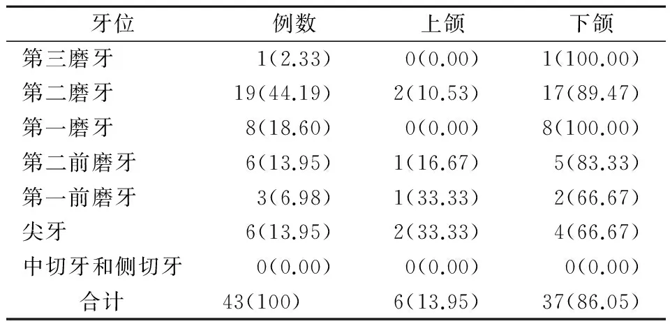
表2 43个萌出前牙冠内吸收牙位及颌位发生情况(n,%)
2.2 症状[12-20]
该病一般无自觉症状,通常在X线检查时偶然发现。本调查结果发现,2例有间断性钝痛, 1例有继发性疼痛, 1例发生软组织肿胀, 1例发生不可复性牙髓炎,其余病例未描述是否有症状,或未提及。
2.3 治疗方法[12-20]
43例萌出前牙冠内吸收中有12例(27.91%)因正畸治疗需要拔除,其中前磨牙1例,第一磨牙1例,第二磨牙10例;有23例(53.49%)采取手术暴露牙冠或等待患牙萌出后进行充填治疗,其中1例采取萌出后预成冠修复;有8例(18.60%)未提及治疗方法。
3 讨论
根据文献该病的命名有5种:萌出前牙冠内吸收(pre eruptive intracoronal resorption)[1]、萌出前牙冠内透射影像(pre eruptive intracoronal radiolucency)[6]、萌出前牙冠内缺损(pre eruptive intracoronal defect)[17]、萌出前牙冠内腐蚀(pre eruptive intracoronal lesion)[21]、萌出前龋(pre eruptive intracoronal caries)[22]。以pre eruption intracoronal resorption为关键词进行文章发表的最多,有8篇,占40%,其中萌出前“龋”受到质疑,原因在于该病发生在牙齿萌出前(牙齿表面有牙槽骨或牙龈覆盖),牙冠尚未暴露在口腔中,因此牙冠的缺损或吸收与龋病的病因不符。
不同学者对该病的流行病学情况调查结果不同,分析原因在于调查对象人数和年龄范围不一致,该病为萌出前冠内吸收,牙齿萌出的时间主要在替牙列阶段,如果调查年龄范围跨度大,发病率必然较低,而在好发年龄段内(5~20岁)调查发病率相对较高是合理的。
本调查结果显示,43个患牙治疗率高于拔除率,提示,对于萌出前牙冠内吸收的病例,根据牙位及钙化阶段预测其萌出时间,如不涉及正畸关闭间隙或牙冠缺损过大而需要拔牙的情况,建议进行密切观察,待患牙萌出后及时进行间接盖髓充填治疗或预成冠修复,如果患牙临近萌出或边缘嵴已经暴露,应及时切除冠方部分牙龈组织,暴露牙冠进行治疗。
萌出前牙冠内吸收在临床中应引起足够的重视,尤其是正畸科医生,在临床工作中很可能最先接触到这类患者,并建议儿童牙科医生在替牙列阶段常规进行全口曲面断层摄影,注意好发牙及好发部位,及时发现该病并给予适当的治疗。
[1]葛立宏.儿童口腔医学[M]. 4版 . 北京:人民卫生出版社, 2012:87.
[2]Bille MLB, Kvetny MJ, Kjr I. A possible association between early apical resorption of primary teeth and ectodermal characteristics of the permanent dentition[J].EurJOrthod, 2008, 30(4):346-351.
[3]Bhatt N, Holroyd I. Generalized idiopathic root resorption: a case report[J].IntJPaedDent, 2008, 18(2):146-153.
[4]Weltman B, Vig KW, Fields HW,etal. Root resorption associated with orthodontic tooth movement: a systematic review[J].AmJOrthodDentofacialOrthop, 2010, 137(4):462-476.
[5]Rankow H, Croll TP, Miller AS. Preeruptive idiopathic coronal resorption of permanent teeth in children[J].JEndod, 1986, 12(1):36-39.
[6]Seow WK, Lu PC, McAllan LH. Prevalence of pre- eruptive intracoronal dentin defects from panoramic radiographs[J].PediatrDent, 1999, 21(6):332-339.
[7]Seow WK, Wan A, McAllan LH. The prevalence of pre- eruptive dentin radiolucencies in the permanent dentition[J].PediatrDent, 1999, 21(1):26-33.
[8]Wang Y, Chen J, Liu H. Prevalence of preeruptive intracoronal radiolucency in Chinese children from panoramic radiographs[J].ChinJDentRes, 2013,16(2):153-156.
[9]Al- Batayneh OB, AlJamal GA, AlTawashi EK. Pre- eruptive intracoronal dentine radiolucencies in the permanent dentition of Jordanian children[J].EurArchPaediatrDent, 2014,15(4) :229-236.
[10]O¨zden B, Acikgoz A. Prevalence and characteristics of intracoronal resorption in unerupted teeth in the permanent dentition: retrospective study [J].OralRadiol, 2009, 25(1):6-13.
[11]Nik NN, Abul Rahman R. Pre- eruptive intracoronal dentin defects of permanent teeth[J].JClinPediatrDent, 2003, 27(4):371-375.
[12]Blackwood HJJ. Resorption of enamel and dentine in the unerupted tooth[J].OralSurgOralMedOralPathol, 1958, 11(1):79-85.
[13]Skaff DM, Dilzell WW. Lesions resembling caries in unerupted teeth[J].OralSurgOralMedOralPathol, 1978,45(4):643-646.
[14]Kalamchi S. External resorption of an unerupted third molar[J].OralSurgOralMedOralPathol, 1983,56(3):338.
[15]McNamara CM, Foley T, O′Sullivan VR,etal. External resorption presenting as an intracoronal radiolucent lesion in a pre~eruptive tooth[J].OralDis, 1997, 3(3):199-201.
[16]Davidovich E, Kreiner B, Peretz B. Treatment of severe pre- eruptive intracoronal resorption of a permanent second molar[J].PediatrDent, 2005, 27(1):74-77.
[17]Klambani M, Lussi A,Ruf S. Radiolucent lesion of an unerupted mandibular molar[J].AmJOrthodDentofacialOrthop, 2005, 127(1):67-71.
[18]Hata H, Abe M, Mayanagi H. Multiple lesions of intracoronal resorption of permanent teeth in the developing dentition: a case report[J].PediatrDent, 2007, 29(5):420-425.
[20]Kakade A,Juneja A. An unusual association of extraoral sinus tract with unerupted permanent tooth[J].PediatrDent, 2013 ,35(3):284-287.
[21]O′Neal KM, Gound TG, Cohen DM. Preeruptive idiopathic coronal resorption: a case report[J].JEndod,1997,23(1):58-59.
[22]Moskovitz M, Holan G. Pre- eruptive intracoronal radiolucent defect: a case of a nonprogressive lesion[J].JDentChild,2004,71(2):175-178.
Pre-eruptive intracoronal absorption: a literature review of 43 cases
ZHAO Dong- fang, ZHANG Yuan- yuan, GUO Qing- yu
(DepartmentofPediatricDentistry,StomatologicalHospitalofXi'anJiaotongUniversity,Xi′an710004,China)
AIM: To investigate the characteristics and treatment methods of pre- eruptive intracoronal absorption. METHODS: A literature research of case reports and epidemiological investigations about pre- eruptive intracoronal absorption was done in Pubmed database, using the key words of preeruptive(pre eruptive) intracoronal radiolucency,preeruptive(pre eruptive) intracoronal defect,preeruptive(pre eruptive) intracoronal lesion,preeruptive(pre eruptive) intracoronal caries,and preeruptive(pre eruptive) intracoronal resorption. RESULTS: A total of 34 case reports (1978~2013) and 6 articles of epidemiological investigation were found. The cases were usually found during routine radiographic examinations of children at the age of 6~20 years old with otherwise good oral health. Few cases of toothache had been reported. In the 43 cases, 44.19% were found in the second permanent molar, followed by the first permanent molar (18.60%) and the second permanent molar and canine (13.95%), mostly in the lower jaw (86.05%). Most of the cases were clinically treated. CONCLUSION: In the mixed dentition stage, panoramic radiograph should be taken to screen pr- eruptive intracoronal absorption. Early detection and early treatment is important.
pre- eruption; intracoronal resorption; intracoronal defect; pre- eruption caries
2015-01-25;
2015-07-16
赵东方(1973-),女,汉族,吉林人。硕士,副主任医师
赵东方, E-mail: zjz158297@163.com
R781.3
B
1005-2593(2015)10-0618-03
猜你喜欢
中国典型病例大全(2022年11期)2022-05-13
北京大学学报(医学版)(2022年2期)2022-04-11
当代医药论丛(2021年22期)2021-11-29
中国典型病例大全(2021年5期)2021-06-20
锦州医科大学报(2021年12期)2021-03-19
现代口腔医学杂志(2021年1期)2021-03-12
中国保健营养(2019年7期)2019-10-21
华西口腔医学杂志(2013年5期)2013-11-11
华西口腔医学杂志(2013年1期)2013-05-10