清心Ⅱ号对病毒性心肌炎心肌纤维化内皮素1及转化生长因子β1表达的干预作用
2016-10-19 20:47:57刘旺王娟徐百鸿程志清
中国医药导报
2016年6期
刘旺 王娟 徐百鸿 程志清
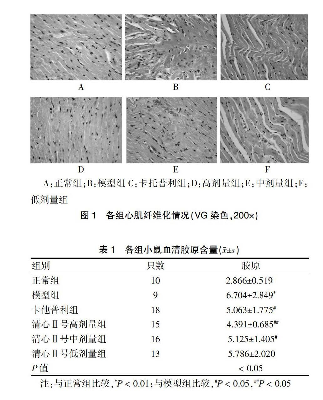
[摘要] 目的 研究清心Ⅱ号对病毒性心肌炎心肌纤维化内皮素1(ET-1)及转化生长因子β1(TGF-β1)表达的干预及抗病毒作用。 方法 150只清洁级体重14~16 g健康雄性BALB/c小鼠,采用随机数字表法分出10只作为正常组;其余140只作为造模组,采用腹腔注射含不同浓度柯萨奇病毒B3(CVB3)半数组织培养感染剂量为10-5的病毒液3次,每次每只2 mL,首次1∶2000浓度CVB3病毒液,2周后腹腔接种1∶1600浓度CVB3病毒液,4周后腹腔注射1∶800浓度CVB3病毒液。60 d后模型构建成功,存活小鼠94只。随机选取90只,分为清心Ⅱ號高、中、低剂量组各20只,卡托普利组20只,模型组10只。正常组和模型组给予生理盐水灌胃,治疗组分别给予卡托普利和清心Ⅱ号高、中、低剂量灌胃。实验结束后颈椎脱臼法处死动物,采集心脏组织液氮速冻。分别应用免疫组化方法检测TGF-β1含量,放免法检测各组血浆ET-1含量,RT-PCR技术检测CVB3 RNA,VG染色观察各组纤维化形成情况,并检测胶原阳性面积占测量面积的百分比,即容积分数。 结果 VG染色后正常组含少量胶原纤维,分布于血管周围;模型组心肌细胞排列紊乱,胶原纤维含量与正常组比较增生明显(P < 0.05);TGF-β1、ET-1、CVB3 RNA模型组及各治疗组均高于正常组(P < 0.05);治疗后各治疗组胶原纤维较模型组减少(P < 0.05),清心Ⅱ号各治疗组随剂量增加胶原纤维逐渐减少;各治疗组TGF-β1、ET-1、CVB3 RNA与模型组比较均相应减低(P < 0.05),清心Ⅱ号高剂量组TGF-β1与正常组比较,差异无统计学意义(P > 0.05);对减少ET-1,清心Ⅱ号高剂量组与卡托普利组疗效相当(P > 0.05)。 结论 血浆ET-1与TGF-β1在病毒性心肌炎心肌纤维化形成过程中发挥重要作用,清心Ⅱ号具有抗病毒、抗心肌纤维化作用。
[关键词] 内皮素1;转化生长因子β1;病毒基因检测;VG染色;心肌纤维化
[中图分类号] R285 [文献标识码] A [文章编号] 1673-7210(2016)02(c)-0009-05
Intervention effect of qinxin Ⅱ recipe on expression of ET-1 and TGF-β1 in mouse of viral myocarditis myocardial fibrosis
LIU Wang1 WANG Juan2 XU Baihong3 CHENG Zhiqing4
1.Department of General, Tumor Hospital of Hangzhou, Zhejiang Province, Hangzhou 310003, China; 2.Department of Infection, Children's Hospital of Hangzhou, Zhejiang Province, Hangzhou 310014, China; 3.Department of Cardiology, Puyang Hospital of Traditional Chinese Medicine, He′nan Province, Puyang 457001, China; 4.College of Basic Medicine, Zhejiang University of Traditional Chinese Medicine, Zhejiang Province, Hangzhou 310053, China
[Abstract] Objective To study intervention effect of qinxin Ⅱ recipe on expression of endothelin-1 (ET-1) and transforming growth factor-β1 (TGF-β1) in mouse of viral myocarditis myocardial fibrosis and ant-virus effect. Methods The 150 health male BALB/c mice (weight: 14-16 g) were randomly allotted into two groups: the normal group (n=10) and the model constructed group (n=140). The mice of the model constructed group were injected coxsackie virus B3 (CVB3) of median tissue culture infectious dose 10-5 poison by intraperitoneal. 2 mL CVB3 poison was injected by intraperitoneal for each mouse, repeating three times. The injected concentration and time were as following: 1:2000 of CVB3 at beginning, 1:1600 of CVB3 after two weeks and 1:800 of CVB3 4 weeks later. The model was constructed successfully after 60 days and 94 mice were live. 90 mice were selected randomly from 94 mice for following three groups: model group (n=10) and captopril group (n=20) and qinxin Ⅱ (high, medium and low dose) groups (20 mice in each group). The normal group and model group were fed normal saline by gavage, treatment groups were fed captopril and qinxin Ⅱ groups (high, medium and low dose), respectively. The mice were killed by cervical dislocation after experiment, heart tissue was collected and snap-frozen in liquid nitrogen. TGF-β1 and ET-1 contents in heart were detected using immunohistochemical method and radioimmunoassay, respectively; CVB3 RNA was assayed by RT-PCR; Fibrosis formation was observed by VG staining and percentage of positive area (volume fraction) of collagen was also detected. Results The results showed that collagen fiber in heart of mice in the normal group was detected by VG staining, distributing around the blood vessels; arrangement of myocardial cell in heart disorder in model group and collagen fiber in heart of mice in the model group was significant hyperplasia compared with those of normal group (P < 0.05). TGF-β1, ET-1 and CVB3 RNA in heart of each treatment group and model group were all higher than those of the normal group (P < 0.05). After treatment, the collagen fiber in heart of all treatment groups was lower than model group (P < 0.05), and with increasing dose of qingxin Ⅱ, the collagen fiber in heart of treatment group was decreased gradually. TGF-β1, ET-1 and CVB3 RNA in heart of treatment group cured by qingxin Ⅱ recipe were reduced compared to model group. TGF-β1 in heart of high qingxin Ⅱ recipe almost was close to normal group, there was no statistically significant difference (P > 0.05). For reducing ET-1, curative effect of high qingxin Ⅱ recipe group was almost same to captopril group (P > 0.05). Conclusion ET-1 and TGF-β1 of plasma play an important role in process of viral myocarditis myocardial fibrosis, and qingxin Ⅱ may have a role in anti-virus and anti-myocardial fibrosis.
[Key words] ET-1; TGF-β1; Virus gene detection; VG staining; Myocardial fibrosis
病毒性心肌炎是临床常见病、多发病。近年来发病率有增高趋势,很多患者由于症状不典型未引起重视,且由于本病为病毒性疾病,西医目前无特效抗病毒药物,因而无特效疗法,所以导致患者长期得不到有效治疗,经久不愈,给患者的生活以及精神带来极大的痛苦。病毒……
登录APP查看全文
猜你喜欢
童话王国·奇妙逻辑推理(2024年5期)2024-06-19 16:03:38
课堂内外·初中版(科学少年)(2023年10期)2023-12-10 00:43:06
全科护理(2022年10期)2022-12-26 21:19:15
中国合理用药探索(2022年1期)2022-11-26 00:22:32
乡村科技(2021年33期)2021-03-16 02:26:54
国际放射医学核医学杂志(2021年10期)2021-02-28 08:41:58
数学物理学报(2020年2期)2020-06-02 11:29:24
光学精密工程(2016年6期)2016-11-07 09:07:19
核科学与工程(2015年4期)2015-09-26 11:59:03
中国合理用药探索(2014年11期)2014-03-11 20:30:20