Apoptosis of Pancreatic Beta Cells in Pregnant Insulin-resistant Rats Fed with High-fat Diet
2019-10-29 07:06LiliZHANGYajuanHUANGShengGE
国际感染病学(电子版) 2019年2期
Lili ZHANG, Yajuan HUANG, Sheng GE
1.Department of Obstetrics and Gynecology, North District of Suzhou State Hospital Affiliated to Nanjing Medical University, Suzhou Jiangsu 215000, China; 2.Department of Obstetrics and Gynecology, Sixth People's Hospital Affiliated to Shanghai Jiaotong University, Shanghai 200233, China; 3.Department of Nutrition, Sixth People's Hospital Affiliated to Shanghai Jiaotong University, Shanghai 200233, China
ABSTRACT:Objective The aim of this study is by observing the number change of islets beta cells in gestational rats exposed to high fat diet, tofurther reveal the mechanism of gestational diabetes mellitus. Methods Female Wistar rats were exposed to high fat diet for five weeks, and then became pregnant.During pregnancy dynamically detected indicators of glucose and fat.Until the third trimester of pregnancy evaluated the sensitivity of insulin and glucose tolerance.After executed rats, selected pancreatic tail tissue and fixed, further slides were stained with insulin antibody by immunohistochemistry to confirm the location of beta cells.Image analysis system determined mean area stained positive cells in each islet, which stood for total number of beta cells.The apoptotic beta cells in islet were detected and quantified by the Tunel technology to calculate apoptosis ratio. Results The level of free fatty acids in rats exposed to high fat diet was significantly higher than the control groups, and insulin resistance was more serious.Compared mean stained positive area among each group, the largest was gestational rats fed high fat diet, and gestational rats was larger than virgin rats, but the difference had no statistical significance.About apoptoticratio of beta cells was higher in diet intervened rats, gestational rats were higher than virgin rats.The same trend happened in the number of positive cells, but discrepancy was not remarkable. Conclusion Based on insulin resistance, apoptosis of pancreatic beta cellsincreased in gestational ratstaking high fat diet, through changing the number of beta cells to down regulate the pancreas endocrine function.That may be the mechanism of gestational mellitus.
KEY WORDS:Insulin resistance; Lipotoxicity; βcells; Apoptosis; Disease model, animal; Rats, wistar; High fat diet; Diabetes mellitus, gestational
1 Introduction
Gestational diabetes mellitus (GDM) is a special complication during pregnancy, but the mechanism has not always been very clear.Plenty of researches have convinced that insulin resistance is pathophysiology foundation.Pancreas as an endocrine organ, its function is bounds with the number of beta cell closely.Based on previous experience of successfully induced insulin resistance rat model[1], further by immunohisto chemistry to study apoptosis and the number change about beta cells, therefore we may obtain precious conference about the mechanism of GDM.
2 Materials and Methods
2.1 Experimental animals and main reagents Healthy nine weeks years old femaleWistar rats 40, weight 160-180 gram [from Chinese Academy of Sciences experimental animal center, license number: SCXK(shanghai) 2002-0010].
Blood glucose meter and test papers form Roche company, Free fatty acids reagent form Nanjing JianCheng company, Shanghai biological company productstriglycerides (TG) and cholesterol (TC)reagents, insulinfrom Novo Nordisk in Denmark.Terminal deoxynucleotidyl transferase, Biotinylated digoxin antibody, DAB chromogenic reagent,ProteinaseKall bought from DAKO company.
2.2 Method Once adopting environment, female Wistar rats were divided into normal diet group and high fat diet group, each group had 20 number separately.After fed according dietfive weeks, and then made them pregnancy, every group again divided into two subgroup, thus high fat diet pregnant rats (HP) and high fat diet virgin rats (HV), normal diet pregnant rats(NP) and normal diet virgin rats (NV).Dynamically detected indicators of FFA, T, TG and TC, when the day of seventhand the day of twentieth pregnancy,until 18-20 day we evaluatedglucose tolerance and insulin sensitivity test.After executed rats, selected randomLy pancreatic tail tissue and fixed from each group, then paraffin embedded tissue slice thickness about 5-6 μm, further slides were stained with insulin antibody to confirm the location of beta cells, marked two resistance, used DAB-H2O2to make clear.we applied image analysis system, selected 3-5 islets in every section which stained clearly, each group was selected 5 pathological sections at least.By determined area which stained brown in each islet to stand for the number of beta cells and pancreatic secretory function, at last compared mean value with groups.Tunel technology was used to detect apoptotic cells.Professional pathologist had been blind reading.
The nuclear of apoptotic cell was stained deep brown, and this positive cell defined as apoptotic cell.Ratio of apoptoticbeta cells in each islet=the number of apoptoticbeta cells/total number of beta cells×100%.
2.3 Statistical analysis Statistical analyses were done using SAS statistical software, data are expressed as mean±SEM,P<0.05 was considered statistically significant.
Depending on the experimental design, the area value about positive beta cells in islet tail after logarithmic transformation into normal data, two group were compared witht-test, analysis of variance between multiple groups, 22 comparison SNK test for multiple groups.The ratio of beta cell compared with χ2test.Repeated measurement data analysis was applied to insulin and glucose tolerance.
3 Results
3.1 Values changes of blood sugar, free fatty acid,triglycerides and cholesterol compared with normal diet group, after exposed to corresponding diet five weeks the values of rats taking high fat diet increased,and the difference was significant.Except normal diet groups during early pregnancy, other groups had difference, high fat diet group was more obvious.When the day of 20th, compared with normal diet group the blood sugar value of each group had statistically significant, but this trend was not seen in cholesterol value between normal diet groups.And the values of free fatty acid, triglycerides which had no obvious difference between normal diet and high fat diet group before pregnancy, again increased in early pregnant time, until the third trimester of pregnancy there was a remarkable discrepancy between pregnant rats and virgin rats, especially in high fat diet pregnant rats.(Table 1 and Table 2).
3.2 Insulin sensitivity Insulin resistance test results:the glucose values at 0 and 120 minutes had no obvious discrepancy, but at 60 minutesdiscrepancy was evident when compared with other three groups.When at 90 minutes, other groups all had difference, except high fat diet virgin rats compared with normal diet pregnant rats.The area under curve appeared same trend at 15 and 30 minutes, that was: there was a statistically significant about blood sugar value between normal diet pregnant rats and virgin rats, but the first value was higher than the latter, high fat diet group was much higher than normal diet group, and high fat diet pregnant was higher than high fat diet virgin rats.(Table 3).
3.3 Number of pancreatic beta cells in rats In islet cytoplasm was stained brown grain, which was beta cell.The positive brown area stands for the insulin expression.Image analysis system: the area of positive staining in every islet were: NV-1.57±0.99, NP-1.81±1.60,HV-1.77±1.34, HP-2.77±0.65 (×10-3mm2).High fat diet pregnant group was largest, pregnant rats was larger than virgin rats, but difference between groups was not remarkable (Figure 1).
3.4 Ratio of beta cell apoptosis The total number of beta cells in islet, pregnant rats was more than virgin rats, and the number of beta cell apoptosis high fat diet group was more than normal diet group, especiallypregnant rats was more than virgin rats.The same trend was found in apoptosis ratio, but ratio was not dramatic.(Table 4, Figure 2).

Table 1 The indicators of glucose and fat before pregnancy [n, (mmol/L)]

Table 2 Comparison of glucose and fat indicators during pregnancy [n, (mmol/L)]

Table 3 Comparion of glucose value at different time
4 Discussion
4.1 Insulin resistance and Gestational Diabetes mellitus During normal pregnancy insulin resistance is constantly aggravating accompaniedbygestational week increasing, reaches peak until third trimester of pregnancy.After deliver placental discharges,plenty of hormones and enzymes retreat which resist insulin, and the level of insulin resistance recovers normal quickly as no gestational state, it is so called“Physiological insulin resistance”.Although insulin resistance exists in gestational period commonly, about 3%-7%[2]pregnant women occurred GDM, therefore GDM patients have more serious “Pathological insulin resistance”.In our study, rats taking high fat diet weight increased, the level of FFA, TG and TC all obviously raised, visceral fat gathered, insulin resistant was evident.These features performmore obviously in high fat diet pregnant rats.
4.2 The function of pancreatic beta cells and GDM Many researches have convinced that “Pathological insulin resistance” is the physiological foundation of occurring GDM, but body could mobilize pancreaticreserve function by the means of increasing number and enlarging beta cell, hence increase insulin production to meet the need of body, thus keeps the blood sugar balance dynamically[3-5].When disfunction of beta cells, the burden of insulin resistance cannot be conquered, GDM happens.Contrast to NGT group,islet beta cell function index (HOMA-β) in GDM group,30minutes insulin net value added/glucose net value added (ΔI30/ΔG30) ratio after glucose burden and insulin secretion index all decreased greatly.GDM groupfirst-phase and second-phase insulin secretion index declined when compared with NGT, gradually decreased along with condition aggravating[6], which give a notion that insulin resistance of GDM women evidently heavier, the early-phase secretion function damaged, and had a relation with the degree of illness state.Cianni G and so on who had a study on GDM and GIGT patients found that two group’ s insulin sensitivity declined and HOMA-βcell index of GDM was equal to GIGT but lower than NGT.From NGT-GIGT-GDM, pancreatic beta cell function progressive declined accompanied by insulin resistance aggravating[7,8].Xiang and so on evaluated according to first-phase insulin reaction to vein glucose tolerance test and early-phase insulin reaction to oral glucose tolerance test, that in women having GDM history compensation function of beta cell reaction to insulin resistance had declined 67%, and beta cell function declined about 34%-41%.Hence based on insulin resistance, the disfunction of pancreatic secretion is the real reason of occurring GDM.The declining of beta cell function become key links to GDM.

Table 4 Comparison ratio of beta cell apoptosis

Figure 1 Immunohistochemistry: observed 400 times under microscope with hematoxylin staining.P1=positive staining area of NV group, P2=positive staining area of NP group, P3=positive staining area of HV group, P4=positive staining area of HP group.
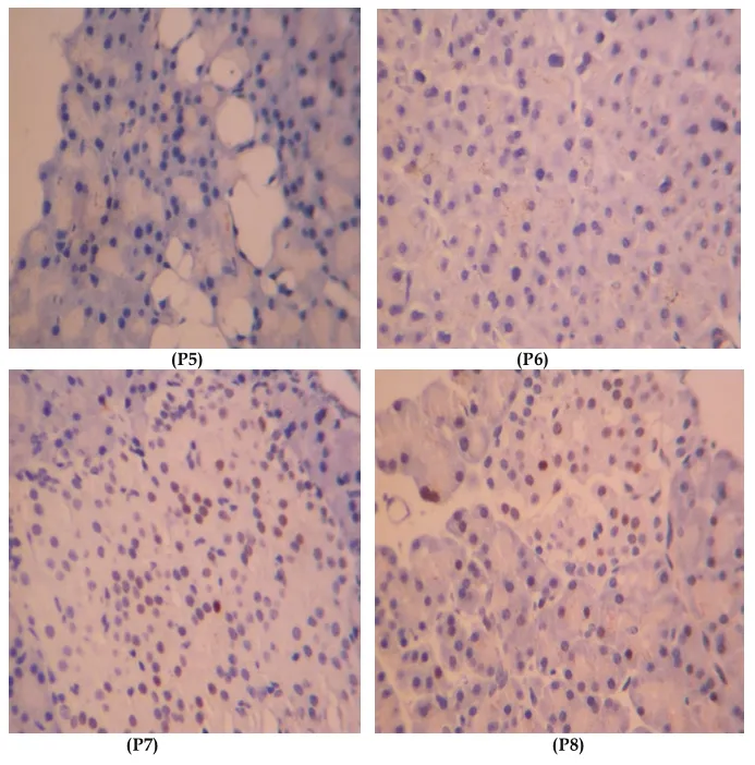
Figure 2 Tunel Method: observed apoptosis 400 times under microscope.P5=positive staining area of NV group, P6=positive staining area of NP group, P7=positive staining area of HV group, P8=positive staining area of HP group.
4.3 Beta cell function and the number of beta cell The microscopic expression of beta cell functionglucose stimulating insulin secretion, keeps a close relation with microcosmic volume and number of beta cell, Which influenced by insulin requirement,insulin resistance and pregnancy.Pick A found that in female Zucker fat diabetic rats beta cell volume obviously enlarged than the contrast.In early seventy age researches have confirmed that the pancreatic beta cell volume of delivery rats was bigger than the virgin rats, islet of gestational rats out of body could excrete more insulin.By enlarge volume is one of beta cell compensation ways, and this way is quick, instant and reversible, but enlarged cell is prone to apoptosis[9].Hypertrophy ultimately decrease number of beta cell.
The number of beta cell compensatory increases,after labor it recovers to normal level again, this regulation mainly through copy and apoptosis to realize[10,11].GDM females exist more serious pathological insulin resistance, it is supposed that the number of beta cell decreased, but lack of relative clinical researches.Butler-AE and so on found that in autopsy, obesity patients with increasing fasting bloodglucosetheir beta cell number decreased 40% compared with obesity patients without diabetes, obesity patients with diabetes decreased 63%, diabetes without obesity patients their beta cell number decreased 41%compared to patients without obesity and diabetes.Apoptotic ratio of beta cell in diabetes patients with low weight and no obesity increased 10 times and 3 times separately.And pointed that from glucose damaged to diabetes, the number of beta cell decreased because of beta cell apoptosis[12,13].Hence the function of beta cell deregulation had link with beta cell apoptosis.
Today about the possiblemechanism of beta cell apoptosis, research findingsin vivoandin vitrohave found that elevating FFA could birth lipotoxicity,through mitochondria injury, reticuloendothelial stress,oxidative stress and so on ways to start up apoptosis gene transcription, finally make beta cell apoptosis.
Based on previous experience of successfully induced insulin resistance rat, in our study, rats taking high fat diet the level of FFA, TG and TC all obviously raised, these features were more obviously during pregnancy.TG as the main source of FFA and FFA as a main role of lipotoxicity, all elevated more evidently.Rats taking high fat diet insulin sensitivity declined,insulin resistance aggravated, occurred pathological insulin resistance the same as human gestational diabetes mellitus, in a state of pre-diabetes.
By immunohistochemistry staining islet betacells, positive stained area representing of insulin expression, the area of high fat diet pregnant group was the largest, the total number of pancreatic beta cells in pregnant rats was more than virgin rats, this was in accordance with the area under insulin tolerance test curve.Further convinced that hyperinsulinemia is the symbol of insulin resistance.About apoptosis beta cells of high fat diet rats was more than virgin rats, frequency of apoptosis was similar to positive cells change, but discrepancy was not dramatically.Because of the small sample and limit sensitiveness of study way, in our study beta cell apoptosis had no statistical significance, but it provided us a tendency that high fat diet pregnant rats having serious insulin resistance beta cell apoptosis increased.Also supply a notion that based on insulin resistance beta cells might be apoptotic under the influence of FFA lipotoxicity.2 type diabetes mellitus patients without pregnancy andin vitrotests all have convinced that.
4.4 Unsolved problems In Summary, at the basis of insulin resistance, beta cell apoptosis decreases the value of beta cell, further influences the function of beta cell, therefore plays a pushing role to GDM occurrence.Researches about beta cell’s number are still according the achievements in 2 type diabetes mellitus, and on the basis of animal tests andin vitrotests outcomes,hence GDM still need to deeply explore.If beta cell decreases indeed, confirm placenta hormone plays what role in the mechanism of GDM, which seems to explain blood glucose recover to normal level after labor, but all this unknown.Study on function and value of beta cell may open a wider world to explore mechanism of GDM.Improving adaption of beta cell to pathological state, reducing insulin requirement, protecting beta cell becomes a new visual angle.