
冠状动脉CT血管成像在自发冠状动脉壁内血肿患者随访中的应用
2021-08-23 18:20:18沈海明
中国典型病例大全
2021年8期
沈海明

摘要:目的:研究冠状动脉CT血管成像在自发冠状动脉壁内血肿的应用价值。方法:选取2019年3月~2021年3月期间我院收治的自发冠状动脉壁内血肿患者21例作为观察对象,随访后采用回顾性分析方法,对所有患者的初始冠脉造影、冠状动脉CT血管成像、病变处冠脉管腔直径进行对比分析。结果:前降支患者为10例,数量最多;2型自发冠状动脉壁内血肿临床中最为常见;13例采用保守治疗方式,另外5例采用球囊扩张,3例采用支架植入术;无心血管不良事件发生;随访后,1例患者行支架植入术后一年支架内呈狭窄状态,行药物球囊治疗术。结论:冠状动脉CT血管成像是对自发冠状动脉壁内血肿较为有效的诊断方法,虽然有一定的不足之处,但仍是临床检测的首选方法。
关键词:冠状动脉CT血管成像;冠状动脉;壁内血肿
Abstract:Objective: To explore the value of coronary computed tomography (CT) angiography in the application of spontaneous intramural coronary artery hematoma. Methods: Twenty-one patients with spontaneous intramural coronary hematoma admitted to our hospital between March 2019 and March 2021 were selected as observation subjects, and after follow-up, the initial coronary angiography, coronary CT angiography, and coronary lumen diameter at the lesion of all patients were compared and analyzed by retrospective analysis.Results: 10 cases, the largest, had spontaneous intramural coronary hematoma in anterior descending branches; type 2 spontaneous intramural coronary hematoma was the most common in the clinic; 13 cases were treated by conservative treatment, another 5 by balloon dilation and 3 by stenting; no adverse cardiovascular events occurred. After follow-up, 1 case had in-stent restenosis one year after stenting and underwent drug balloon therapy.Conclusion: Coronary CT angiography is a more effective diagnostic method for spontaneous intramural coronary hematoma. Despite certain shortcomings, it is still the preferred method for clinical detection.
Keywords: Coronary CT angiography; coronary artery; intramural hematoma
【中图分类号】R543 【文献标识码】A 【文章编号】1673-9026(2021)08-102-01
自发冠状动脉壁内血肿(spontaneous coronary artery hematoma,SCAH)是一种特殊类型的自发性冠状动脉夹层(spontaneous coronary artery dissection,SCAD),主要是由血液在冠状动脉血管壁中层或中层与外弹力膜间集聚膨大形成,近年来在临床检测中较为常见[1]。冠状动脉CT血管成像是当前临床较为先进的冠心病诊断、随访手段,受到了广大医护工作人员和患者的认可,但应用于SCAH患者的诊断、随访中应用效果尚不明确。本文选取了21例SCAH患者作为观察对象,分析冠状动脉CT血管成像的实际应用价值。现报道如下。
1资料与方法
1.1一般资料
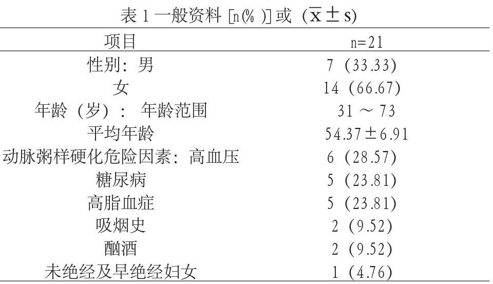
选取2019年3月~2021年3月期间我院收治的SCAH患者21例作为观察对象,所有患者均接受冠状动脉CT血管成像,并术后进行随访,对所有患者病例资料进行回顾性分析。……
登录APP查看全文
