Rutaecarpine ameliorates pressure overload-induced left cardiac structure changes and function abnormality in rats
2018-05-24 01:24,,,,,,
遵义医科大学学报 2018年2期
, , , , , ,
(Key Laboratory of Basic Pharmacology of Ministry of Education and Joint International Research Laboratory of Ethnomedicine of Ministry of Education,Zunyi Medical University,Zunyi Guizhou 563099,China)
Cardiovascular disease is one of the three major non-epidemic diseases in the world which greatly impairs the quality of life of the patients.Myocardial hypertrophy is the most common pathological changes associated with cardiovascular diseases.chemical stimulation,stress stimulation,or other causes could lead to myocardial hypertrophy,then the heart develops the adaptive structural changes,which eventually leads to malignant arrhythmia,heart failure,and even sudden death[1-4].Cardiac hypertrophy mainly occurs in the case of long-term stress overload and the common causes are mainly from high blood pressure.At the early stage of these diseases,the heart is able to maintain normal cardiac function and normal blood circulation through its own compensatory mechanism.With the progression and the aggravation of the disease,the hypertrophic myocardium increases oxygen demand and decreases the myocardial contractility,ultimately resulting in the irreversible myocardial hypertrophy[5-6].
Rutaecarpine is one of the main active alkaloids of Evodia rutaecarpa(Wu-Chu-Yu)with a variety of biological activities[7-8].Wu-Chu-Yu is a well-known herbal drug used for hypertension.Rutaecarpine and evodiamine are main bioactive components of the herb in the treatment of cardiovascular diseases[9].Its pharmacological actions involve in the cardiovascular,digestive and nervous systems,and rutaecarpine shows anti-inflammatory,blood pressure lowering,anti-thrombotic and cardiovascular protective effects[10].Our Laboratory has found that rutaecarpine is effective against AngII-induced vascular smooth muscle cell proliferation[11],and protects against ACC-induced cardiotrophy[12].Rutaecarpine is relatively less toxic[13],but could affect some drug processing genes to produce potential herb-drug interactions[14].
Ultrasound is novel technology and has been widely applied in clinic because of its unique dynamic monitoring and noninvasive characteristics.The high resolution ultrasound imaging system(HRUIS)can reflect the situation of rat cardiac hypertrophy.However,little is known about ACC-induced cardiac structure and functional changes,and the protective effects of rutaecarpine.This gap should be filled.
In this study,we established ACC-induced myocardial hypertrophy rat model and investigated the effect of rutaecarpine on left cardiac structure and function using HRUIS,together with physiology(blood pressure),pathology(H&E staining),and molecular biology(ANF mRNA expression).
1 Materials and methods
1.1 Experimental materials
1.1.1 Drugs and reagents Rutaecarpine ≥98% was purchased from Nanjing Guangrun Biological Products Co.Ltd.(batch number:GR-133-160402).Rutaecarpine was dissolved in double distilled water and 1%Tween20.Trizol and reverse transcription kit were purchased from Dalian Bao Biological Company.SYBR-Green Supermix was purchased from Bio-Rad.The PCR primers were synthesized and purified by Shanghai Bioengineering Co.,Ltd.,other reagents were of domestic analysis and pure.
1.1.2 Animals Male SD rats(Specific pathogen Free,280±20 g)were purchased from Da-ping Experimental Animal Center of the Third Military Medical University(Certificate # SCXK 2012-0005).Rats were housed in standard cages with controlled temperature(22 ± 2℃)and a 12/12 h light/dark cycle and received standard diet and water ad libitum.
1.1.3 Main instruments and equipment Kent Scientific CODA Eight channel noninvasive sphygmomanometer(Kent Scientific,USA)and High resolution ultrasound imaging system(HRUIS,Visualsonics,Canada)were detected for rats.
1.2 Experimental methods
1.2.1 Model of myocardial hypertrophy SD rats were acclimated for 1-week in a temperature and humidity controlled facility with a standard 12-h light schedule and the body weights were recorded.After intraperitoneal injection of 7% chloral hydrate(0.5 mL/100 g),an incision was made under the sternum incision long the linea alba(about 1.8 cm),opened the abdomen,the left kidney was found adjacency right inferior vena cava,isolated from the abdominal aorta from pulsating below the branch.The abdominal aorta was ligated with 0.5 cm 8 needle in parallel to the abdominal aorta after the thread is worn(rats of Sham group were received the same surgical operation without ligation).Then the needle was pulled out the left kidney the abdominal aorta was partially constricted and the internal reposition of the abdominal cavity was closed after the closure of the abdominal cavity.Each rat was given a continuous 3 day injection of 120,000 units of penicillin to prevent infection
1.2.2 Rutaecarpine administration After the establishment of the model for 4 weeks later,the blood pressure of the tail artery was measured.Eighteen model rats were randomly divided into three groups:AAC group,high dose of rutaecarpine group(Rut-H,20 mg/kg)and low dose of rutaecarpine group(Rut-L,10 mg/kg)and the Sham group(n=6).AAC group and Sham group were administered the same volume of distilled water.All animals were intragastrically administered for once a day for four weeks in the Key Laboratory of Basic Pharmacology of Ministry of Education,Zunyi Medical University Co.Ltd.(Certificate #syxk2012-003).
1.2.3 Blood pressure measurement of noninvasive tail artery in rats The Kent Scientific CODA noninvasive blood pressure measuring instrument was used to detect the blood pressurt.The instrument was preheated 30 min with infrared temperature adjustment.A series of blood pressures were measured before and after a gas tightness of channel.The rats were loaded into a size-suitable holding box and the tail was exposed from the root.The box was put it in the thermostatic state of the pressure measurement station fixed groove.The distance from the root of the rat tail about 1cm had the compression set,slightly adjusted the position of the pressure sleeve so that the tail of rat tight was fit with it.The measurement was performed in a quiet and silent room.
1.2.4 High resolution ultrasound imaging system detection of left ventricular hypertrophy and changes Rats were weighed and anesthetized with 7% chloral hydrate intraperitoneally,spare skin epilating,the rat supine was fixed in the center conso.The chest smeared a sufficient amount of coupling agent,with high resolution MS-250 probe(21 MHz)vertically scaned the heart slightly,and images were acquired to the left for 30 degrees.Using B-Mode and M-Mode to checked echocardiography.Under M-mode test,were randomly measured 4 cardiac cycle parameters and calculated these means.Finally,the systolic(s)and diastole(d)of left ventricular anterior wall(LVAW)thickness,left ventricular posterior wall(LVPW)thickness and left ventricular internal diameter(LVID),and average cardiac stroke volume(SV)were measured.Ventricular volume at the end of the left ventricular systole(LV Vol.s),ventricular volume at the end of left ventricular diastolic(LV Vol.d),average cardiac stroke volume(SV)were recorded and fitted the data with the computer.
1.2.5 Determination of left ventricular hypertrophy index After ultrasound examination,immediately open the chest and take out the heart.The blood in the heart was cleaned by physiological saline.The heart was dried with a filter paper and weighed.The left ventricle(LV)was isolated and weighed.The left ventricular hypertrophy index(LVHI)was calculated according to formula:LVHI = LVW/BW.
1.2.6 Histopathological examination of left ventricle Myocardial tissue was quickly removed and fixed with 10% formaldehyde solution for 48 hours,then dehydrated with graded alcohol,and embedded in paraffin.Tissues were cut in 5μm-thick sections and stained with H&E staining for morphological analysis with optical microscopic(BX-43,OLYMPUS Co.,Ltd.,Tokyo,Japan).
1.2.7 Detection of ANF mRNA level in the left ventricular tissue by qRT-PCR Total RNA from myocardial tissue was extracted by Trizol.Primers(Tab 1)were designed according to the gene sequences of rats in Genbank.The cDNA were synthesized by the reverse transcriptase kit.Using SYBR Green fluorescence technology,amplification was performed on the Bio-Rad CFX96 fluorescence quantitative PCR instrument according to the instruction of the kit.
Tab1PrimerssequenceofqRT-PCR
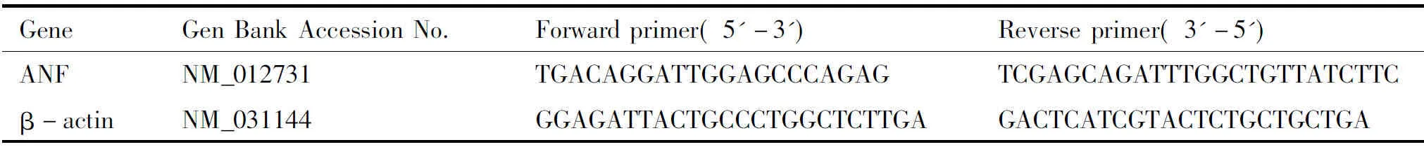
GeneGenBankAccessionNo.Forwardprimer(5'-3')Reverseprimer(3'-5')ANFNM_012731TGACAGGATTGGAGCCCAGAGTCGAG-CAGATTTG-GCTGTTATCT-TCβ-actinNM_031144GGAGATTACTGCCCTGGCTCT-TGAGACTCATCG-TACTCTGCT-GCTGA
1.2.8 Statistical analysis Data were presented as the mean ± standard deviation and analyzed using a one-way ANOVA followed by Duncan′s multiple range test and Dunnett’s T3(P<0.05)using SPSS 19.0 Software(SAS,Raleigh,NC,USA).
2 Results
2.1 Effect of rutaecarpine on blood pressure in AAC rats After the AAC surgery,the blood pressure of 95% rats was significantly increased,rats after and higher than Sham group(P<0.01).Compared with AAC group,BP of rats in the Rut-L and Rut-H remarkably descended(P<0.05),Compared with Rut-L group,Rut-H group showed higher efficacy in decreasing the blood pressure(Tab 2).
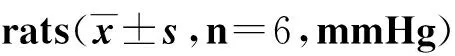
GroupsBloodpressurebeforemodelestablishmentBloodpressure4weeksofmodelestablishmentBloodpressure4weeksofrutaecarpineSham106±8.0103±10.2110±8.17 AAC103±10.1159±18.0##168±21.4##AAC+Rut-L 99±11.1156±20.2##136±10.4*AAC+Rut-H112±9.1152±18.1##135±11.3*
##P<0.01 vs Sham;*P<0.05 vs ACC.
2.2 Effect of rutaecarpine on the left ventricular hypertrophy index(LVHI)in AAC rats Compared with Sham group,the LVHI was increased significantly in AAC group(P<0.05).After Rut treatment for 4 weeks,the LVHI was significantly inhibited in the rutaecarpine group(P<0.05).Those results suggested that rutaecarpine could efficacy inhibit the hypertrophy of myocardium after AAC(Tab 3).
2.3 Effects of rutaecarpine on LVAW,LVPW and LVID in AAC rats Under the ultrasonic B-mode,thickness of left ventricular anterior and posterior wall were thickened,and left ventricular diameter was narrowed in AAC group(Fig 1).Result of calculation showed that the LVAW s,LVPW s,LVAW d and LVPW d notable increased,while LVID s and LVID d decreased in ACC group(P<0.05).After rutaecarpine treatment,the LVAW s,LVPW s,LVAW d and LVPW d significantly descend in Rut-L and Rut-H group(P<0.05),and the LVID s and LVID d were obviously increased(P<0.05).The results showed that the left ventricular hypertrophy was effectively inhibited after rutaecarpine treatment see Tab 4.
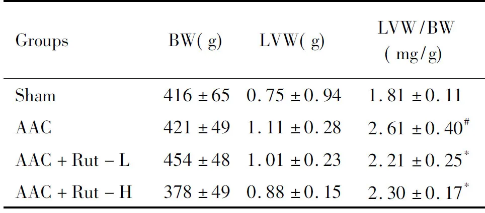
GroupsBW(g)LVW(g)LVW/BW(mg/g)Sham416±650.75±0.941.81±0.11AAC421±491.11±0.282.61±0.40#AAC+Rut-L454±481.01±0.232.21±0.25*AAC+Rut-H378±490.88±0.152.30±0.17*
##P<0.05 vs Sham; *P<0.05 vs ACC.
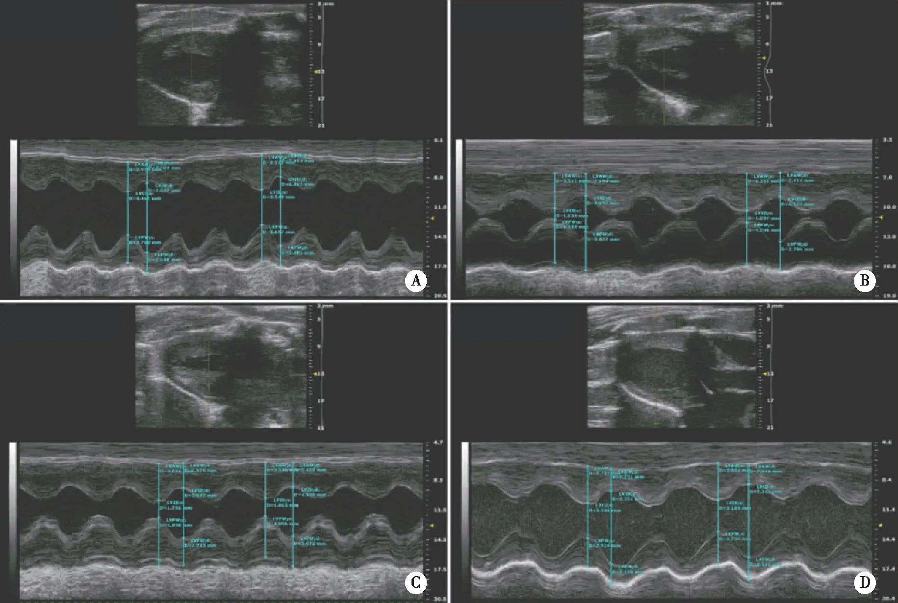
A:Sham;B:AAC;C:AAC+Rut-L;D:AAC+Rut-H.Fig 1 The effect of rutaecarpine on myocardial hypertrophy in AAC rats(n=6)

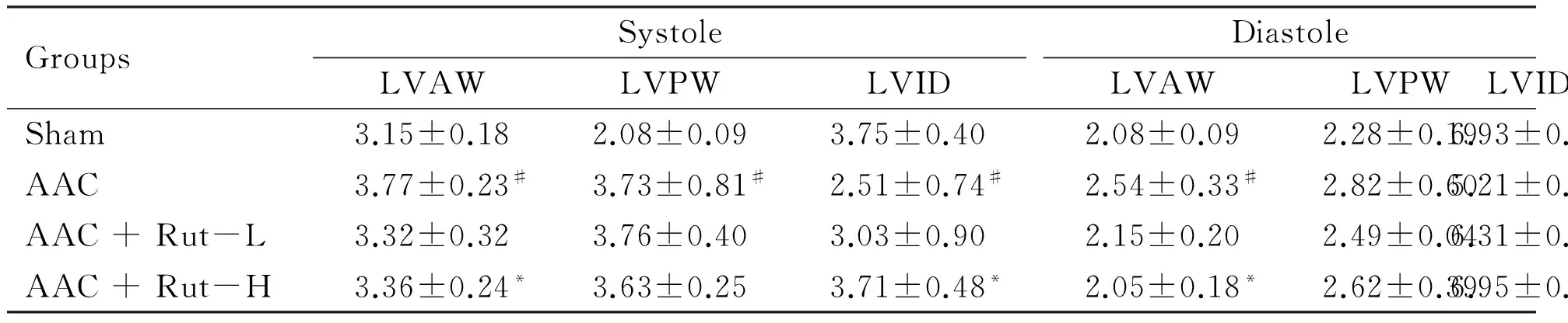
GroupsSystoleLVAWLVPWLVIDDiastoleLVAWLVPWLVIDSham3.15±0.182.08±0.093.75±0.402.08±0.092.28±0.196.93±0.53AAC3.77±0.23#3.73±0.81#2.51±0.74#2.54±0.33#2.82±0.605.21±0.79#AAC+Rut-L3.32±0.323.76±0.403.03±0.902.15±0.202.49±0.046.31±0.59AAC+Rut-H3.36±0.24*3.63±0.253.71±0.48*2.05±0.18*2.62±0.396.95±0.43*
#P<0.05 vs Sham;*P<0.05 vs ACC.
2.4 Effects of rutaecarpine on LV Vol.and SV in AAC rats Compared with Sham group,the LV Vol.s,LV Vol.d and the SV were significantly decreased in AAC group(P<0.05;P<0.01).After 4 weeks of rutaecarpine administration,the LV Vol.d,LV Vol.s and SV were significantly increased both in Rut-L and Rut-H group(P<0.05;P<0.01).Those outcomes suggested that rutaecarpine could effectively improve the cardiac systolic and diastolic function of ACC rats(Tab 5).
2.5 Effect of rutaecarpine on myocardial structure in AAC rats H&E staining showed that the AAC group myocardial cells were red stained,and arranged in disorder with widening of cell gap and partial rupture of muscle fibers.After rutaecarpine intervention,cardiac myocytes were closely arranged,structure integral,with dyed homogeneity of cytoplasm(Fig 2).

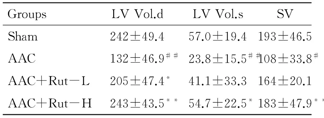
GroupsLVVol.dLVVol.sSVSham242±49.457.0±19.4193±46.5AAC132±46.9##23.8±15.5##108±33.8#AAC+Rut-L205±47.4*41.1±33.3164±20.1AAC+Rut-H243±43.5**54.7±22.5*183±47.9**
#P<0.05 vs Sham;##P<0.01 vs Sham;*P<0.05 vs ACC;**P<0.01 vs ACC.
2.6 Effect of rutaecarpine on myocardial ANF mRNA in AAC rats Compared with Sham group,the expression of ANF mRNA in AAC group increased significantly(P<0.01).The ANF expression showed significantly reduction in Rut-L and Rut-H group(P<0.01,Fig 3).
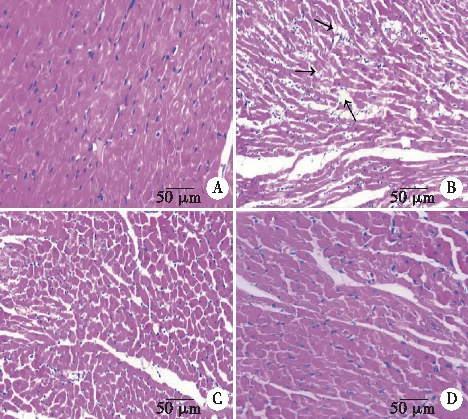
A:Sham;B:AAC;C:AAC+Rut-L;D:AAC+Rut-H. Fig 2 The effect of rutaecarpine on myocardial structure in AAC rats(×200)
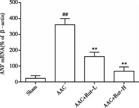
##P<0.01 vs Sham;**P<0.01 vs ACC.Fig 3 The effect of rutaecarpine on myocardial ANF mRNA in AAC
3 Discussion
Cardiac hypertrophy is the adaptive response of the heart to pressure or volume overload,myocardial infarction and other cardiovascular stimuli[4].In this study,abdominal aorta constriction was used to induce aortic pressure increase.Four weeks after ACC,the tail artery pressure significantly increased.Before sacrificed,the left ventricular structure and function was detected by ultrasonography.The detection of results showed that the thickening of left ventricular wall,the left ventricular internal diameter and functional decline of left ventricular,and also showed that the myocardial hypertrophy model was successfully established.Meantime,the outcomes of LV Vol and SV demonstrated that the left ventricular ejection function was severely damaged in ACC group.To our knowledge,this is among the first reports for successfully use of high resolution ultrasound imaging system(HRUIS)to illustrate left heart hypertrophy induced by ACC.
Rutaecarpine is one of the main effective components of the Traditional Chinese Medicine Tetradiumruticarpum.Rutaecarpine was metabolized by liver microsomal enzymes like CYP3A4,CYP1A2,CYP2C9 to oxidation metabolites[15].The study reported that the oral absorption of the Wu-Chu-Yu is rapid,and the bioavailability and purity are closely related[9].Studies have shown that Rutaecarpine has protective effects on myocardial injury in hypoxic environment and right ventricular remodeling caused by pulmonary hypertension[16].Rutcaecarpine is effective against ACC-induced cardiotrophy[12],but this study is the first reports using HRUIS to dynamically visualize its beneficial effects on heart structure and function.
Compared with the pathological indexes of conventional ventricular hypertrophy,Ultrasonic examination is a more direct reflection to the degree of ventricular hypertrophy,late enlargement of the heart cavity,and the decrease of cardiac pump blood capacity.In addition,the degree of damage can be quantified by data processing.At the same time,the results of the left ventricular hypertrophy index and the observation of pathological changes showed that the results of ultrasonography were consistent with the index of ventricular hypertrophy and histopathology.The accuracy and stability of HRUIS are further explained.With the continuous improvement of research programs,it is particularly important to dynamically observe the pathological progress of some diseases and to study the pathogenesis of the disease.In addition,the effects of drug intervention on different time periods of the same individual can be observed.It is convenient for the prognosis of the disease and the later detection.Therefore,the ultrasonic testing technology is expected to be an effective technical means in the follow-up experimental research.
The LV Vol.s,LV Vol.d and SV were applied in clinical to measurement of cardiac function indexes which played a vital role in disease therapeutic evaluation and prognosis estimation[17-18].Therefore,the functional state of the heart can be intuitively reflected by the detection of the above indicators.Ultrasonography outcomes suggested that left ventricular hypertrophy has formed and left ventricular systolic and diastolic function has arisen abnormal(Tab 4,Tab 5).Down-regulation of ANF expression could reduce myocardial hypertrophy,fibrosis and ventricular remodeling[19].Results showed that rutcaecarpine significantly reduced the level of blood pressure and ANF mRNA.The HRUIS outcomes indicated that rutcaecarpine remarkably improved function of myocardial hypertrophy,myocardial systolic and diastolic function.
In conclusion,rutcaecarpine can significantly ameliorate the myocardial hypertrophy and cardiac damage in SD rats induced by ACC.The application of HRUIS in the evaluation of ACC-induced cardiac hypertrophy and the therapeutic effects is a success and novel,and could open a research avenue in cardiopharmacology research.
[References]
[1] Schirone L,Forte M,Palmerio S,et al.A review of the molecular mechanisms underlying the development and progression of cardiac remodeling[J].Oxidative Medicine and Cellular Longevity,2017,2017(4):1-16.
[2] Matsuda J,Kitamura M,Takayama M,et al.Chronic phase improvements in electrocar-diographic and echocardiographic manifestations of left ventricular hypertrophy after alcohol septal ablation for drug-refractory hypertrophic obstructive cardiom-yopathy[J].Heart & Vessels,2017(356):1-9.
[3] Ding B,Price R L,Goldsmith E C,et al.Left ventricular hypertrophy in ascending aortic stenosis mice:anoikis and the progression to early failure[J].Circulation,2000,101(24):2854-2862.
[4] Ku H C,Lee S Y,Wu Y A,et al.A model of cardiac remodeling through constriction of the abdominal aorta in rats[J].Journal of Visualized Experiments Jove,2016,2016(118):54818-54822.
[5] Chen Y F,Lee N H,Pai P Y,et al.Tanshinone-induced ERs suppresses IGFII activation to alleviate Ang II-mediated cardiac hypertrophy[J].Journal of Receptor & Signal Transduction Research,2017,37(5):493-499.
[6] 邓江,邓雪松,罗洁,等.当归提取物对大鼠左心室肥厚的保护作用[J].中草药,2013,44(6):721-726.
[7] Lee S H,Son J K,Jeong B S,et al.Progress in the studies on rutaecarpine[J].Molecules,2008,13(2):272-300.
[8] Jia S J,Hu C P.Pharmacological effects of rutaecarpine as a cardiovascular protective agent[J].Molecules,2010,15(3):1873-1881.
[9] Xu S,Peng J,Li Y,et al.Pharmacokinetic comparisons of rutaecarpine and evodiamine after oral administration of Wu-Chu-Yu extracts with different purities to rats[J].Journal of Ethnopharmacology,2012,139(2):395-400.
[10]Yi H H,Rang W Q,Deng P Y,et al.Protective effects of rutaecarpine in cardiac anaphyla-ctic injury is mediated by CGRP[J].Planta Med,2004,70(12):1135-1139.
[11]张婧怡,林淑娴,侯化化,等.吴茱萸总碱抑制大鼠左室肥厚及对钙调神经磷酸酶信号通路的影响[J].遵义医学院学报,2014,37(2):151-155.
[12]吴芹,邓江,罗洁,等.吴茱萸次碱对大鼠压力超负荷致心肌肥大的影响[C].中国成人医药教育论坛,2010:15-17.
[13]林淑娴,任丽娜,孙安盛.吴茱萸碱、吴茱萸次碱和吴茱萸总碱的小鼠急性毒性[J].遵义医学院学报,2015,38(2):146-149.
[14]Zhu Q N,Zhang D,Jin T,et al.Rutaecarpine effects on expression of hepatic phase-1,phase-2 metabolism and transporter genes as a basis of herb-drug interactions[J].Journal of Ethnopharmacology,2013,147(1):215-219.
[15]Sang K L,Lee J H,Yoo H H,et al.Characterization of human liver cytochrome P450 enzymes involved in the metabolism of rutaecarpine[J].Journal of Pharmaceutical & Biomedical Analysis,2006,41(1):304-309.
[16]Liu W,Deng J,Ding W,et al.Decreased KCNE2 expression participates in the development of cardiac hypertrophy by regulation of calcineurin-NFAT(nuclear factor of activated T cells)and mitogen-activated protein kinase pathways[J].Circ Heart Fail,2017,10(6):3960-3980.
[17]Ünlü S,öahinarslan A,Gökalp G,et al.The impact of volume overload on right heart function in end-stage renal disease patients on hemodialysis[J].Echocar-diography,2017,35(3):314-321.
[18]Perny J,Kimmoun A,Perez P,et al.Evaluation of cardiac function index as measured by transpulmonary thermodilution as an indicator of left ventricular ejection fraction in cardiogenic shock[J].Biomed Research International,2014,2014:598029-598035.
[19]冯秋婷,钱晓军,徐欣,等.氨氯地平通过影响心房钠尿肽抑制心肌细胞的重构[J].实用医学杂志,2014,30(15):2360-2363.
猜你喜欢
浙江医学教育(2022年4期)2022-09-23
北京航空航天大学学报(2022年8期)2022-08-31
北京航空航天大学学报(2022年5期)2022-06-06
——以吴茱萸为例
内蒙古科技与经济(2021年23期)2022-01-18
北京航空航天大学学报(2021年4期)2021-11-24
北京航空航天大学学报(2021年9期)2021-11-02
生物骨科材料与临床研究(2021年3期)2021-06-25
右江民族医学院学报(2021年1期)2021-03-18
世界最新医学信息文摘(2021年25期)2021-01-06
同济大学学报(医学版)(2019年4期)2019-09-12