
MSCT对薄壁空洞肺结核和薄壁囊腔类肺癌的影像学特征比较
2021-03-22 19:27:16黄光辉
中国医学创新
2021年30期
黄光辉

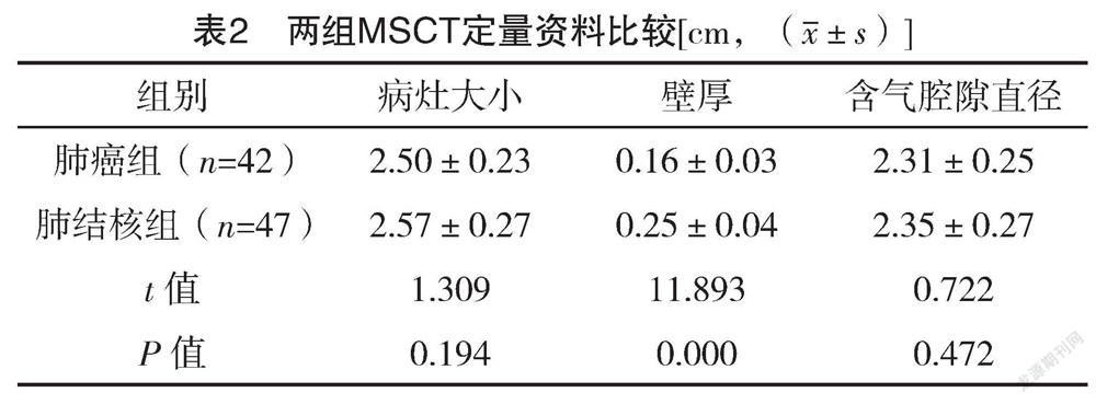
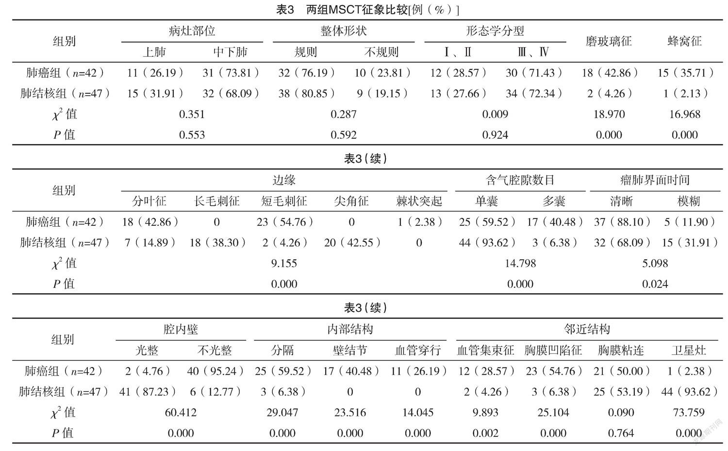
【摘要】 目的:研究比較MSCT对薄壁空洞肺结核和薄壁囊腔类肺癌的影像学特征。方法:选取2018年1月-2020年1月收治的47例薄壁空洞肺结核以及42例薄壁囊腔类肺癌患者进行研究,分别记作肺结核组与肺癌组。比较两组临床资料和MSCT影像学特征。结果:肺癌组年龄(58.74±6.32)岁,明显高于肺结核组的(46.11±4.80)岁,差异有统计学意义(P<0.05);两组性别、BMI、糖尿病以及高血压比较,差异均无统计学意义(P>0.05)。肺癌组壁厚为(0.16±0.03)cm,明显低于肺结核组的(0.25±0.04)cm,差异有统计学意义(P<0.05);两组病灶大小、含气腔隙直径比较,差异均无统计学意义(P>0.05)。肺癌组MSCT征象中磨玻璃征、蜂窝征、边缘分叶征、边缘短毛刺征、含气腔隙数目为多囊、瘤肺界面清晰、腔内壁不光整、内部结构分隔、内部结构壁结节、内部结构血管穿行、邻近结构血管集束征、邻近结构胸膜凹陷征例数占比分别为42.86%、35.71%、42.86%、54.76%、40.48%、88.10%、95.24%、59.52%、40.48%、26.19%、28.57%、54.76%,均明显高于肺结核组的4.26%、2.13%、14.89%、4.26%、6.38%、68.09%、12.77%、6.38%、0、0、4.26%、6.38%,而邻近结构卫星灶人数占比为2.38%,明显低于肺结核组的93.62%,差异均有统计学意义(P<0.05)。结论:薄壁空洞肺结核和薄壁囊腔类肺癌患者在MSCT上磨玻璃征、蜂窝征、边缘分叶征、边缘短毛刺征、含气腔隙数目、瘤肺界面、腔内壁、内部结构、邻近结构血管集束征、邻近结构胸膜凹陷征以及卫星灶等方面差异明显,可能具有较高的临床诊断价值。
【关键词】 薄壁空洞肺结核 薄壁囊腔类肺癌 多层螺旋CT
Imaging Features Comparison of MSCT for Thin-walled Cavitary Pulmonary Tuberculosis and Thin-walled Cystic Lung Cancer/HUANG Guanghui. //Medical Innovation of China, 2021, 18(30): -143
[Abstract] Objective: To study the imaging features comparison of MSCT for thin-walled cavitary pulmonary tuberculosis and thin-walled cystic lung cancer. Method: A total of 47 patients with thin-walled cavitary pulmonary tuberculosis and 42 patients with thin-walled cystic lung cancer admitted to our hospital from January 2018 to January 2020 were selected for the study, which were respectively classified as tuberculosis group and lung cancer group. The clinical data and imaging features of MSCT were compared between the two groups. Result: The lung cancer group was (58.74±6.32) years old, which was significantly higher than the pulmonary tuberculosis group (46.11±4.80) years old, the difference was statistically significant (P<0.05). There were no significant differences in gender, BMI, diabetes and hypertension between the two groups (P>0.05). The wall thickness of lung cancer group was (0.16±0.03) cm, which was significantly lower than that of the pulmonary tuberculosis group (0.25±0.04) cm, the difference was statistically significant (P<0.05). There were no significant differences in the size of lesion and the diameter of air-bearing cavity between the two groups (P>0.05). In the MSCT signs of lung cancer group, the proportion of ground glass sign, honeycomb sign, marginal lobulation sign, marginal short hair prick sign, number of air containing cavities was polycystic, clear tumor lung interface, uneven cavity inner wall, internal structure separation, internal structure wall nodules, internal structure vascular passage, adjacent structure vascular bundle sign and adjacent structure pleural depression sign were 42.86%, 35.71%, 42.86%, 54.76%, 40.48%, 88.10%, 95.24%, 59.52%, 40.48%, 26.19%, 28.57%, 54.76% were significantly higher than 4.26%, 2.13%, 14.89%, 4.26%, 6.38%, 68.09%, 12.77%, 6.38%, 0, 0, 4.26% and 6.38% in pulmonary tuberculosis group, while the number of satellite foci in adjacent structures accounted for 2.38%, which was significantly lower than 93.62% in pulmonary tuberculosis group, the differences were statistically significant (P<0.05). Conclusion: There are obvious differences between patients with thin-walled cavitary pulmonary tuberculosis and thin-walled cystic lung cancer in MSCT, such as grinding glass chippings, honeycomb sign, marginal lobulation sign, marginal short hair prick sign, number of air containing cavities, tumor lung interface, cavity inner wall, internal structure, adjacent structure vascular bundle sign, adjacent structure pleural depression sign and satellite foci, which may have high clinical diagnostic value.
[Key words] Thin-walled cavitary pulmonary tuberculosis Thin-walled cystic lung cancer Multislice spiral CT
First-author’s address: Chaoyang Central Hospital, Chaoyang 122000, China
doi:10.3969/j.issn.1674-4985.2021.30.033
肺癌属于临床上较为常见的一种呼吸系统恶性肿瘤,相关调查数据显示,肺癌是全球范围内发病率最高的恶性肿瘤,且有病死率较高的特点,已成为严重威胁人类生命健康安全的重大疾病之一[1-2]。……
登录APP查看全文
