为人类健康服务的光学技术
2022-01-06 05:25储扎克ZacharySmith杨云舟
国际人才交流 2021年11期
文 / 储扎克(Zachary J. Smith,美) 译 / 杨云舟
什么是科学研究?科学研究有其遵循的一定流程。首先,我们需要提出一个想法来构成初步的假设;其次,我们需要制定一个研究方案来验证假设,精心设计并开展试验;最后,通过分析试验得出的数据,我们能够揭示最初的假设是否正确。如果假设错误,我们就必须回到第一步,修改假设,进行“再研究”,再将流程循环到底。其实,科学研究的方法可以越过纯粹科学的边界,应用到生活中的许多方面。它实际上是帮助我们应对和解决生活难题的一个视角。正因如此,我们发现,有着严谨诚实的态度并且秉持以公众利益为重的科学,掌握着世间所有问题的答案,也拥有能够改变世界的力量。
那做科学家又是什么样的呢?他是一个像谢尔顿·库珀(美剧《生活大爆炸》中主角之一)那样的人,还是一个整天做实验的实验室小白鼠?其实,医院里的医生也是科学家,他们采集样本,进行测试,以验证他们的假设——医疗诊断——是否正确。某些政府领导人也是科学家,他们在不同研究领域都有着各自的科研成就,科学训练帮助他们更好地领导、治理我们的社会。
我和我的研究团队将目光定格在了一个特殊的领域:生物医学光子学。这一方向研究的基本思路是利用光子的力量来解决生物和医药领域的核心问题。目前,我们的研究主要有3个分支:无标记显微技术、纳米尺度拉曼光谱和成像,以及资源匮乏地区专用的医疗诊断技术。
What is scientific research? It has its own internal cycle where the first step is to bring forward an idea, which will constitute your hypothesis. To examine this hypothesis you will need to come up with a research protocol and carry out well-designed experiments. Experiments produce data to be analyzed, thus revealing if the initial hypothesis is correct or not. And if not, we must return to the first step, revise our hypothesis, and “reresearch” until the whole cycle is completed. This method of scientific research can also be applied to many other aspects in life beyond the boundaries of strict science. The scientific method is actually a perspective through which to look at any problem we are faced with in life. It is with this idea we can see that science, carried out faithfully and in the public interest, possesses the answer to all our worldly problems and has the power to change the world.
And what does it mean to be a scientist? Does it mean someone like “Sheldon Cooper”, or a lab rat spending all day carrying out experiments? It can also be a doctor in a hospital, who runs tests on collected samples to see if their hypothesis—their diagnosis- is correct or not. It can also be one of our government leaders, among whom there are many accomplished scientists specialized in different areas of studies. Scientific training has helped them become more proficient as leaders of our society.
As for me and my team, our research fixes the eyes on one particular field: biomedical optics. The basic idea of this study is utilizing the power of light to solve problems crucial to our society that have risen in the realm of biology and medicine. Our research has three major branches, which are namely label-free microscopy, nanoscale Raman spectroscopy and imaging, and point of care technologies for resource-limited settings.
无标记显微技术
我们首先来看无标记显微技术。当低头看向自己的双手时,我们可以分辨出它们的颜色和背景是不一样的。这种颜色的区别使我们能够将特定事物与附近的东西区分开来。但一个自然状态下的细胞是无色的,导致研究人员要想对它直接进行观测会十分困难。所以,如果我们想锁定某个分子并对其进行观察,就要首先赋予它一种颜色——一种荧光标记——使它变得易于区分。举个例子,科学家可以给艾滋病毒打上荧光标记。感染了艾滋病毒的细胞会和健康的细胞进行信息交换,为病毒创造一条通道,让它能够移动到正常细胞内并感染正常细胞。如果成功地给病毒贴上了一个荧光标记,研究人员就可以顺利观测到艾滋病毒在细胞间的移动现象,并进一步对其进行研究。
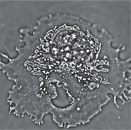
运用新的技术,我们能够清晰地看到细胞的内部结构
The Label-free Microscopy Technology
The first is the label-free microscopy technology. Looking down at our hands, we can tell they have a different color than the background, which enables us to tell them apart from things nearby. But a cell in its natural form is colorless, difficult for researchers to observe. So, should we wish to find a certain molecule and look at it, we first have to give it a color—a fluorescent label—thus making it discernible. For example, we can give a fluorescent label to an HIV virus. A cell infected with HIV will exchange information with a healthy cell, creating a tunnel for the virus to move to and infect the normal cell. If labeled with fluorescence, the cell-to-cell movement of the HIV virus will become observable for researchers.
As we can see from the example above, a fluorescent label is a powerful tool that allows us to see many things that have eluded our eyes before. Then why are scientists researching a “labelfree” microscopy technology? First, the addition of a fluorescent label to a cell is challenging in itself—in fact, it might just kill the cell in the first place. Second, it is still not an easy task to observe the cell even with a fluorescent label. In order to capture better images, researchers will have to hit the labeled cell with a powerful laser beam, which will possibly overheat the cell or make it release toxic free radicals. Finally, even if researchers have succeeded in both labeling and keeping the cell alive, they still will face critical difficulty in observing it, since fluorescent labels can “break”, a process we call “photo-bleaching”, where the signal we receive gets weaker and weaker over time. Thus, the first image is very high quality, but the 10th or 20th image may be impossible to recognize. One of the solutions to this problem is to use a gentler laser, producing a poorer image of cells. Then we can take these poor-quality images and use a computer algorithm to help us enhance the quality, thus obtaining a relatively clearer image while protecting the cell. However, this method still has its limitations, and we nevertheless need to find other ways to observe the cell.
从这个例子中我们可以看出,荧光标记是一个非常有用的工具。通过这项技术,我们能够看到很多以前无法看到的东西。那么,为什么科学家们还要研究一种“无标记”的显微技术呢?原因在于,首先,给细胞打上荧光标记本身就是一项挑战。这一步骤很可能会直接杀死细胞。其次,即使有了荧光标记,要观察细胞仍然不是一件容易的事。为了捕捉到更好的图像,研究人员必须用强大的激光束照射被标记的细胞。这可能使得细胞过热,或者释放出有毒的自由基。最后,即使研究人员既成功地给细胞打上了标记,又让细胞存活了下来,他们在观察细胞时仍然会面临关键性的困难。这是因为荧光标记可能会“破损”,这个过程被称为“光漂白”,即随着时间的推移,我们接收到的信号会越来越弱。因此,第1张图像的质量通常会非常好,但第10张或第20张图像可能会变得无法识别。解决这个问题的方法之一是使用较温和的激光。较温和的激光生成的细胞图像质量较差,但我们可以利用计算机算法来帮助我们提高这些照片的成像质量,从而在保护细胞的同时获得相对更清晰的图像。但是,这种方法仍然有其局限性,科学家们还是需要寻找其他的方法来观察细胞。
To observe cells without fluorescent labels, scientists chose to utilize the outstanding work of Frits Zernike, the phase contrast microscope, (for which Zernike earned the Nobel Prize) and modify it to our demands. Beams of light that go through the cell move slower than those that move through the water surrounding the cell. This speed difference changes a property of light called its “phase”. Making use of this speed difference, the phase contrast microscope allows us to see colorless cells that are otherwise invisible. Yet the resolution of images coming from a phase contrast microscope is limited, and the information is “qualitative”, meaning that the brightness or dimness of certain parts of the cell cannot be exactly mapped to the cell’s real structure. Scientists need more to see better and as well deeper into a cell’s internal structure, and to make the measurement “quantitative”.
为了能够在没有荧光标记的情况下观察细胞,科学家们选用了弗里茨·泽尔尼克发明的相衬显微镜(泽尔尼克也因此获得了诺贝尔奖),并根据需要对其进行了一些改进。一束光在穿过细胞时的速度会比穿过细胞周围的液体时要慢,这种速度差异会影响到光的一种被称为“相位”的属性。利用速度差,相衬显微镜可以让观测者看到原本难以看见的无色细胞。然而,相衬显微镜只能呈现有限分辨率的图像,而且,这些图像都是“定性”信息。也就是说,图像上细胞某些部位的明暗并不能准确反映出细胞内部的真实结构。科学家们需要更适合的技术来更全面、深入地观察细胞的内部结构,并使测量出的数据变得“定量”。
The devising of the ultra-oblique illumination high resolution phase contrast microscope technology affords us with new insights into cells that are alive and functioning. We can now clearly visualize most of the cell’s internal structure, including lipid droplets, nucleus, mitochondria, vesicles, and a very tiny web-like structure called the endoplasmic reticulum. We can also notice many things that haven’t yet to be found up to this moment. For example, with this new technology, we have found out that many of the mitochondria within certain cells are always spinning, as if the mitochondria are dancing. This movement costs the cell energy, but cells are very conservative and don’t like to waste any energy. Therefore, this mitochondrial dancing must have an important reason behind it. Now we need to use the scientific method, and devise a hypothesis we can test through experiment to discover the answer to this question. Since the phase image does not “photobleach”, and the imaging does not hurt the cell, scientists now also have all the time in the world to look at the endoplasmic reticulum, an important organelle, and study the biological function behind its incessant shaking motion.
超斜照明高分辨率相衬显微镜技术的出炉使得我们掌握了观测活体正常细胞的全新角度。有了这项技术,科学家可以清晰地看到细胞内部的大部分结构,包括脂滴、细胞核、线粒体、液泡,以及一种非常微小的网状结构——内质网。我们还得以注意到从前没有被发现过的东西。例如,我们发现在某些细胞内,许多线粒体总是在不停旋转,就像跳舞一样。这种运动会消耗细胞的能量,但细胞通常都十分节约,不喜欢浪费任何能量。因此,线粒体的这种“舞蹈”背后一定有着重要的原因。我们需要用科学的方法,提出一个假设,通过实验来检验,由此发现这个问题的答案。又如,内质网存在着一种不间断的摇摆运动。由于新的显微技术不存在“光漂白”效应,也不会伤害细胞,所以科学家们现在有充分的时间去观察内质网不停摇摆背后的生物功能。
拉曼光谱与成像
既然已经能够对细胞内部进行更清晰的观察,科学家们便开始设想,是否可以进一步观察出这些内部结构之间在化学成分上的差异?要满足这个雄心壮志,科学家们需要一种名为拉曼光谱的技术。简单来讲,科学家会将一种颜色的光照射向细胞,而由于光线与不同化学成分之间的相互作用,许多不同颜色的光束会从细胞里折射出来。在光谱仪的帮助下,这些不同的颜色可以被分离成被我们称为“光谱”的信号,它可以告诉我们每种颜色各占多少。每个分子都有自己独特的拉曼信号。而运用拉曼光谱,我们可以分析出在细胞的每个部分存在着什么样的化学分子。这项技术有着多种多样的应用。例如,通过观察一个脂滴,科学家可以判断出里面有什么,以及它是否由健康的脂肪构成。通过进一步分离并辨别细胞摄入的脂肪,我们甚至可以判断出它是一个健康的细胞还是癌细胞。
Raman Spectroscopy and Imaging
With a clearer vision looking inside the cell, scientists also want to know, what are the chemical differences between these internal structures? The technique required for this ambition is called Raman spectroscopy. Basically, scientists shine one color of light onto a cell. Many different colors will come out from it due to the interaction of the light with different chemical components. With the help of a spectrometer, these colors can be separated into a signal we call a “spectrum”, which tells us how much of each color is present. The Raman spectrum can tell us what kind of chemicals are present within each point of the cell, because each molecule has its own unique Raman signal. Therefore, scientists, for example, can look at a lipid droplet and tell what’s inside and if it’s made of healthy fat or not. By separating the fat intake of a cell, we can even tell if it’s a healthy one or a cancer cell.
这项技术还可以从其他角度对癌症进行诊断。目前,“细胞外小体”的研究是生物学中的一个新兴领域,其中最受关注的是“外泌体”。外泌体是一种纳米级大小的信使,它的功能类似细胞之间沟通时所发送的“信件”。外泌体包含蛋白质和核酸,可以向附近的细胞发出指令。但是,不仅正常细胞会释放外泌体,癌细胞也会,而且数量往往比正常细胞多得多。事实上,癌细胞可以利用外泌体发送虚假信息,来欺骗本应消灭它们的白细胞。在拉曼光谱仪下,我们能够研究每一个外泌体的化学成分,并分析它们之间的微小差异。针对这一点,科学家们使用一种叫作“光镊”的工具,挑选出单个的外泌体进行深入研究。我们用这种方法发现,癌细胞和健康细胞的外泌体有着不同结构的表面蛋白。这无疑将为癌症的研究、诊断及可能的新治疗手段提供参考。
This technology can take on cancer diagnosis from other perspectives. A new topic in biology is the study of “extracellular vesicles”, among which one of the most studied is called an “exosome”. The exosome is a nanoscale messenger. It functions like a letter sent between cells for communication purposes. It contains proteins and nucleic acids that can give instructions to nearby cells. Not only do normal cells release these exosomes, but cancer cells also release them, often in much higher numbers than normal cells. In fact, cancer cells can use exosomes to send false information to trick the white blood cells that are supposed to eliminate them. Using Raman spectroscopy, we can study the chemical composition of each individual exosome and see that every exosome is a little bit different from one another. Regarding this, scientists use a tool called optical tweezers to pick up each single exosome and examine them closely. We found that the surface protein of the exosome is different between a cancer cell and a healthy one, which can provide new information for cancer studies, cancer diagnosis, and possibly cancer treatments.
适于资源匮乏地区的医疗诊断技术
除了解决分子层面的生物学问题,光学科学还可以为组织层面的医学问题作出贡献。人们生病了会去“看医生”,但其实是“医生在看你”——医生通过显微镜观察你的组织或血液样本。值得一提的是,近一个世纪以来,医生使用的显微技术几乎没有根本性的变革。也许,科学家们在相关领域通力合作,如让显微镜变得更小、更智能,也能推动医用显微技术的进步。
Care Technologies for Resource-limited Settings
Besides biological questions at the molecular level, optical science can also contribute to medical questions at a tissue level. When we are sick, it is natural for us to “see a doctor”. What really happens is “the doctor sees you”—the doctor looks at the tissue or blood sample of yours through a microscope. The microscopic technology used by doctors, in fact, hasn’t really changed in almost a century. Maybe through the coordinated efforts of scientists, we can improve medical microscopes, for instance, by making them smaller and smarter.
人们希望无论何时何地都能获得高质量的医疗服务。但在现实生活中,患者必须去到特定的地方——医院——才能顺利就诊。医院配备有所有可能需要的专业设备和仪器,它们将帮助医生进行测试,做出可靠、准确的诊断。但这些设备往往体积庞大、价格昂贵,而且需要经过大量的培训才能操作。如果你居住在一个发达国家的城市,这样的资源对你来说可能唾手可得。但那些生活在贫困(如非洲农村)或偏远地区(如西藏地区)的人所面临的情况就大不相同了,在那里资源稀缺且十分昂贵,患者往往需要几天甚至几周的时间才能就医。为了让医疗服务更方便、便捷,科学家们考虑将我们的日常工具改造成医疗设备。手机是否能够承担这样的工作?它有摄像头、麦克风,内置电脑和网络模块。如果再加上一个镜头,它基本上就能变成一台显微镜,足以用来进行疾病观察、细胞计数,甚至成为一台光谱仪。当我们制造出这样一个简单的手机显微镜时,我们发现它的确可以达到很好的显微效果,观察到细胞核和疟疾寄生虫。然而,我们随后发现,尽管手机显微镜具有成为便捷医疗工具的潜力,但它只适合那些具备专业操作技术的人。在手机显微镜的成像方面,由训练有素的实验室研究人员操作出的图像和由医院工作人员或其他没有接受过使用培训的人操作出的图像可能在质量上差异很大。为了更适应医疗卫生从业的需要,我们的低成本显微镜必须更智能,必须对那些不十分熟悉光学科学的人更加友好。于是,我们研发出了一种新型“全自动” 微镜。它可以自动聚焦和扫描微小的微观样品,体积小,便于携带,操作也不需要经过额外培训,还能观察从血液、粪便到组织样品的多种类型的样品。相比之下,虽然普通的自动显微镜可以生成质量稍好的图像,但价格要比我们的显微镜贵很多倍。此外,新型显微镜非常便于组装,材料在网上很容易就能买到,基本上就像一套乐高玩具一样。
It is a universal dream to have access to high quality medical care whenever and wherever we want, which is in stark contrast to the reality where patients must go to certain places—hospitals—for that is where all the professional equipment and machines are. These machines help doctors run tests and make solid diagnoses. But they tend to be large, expensive, and require extensive training to operate. If you are an urban citizen in a developed country, such resources might be not so difficult to get. Things are very different for those live in poorer areas where such resources are scarce and exorbitantly priced, like rural Africa, or a remote district—like Tibet—where it takes days, even weeks, to reach a hospital.
To make medical services more convenient and accessible, scientists are thinking to turn everyday gadgets into medical devices. Can cellphone take up such a job? It has a camera, a microphone, a built-in computer and network modules. With a lens added on, a cellphone can be essentially modified into a microscope that can be used to look at diseases, count cells, and even become a spectrograph. When we developed a simple cellphone microscope, we found we could achieve excellent results, being able to see cell nuclei and malaria parasites. Yet for all the potential it has to become a convenient medical tool, we found out that it only suits those with specialized know-how, as images produced using a cellphone microscope can vary a great deal between those operated by trained lab researchers and those operated by hospital staff or others who have not been trained in the cellphone microscope’s use. In order to truly impact health care, our low-cost microscope must be made smarter and more friendly to those who are not so familiar with optical science. A new type of microscope was then made to satisfy this demand. It is a “fully-automated” microscope that can automatically focus and scan tiny microscopic samples, has a portable size, requires no additional training, and can look at many types of samples, from blood to feces to tissue samples. In comparison, a normal automated microscope can produce slightly better images, but is many times more expensive than our microscope. Ours is easy to put together, basically like a Lego set, and can be set up with materials that can be easily purchased online.

低成本、全自动的显微平台
我们的自动显微镜可以应用于许多不同的医疗场景。它可以协助进行动物诊疗。众所周知,山羊是杂食动物,因此很容易遭到寄生虫的感染。在对山羊进行寄生虫检查时,兽医通常会先从山羊的粪便中取样,与水混合,然后再放在显微镜下分辨并归类样品中的寄生虫卵。如果采取纯手工操作的方法,这套流程会非常累人。医生一般需要花20—30分钟的时间来仔细研究样本。而我们的自动显微镜可以自主对动物的大便样本进行细节拍照,随后利用深度学习技术,自动从图像中挑出虫卵,进行计数,并分辨出这些虫卵是来自蛔虫、壮虫还是其他寄生虫。这样一来,既节省了医生的时间,又可以帮助动物尽快得到正确的药物治疗,减少它们的痛苦。新型显微镜在人类体检中也有用处。当我们去看病时,医生通常会先抽取一小瓶血。通过统计血液中红细胞、白细胞和血小板的数量,医生能够掌握我们的健康状况。对于老年人,尤其是对罹患癌症的老年人来说,定期查血是很重要的一项检查。治疗癌症的药物不仅会杀死癌细胞,还会伤害到身体其他健康的部位,所以医生要时刻监测患者血液中的白细胞水平,避免其降低到临界水平以下。目前,患者只能去医院查血,而每个月可能只查一两次。这就造成了医生在信息掌握上的滞后。一种理想的情况是患者每天在家里就能自己验血。我们的新型、便捷、适合家庭使用的显微镜或许能够为他们提供帮助。患者可以将血液样本与一种特殊液体混合,然后放在我们的显微镜下。显微镜会自动扫描样品,进行分析,然后计数。不同种类的细胞——如红细胞或白细胞——在输出图像中会显示为不同的颜色或亮度。整个系统完全自动,所以即使是由业余的爸爸妈妈们来操作,得出的检测结果也可以和专业的研究人员一样准确、可靠。
Our automated microscope can find application in many different medical scenarios. As one example, we have shown how it can help treat animals. Goats are known to be messy eaters, and are consequently very easily infected by parasites like parasitic worms. The usual procedure for a vet to perform a parasite check on a goat is first taking a sample from its feces, mixing it with water, then putting it under a microscope to tell apart and classify the parasite eggs in the sample. This can be exhausting if done manually. It usually takes 20—30 minutes of the doctor’s careful study. Our automated microscope can take a detailed pictures of a large sample of the animal’s feces and, subsequently, using deep learning technology, pick out eggs from the image automatically, count them, and tell us if they are from ascarids, strongyles or other parasites. This can save the doctor’s time, and help the animal to get the correct drug as soon as possible, reducing its suffering.
This can also be applied to the medical examination for human beings. When seeing a doctor, it is a common practice to have a vial of blood taken. By counting the number of red blood cells, white blood cells and platelets, our blood serves as a window of our health status in the eyes of a doctor. Regular blood checkups are important for the elderly, especially those who have cancer. Drugs used in cancer treatment will not only kill the cancer cells but also hurt the healthy body parts, so doctors will want to monitor the white blood cell level in the patient’s blood to avoid it reducing beyond a critical level. Using current technology, patients have their blood tested only when going to a hospital, which probably only happens once or twice a month. This creates a time-lag of information on the doctor’s side. The ideal for us is to enable the patient to have his or her blood tested right at their own homes on a daily basis. Our new, convenient, household friendly microscope is qualified to help. Blood samples from patients will be mixed with a special liquid that prepares the sample and put under our microscope. Samples will be automatically scanned, analyzed and counted. Different kinds of cells, like red cells or different types of white blood cells in the output image will have different colors or brightness. Because the system is totally automatic, in our experiments the test results obtained by amateur moms and dads can be just as accurate and reliable as those of professional researchers.
即使是要给动物抽血,我们的显微镜也能帮上忙。动物并不了解健康检查的重要性,所以,为了抽血这一过程不刺激到它们或让它们不安,医务人员会尽可能轻柔地从它们身上抽取尽可能少量的血。我们的系统只需要几微升(不到一滴)的血液就足以得到准确的检测结果,因此能够非常好地适应这项工作的特殊条件。
And even when it comes to draw blood from animals, our microscope can get the job done. Animals don’t understand the importance of health check-ups. To not irritate or upset them, medical practitioners would wish to take as little blood as possible from them, and in the most gentle manner. Our system requires only a few microliters (less than one drop) of blood, and it’s sufficient to get an accurate test result.
最后,我们的技术还可以在另一个重要领域——贫血诊断——中发挥作用。贫血是全球公共卫生面临的一大严重威胁,影响着全球约1/3的人口,尤其是那些医疗资源稀缺地区的人。中国有10%—20%的儿童患有缺铁性贫血,通常与营养不良有关;有4300万儿童患有地中海贫血,这是一种遗传性贫血,常见于泰国、越南及中国的广东和广西。这两种不同的疾病对治疗方法有着不同的要求。对于缺铁性贫血,治疗方法可以非常简单——提高铁质摄入量;然而,对于地中海贫血的患者来说则相反,铁的摄入对他们非常危险。正因如此,我们希望能够对社会中的每个公民进行贫血检测。但让全国人民都去医院看病检测是一件不可能的事,所以我们需要研发一种仪器,可以在医院外使用,价格便宜,每个城市都可以有几个这样的仪器。在贫血筛查过程中,第一项任务是找到那些贫血的人;第二项任务是诊断出患者所患的是哪种贫血。人体内红细胞的形状和大小不尽相同,而健康人、缺铁性贫血患者和地中海贫血患者之间的红细胞也不一样。但是,由于缺铁性贫血患者和地中海贫血患者的红细胞大小及形状的差异非常微小,所以,通常需要大型昂贵的设备才能成功区分。这些设备在医院以外的地方很难操作,所以我们采用了一种低成本的光学散射系统来测量红细胞的大小、形状或血红蛋白浓度等参数。通过我们这套系统产生的数据,医生几乎可以100%准确地区分贫血患者和健康人。在缺铁性贫血患者和地中海贫血患者的区分上,这套系统能达到超过90%的准确率。我们希望在未来,这样的技术可以顺利应用于大范围、全人口的贫血筛查。
Last but not least, there is one more application where our technology can pitch in—the diagnosis of anemia. Anemia is a serious threat to public health globally as it’s affecting around one third of the world’s population, especially plaguing those who have the least access to competent medical resources. 10%~20% percent of children in China have iron deficiency anemia, which is related to malnutrition; 43 million in China have thalassemia, a genetic type of anemia, which is commonly found in Thailand, Vietnam, Guangdong and Guangxi in China. These two different diseases should be countered with different treatments. For iron deficiency anemia, the solution can be as simple as boosting up one’s iron intake. Yet on the contrary, iron intake is dangerous for patients with thalassemia. Because of this, we would like to test every citizen for anemia, but it’s not possible to have the whole country go to the doctor to get tested. We need to make an instrument which can be taken outside of the hospital, and is inexpensive enough that every town can have several such instruments. In this screening process the first job is to find those with anemia and the second job is to tell which kind of anemia it is. Blood cells are not the same in shapes and sizes, and they are different among a healthy person, an iron deficiency anemia patient and a thalassemia patient. However, because the size and shape differences are very tiny in iron deficiency and thalassemia, to differentiate blood cells according to their sizes normally requires large and expensive devices that are not easy to operate outside of a hospital. So we have come up with a low cost optical scattering system that can be used to measure parameters of red blood cells like size, shape, or hemoglobin concentration. With data produced from our system, doctors can tell an anemic patient from healthy people with nearly 100% accuracy, and separate iron deficiency anemia patients and thalassemia patients with an accuracy over 90%. We hope in the future such a technology could be used for widespread, whole population screening of anemia.
光的力量可以帮助我们解决许多生物和医学上的问题,助力我们改善现在所处的社会。光学科学的发展是许多杰出科学家努力的结果。每一位科学家都是“站在巨人的肩膀上”贡献自己的微薄之力。在他们的共同贡献下,世界可以变得更加美好。
The power of light can help solve many biological and medical questions, tackle challenges, and change the society we are now living in. The development of optical science is the results of effort from many outstanding scientists. Each scientist “stands on the shoulders of giants”, and tries to add their own small contribution to the greater good. Thanks to their combined contributions, the world can be a better place for all of us.
猜你喜欢
中国生物制品学杂志(2022年3期)2022-11-15
天天爱科学(2022年4期)2022-11-08
山西医科大学学报(2022年9期)2022-11-08
中国生物化学与分子生物学报(2022年3期)2022-09-07
国际心血管病杂志(2022年3期)2022-07-13
新作文·小学高年级版(2020年3期)2020-07-09
家庭医学·下半月(2020年6期)2020-01-07
当代陕西(2019年23期)2020-01-06
健康大视野(2018年17期)2018-12-28
学苑创造·C版(2017年12期)2018-01-17