Study on Color Information Degradation Induced by γ-ray Radiation in CMOS Cameras
2023-12-26 01:03LIKunfangFENGJieWANGHaichuanLIYudongWENLinLIZhenzheGUOQi
原子能科学技术 2023年12期
LI Kunfang, FENG Jie, WANG Haichuan, LI Yudong, WEN Lin, LI Zhenzhe, GUO Qi
(1.Xinjiang Technical Institute of Physics and Chemistry, Chinese Academy of Sciences, Urumqi 830011, China; 2.Xinjiang Key Laboratory of Electronic Information Material and Device, Urumqi 830011, China; 3.University of Chinese Academy of Sciences, Beijing 100049, China;4.Officers College of PAP, Chengdu 610213, China)
Abstract: The γ-rays are widely and abundantly present in strong nuclear radiation environments, and when they act on the camera equipment used to obtain environmental visual information on nuclear robots, radiation effects will occur, which will degrade the performance of the camera system, reduce the imaging quality, and even cause catastrophic consequences. Color reducibility is an important index for evaluating the imaging quality of color camera, but its degradation mechanism in a nuclear radiation environment is still unclear. In this paper, the γ-ray irradiation experiments of CMOS cameras were carried out to analyse the degradation law of the camera’s color reducibility with cumulative irradiation and reveal the degradation mechanism of the color information of the CMOS camera under γ-ray irradiation. The results show that the spectral response of CMOS image sensor (CIS) and the spectral transmittance of lens after irradiation affect the values of a* and b* in the LAB color model. While the full well capacity (FWC) of CIS and transmittance of lens affect the value of L* in the LAB color model, thus increase color difference and reduce brightness, the combined effect of color difference and brightness degradation will reduce the color reducibility of CMOS cameras. Therefore, the degradation of the color information of the CMOS camera after γ-ray irradiation mainly comes from the changes in the FWC and spectral response of CIS, and the spectral transmittance of lens.
Key words:CMOS camera; color reducibility; γ-ray; nuclear radiation
As a new efficient and clean energy, nuclear energy is widely valued, and various countries were also speeding up the construction of nuclear power plants[1]. The working conditions of nuclear power plants is strong nuclear radiation environment, thus its routine maintenance or accident site processing needs to be completed by remote control of nuclear robots[2]. Environmental information is the basis and prerequisite for nuclear robots to complete engineering operations, thus camera imaging equipment is often loaded on nuclear robots to obtain the environmental visual information. However, when the camera is working in a nuclear radiation environment, the γ-rays with high-penetration and high-dose rate[3]acting on camera’s electronic devices will induce radiation effects, resulting in the degradation of the performance of the camera, the reduction of imaging quality, and the increase of visual noise.
A complete camera system mainly includes optical lens, image sensor and motherboard control circuit etc., in which image sensor as the core device of camera imaging, mainly includes CCD image sensor and CMOS image sensor (CIS), compared with CCD, CIS has the advantages of small size, low cost, strong anti-radiation ability, and so on. Therefore, most of the cameras used in the nuclear industry adopt CIS as an imaging device[4-6], also known as CMOS cameras. As the core electronic device in the camera system, CIS will be affected by γ-ray irradiation and produce radiation effect when working in a strong radiation environment[7], which induces degradation of its parameters such as dark current, spectral response and full well capacity, thus affecting the imaging quality of the camera system. In addition, γ-ray irradiation can cause changes in the transmittance of the lens[8], which in turn affects the color information of the camera’s output image. When the accumulated dose of irradiation is not high enough, the change of the motherboard is very small compared with the other two parts[9], thus CIS and lens are the main sources that affect the output image information of the camera during γ-ray irradiation.
In recent years, scholars studied the impact of radiation environment on camera. Yu et al[10]studied and analyzed the impact of transient noise induced by neutron radiation on the resolution of CCD camera, Wang et al[11]analyzed the impact of noise generated by CIS irradiation on camera resolution, Nafradi et al[12]found that the output dark current of CMOS cameras under γ irradiation is related to the total ionization dose, dose rate and exposure time. With the impact of irradiation, the imaging quality of camera reduces, which is not conducive to the identification of target information. To solve this problem, scholars tried to use filtering[13-15], frequency domain transformation[16-17], machine learning[18-19]and other methods to achieve noise reduction of images or videos of nuclear radiation environment. However, the noise reduction methods mentioned above mainly focus on gray high-brightness noise, and the existing researches mainly focus on camera resolution, signal-to-noise ratio and other parameters, but the camera’s output capability of color information is often ignored. As a matter of fact, the degradation of image quality under radiation environment is not only reflected in gray noise, but also the distortion of color information will seriously affect the target recognition of nuclear robots. It is necessary to solve the problem of camera’s color information distortion caused by radiation. In this paper, irradiation experiment of camera system, lens and CMOS image sensor components will be carried out, and the law and mechanism of camera color reducibility degradation after irradiation will be studied and analyzed, to provide theoretical support for camera color correction technology under strong nuclear radiation environment.
1 Experimental detail
1.1 Devices
The camera system (Fig.1) used in the experiment is a modular camera developed by our laboratory, which is composed of an optical lens, CIS and mainboard. The optical lens is connected to the CIS device, and the mainboard circuit is connected to the CIS device through the flexible printed circuit (FPC). The standard test cards were captured by this camera, and then the captured pictures were analyzed with professional software of ImatestMaster to measure the camera’s color reducibility. In which, CIS is the high-performance AR0230 series developed by ON Semiconductor Company with an effective pixel number of 2.1 million, a single pixel size of 3 μm×3 μm, using RGB Bayer array color filter, lens is commercial autozoom, and the mainboard is designed by laboratory.
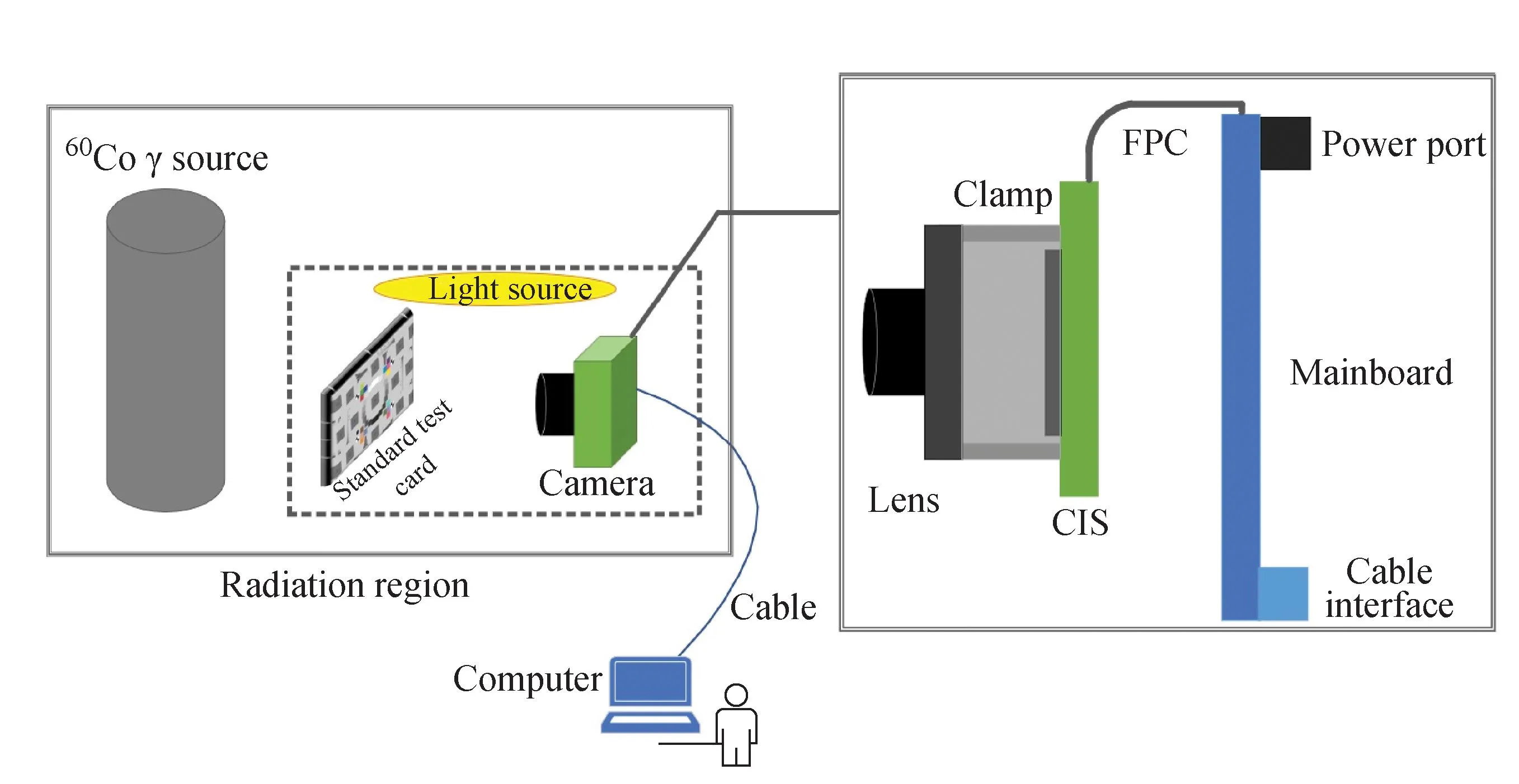
Fig.1 Camera system
1.2 Irradiation conditions
The irradiation experiment was carried out on the60Co-γ radiation source of Xinjiang Technical Institute of Physics and Chemistry, Chinese Academy of Sciences. The camera system and components such as CIS and lens were irradiated respectively. During the irradiation process of the camera system and the combined testing process of different components, the camera system was in online working state, and the irradiation and testing process were carried out at room temperature (24 ℃). The specific experimental setup parameters are listed in Table 1.
2 Test of color reducibility of camera
Color camera is defined as camera that uses CIS with color filter as the core device of imaging system and can reflect the true color of the image object. The process of color imaging system converting the actual color of object into image color is affected by ambient light source, object state, lens parameter, sensor parameter and back-end processing algorithm. Compared with monochrome camera, color reducibility is an important attribute that reflects the imaging quality of color camera. Color cameras are widely used in modern industrial cameras, so the impact of radiation on the color information of the camera system should be considered when acquiring the visual information of the target under strong nuclear radiation environment.
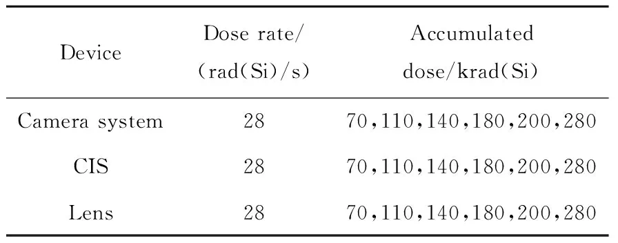
Table 1 Irradiation parameter
There is a certain difference between the color of the image acquired by the camera and the color perceived by the actual object in human eyes. The color reducibility of the camera is the description of the difference between the color of the image and the standard color in the uniform color space, including the two parameters as color deviation and color saturation. Related researchers in 1976 based on natural color characteristics, established the uniform color space CIE1976LAB (L*a*b*) to quantitative description of color deviations. Therefore, the color coordinates of the output image captured by the camera system are converted from RGB space to LAB space coordinates to calculate the values of color deviation and color saturation of output image. The general calculation method of color is to obtain the Euclidean distance between the coordinates of the reference color block in uniform color space and the corresponding spatial coordinates of the color block measured in the actual image. The value of the color deviation can be used to judge the difference between colors. The larger the color deviation value is, the greater the difference between the actual target color perceived by the human eye and the color of the image captured by camera is. Color saturation refers to the purity of the color, and can also be used to describe the vividness of the color.
The steps for calculating color reducibility are as follows. Firstly, the software is used to identify and map the color blocks of the standard test card image, and measure theR,G, andBvalues of the corresponding serial number color blocks. Then, theR,G, andBvalues were converted intoL*a*b*color space via formulas. Finally, the test results ofL*a*b*value of each block is compared with the standard reference formulas (1) to (5) of the target color block to obtain the parameters of average color deviation and color saturation. The standard test card color block of reference is shown in Fig.2. The model based on standardL*a*b*color space is shown in Fig.3, theL*indicates brightness, thea*indicates the color transition from green to red, and theb*indicates the transition from blue to yellow[20].
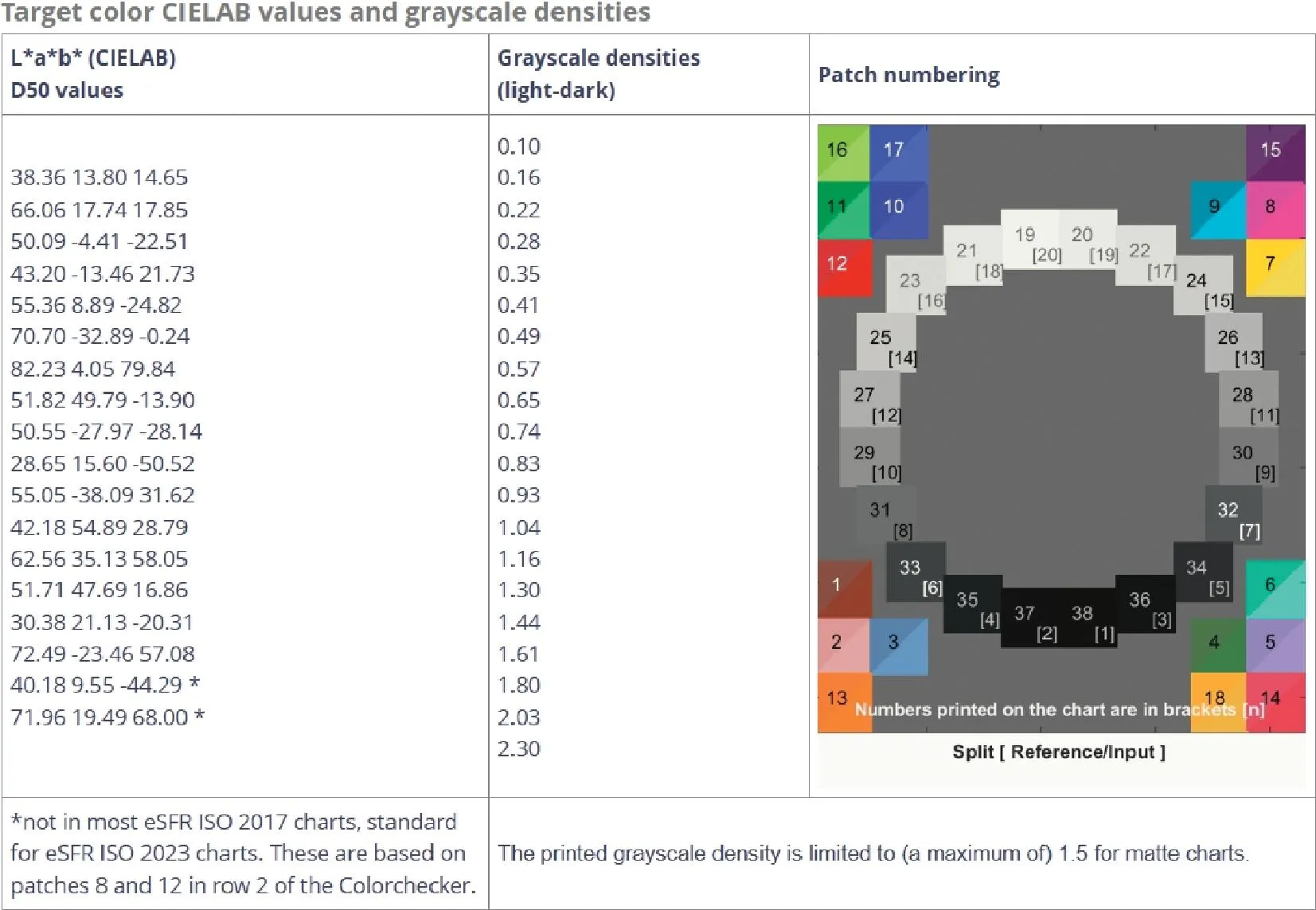
Fig.2 Target color CIELAB values and grayscale densities of standard test card
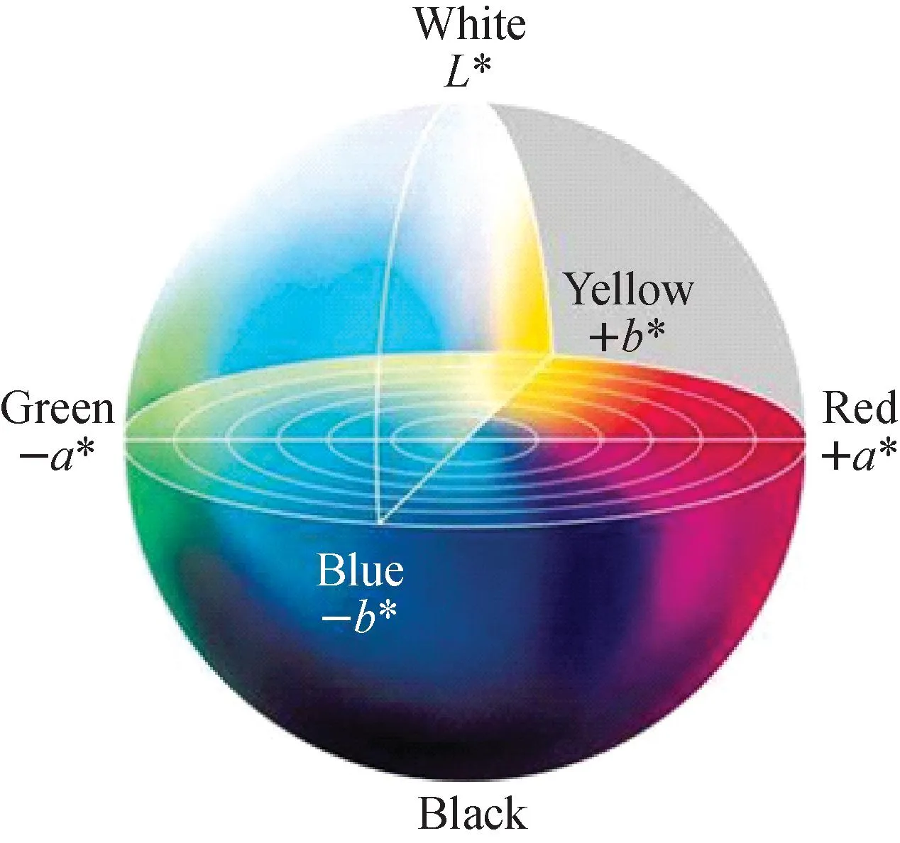
Fig.3 Standard L*a*b* color space model
The calculation formula of the color deviation is shown in equations (1) to (4):
(1)
(2)
(3)
(4)
The average color saturation is calculated by formula (5):
(5)
In which,iis color block label,nis number of color block.
3 Result and discussion
3.1 Degradation of QE(λ) of CIS
Quantum efficiency (QE) is defined as the response of the image sensor to a specific wavelength incident light signal, and the response of the image sensor to different wavelengths (λ) incident light is called spectral response QE(λ), which is to evaluate the ability of CIS to convert the optical signal into an effective electrical signal output. For CMOS cameras, QE(λ) is the ratio of the number of CIS photogenerated carriers to the number of photons collected on the surface under different wavelengths of optical signals in a fixed exposure time[21].
The QE(λ), as a parameter to evaluate the photoconversion ability of CIS for incident photons of different wavelengths, is very important to evaluate the color reducibility of modern cameras. The degradation ratio of CIS’s spectral response curve under γ-ray irradiation is shown in Fig.4. The degradation of parameters under incident light wavelengths of 420, 450, 516, 550 and 630 nm were tested.
It can be seen from Fig.4 that the QE(λ) degradation ratio of CIS varies with wavelength of incident light. It is more degraded in the short-wave band, and the degradation ratio gradually decreases with the increase of wavelength. In addition, when the cumulative dose of irradiation is less than or equal to 200 krad(Si), the degradation of QE(λ) of CIS is almost the same, and there is no significant difference. The degradation of QE(λ) of CIS is more prominent when the cumulative dose of irradiation reaches 280 krad(Si). This suggests that QE(λ) of CIS requires a large cumulative dose to show significant degradation.
The relationship between incident photon energy, incident light wavelength and absorption depth of CIS was analyzed in relevant studies[22], the results show that the shorter the wavelength, the shallower the absorption depth, and the higher proportion of photogenerated carriers generated near the Si-SiO2interface.
After the short-wave incident light enters the epitaxial layer, a large number of photoelectrons are generated near the Si-SiO2interface. At this time, the electron concentration in this region is much higher than the carrier concentration in equilibrium. According to the SRH theory, this region shows a net composite state. With the increase of cumulative dose, the interfacial trap charge density formed by TID effect on the surface of SiO2layer increases. The energy level of the interfacial trap charge is close to the center of the bandgap, which can be used as an effective recombination center, to improve the net recombination rate, reduce the lifetime of photogenerated carriers in this region, directly reduce the diffusion length of carriers, and ultimately reduce the efficiency of collecting photogenerated carriers in the depletion region. As the incident depth of incident light at different wavelength bands is inconsistent, the longer the wavelength of incident light at the interface, the lower the photogenerated carrier, the less affected by the interface trap charge density, and the lower the degradation of QE after irradiation. In addition, the interface trap charge has different charge states, and can form additional potential fields around them. Due to the action of Coulomb potential field, the scattering probability of electrons will change at a certain extent, and affect the electron diffusion coefficient. The rate of net increase of interfacial trap charge density formed on SiO2layer surface in CIS PPD region is inconsistent at different accumulated doses. The difference of QE degradation under different wavelengths of incident light and different doses will directly affect the ability of the camera to restore the color of the object.
3.2 Degradation of FWC of CIS
The full well capacity (FWC) refers to the number of electrons that the pinned photo diode (PPD) of CIS can hold at saturation, which is mainly affected by the PPD capacitance, FD capacitance, and the charge transfer efficiency of PPD to FD. The size of the FWC determines the maximum CIS output voltage, which will affect theL*value of the color deviation in the color reducibility of the camera.
The variation of the FWC of CIS with the increase of cumulative dose during γ-ray irradiation is shown in Fig.5. At different cumulative doses, there are some differences in the variation of FWC: The FWC of CIS barely changes before 140 krad(Si), FWC shows a downward trend after 140 krad(Si), and FWC deteriorates sharply after 200 krad(Si).
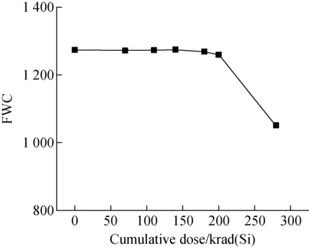
Fig.5 FWC of CIS varying with cumulative dose under γ-ray irradiation
The factors those lead to the decrease of FWC with cumulative dose are the decrease of PPD capacitance and the increase of leakage current due to the decrease of TG channel barrier. Under the γ-ray irradiation oxide trap positive charges are generated and accumulated in the dielectric layer around the PPD, widening the depletion layer width and resulting in a decrease in the capacitance of the PPD. However, compared to the high concentrations of holes in the P+region on the PPD surface, the concentration of oxide trap positive charge generated and accumulated by ionization damage is very low, so the effect on the PPD capacitance is small. Besides, the TID effect leads to the generation of trap positive charges in the STI region near TG, and induced negative charges are generated on the Si-SiO2surface of the shallow trench isolation (STI) layer due to the presence of trap positive charges. The accumulation of these negative charges increases the regional electron concentration and reduces the TG channel barrier. Finally, part of the photoelectrons in PPD can be transferred to FD through the channel side wall without adding voltage to TG. The oxide trap charge generated by the TID effect in the insulation layer can reduce the threshold voltage of the transistor, but the decrease of the threshold voltage is not obvious at a lower cumulative dose, and it has little effect on the minimum potential achieved by the PPD channel after TG turned off. Therefore, FWC does not degrade significantly before 140 krad(Si), and the main reason for the change of FWC caused by γ-irradiation is that the photoelectron escapes PPD due to the decrease of TG channel barrier.
3.3 Degradation of transmittance of lens
At present, the performance of lens usually be assessed by measuring the variation in transmittance. Lens transmittance refers to the percentage ratio of the light flux through the lens to the actual incident light flux. In addition, for color cameras, the change of the spectral transmittance of the lens before and after irradiation will affect the color reproduction capability of camera. In order to accurately detect lens transmittance and spectral transmittance, in this paper, D65 standard light source and CL500A spectral irradiance meter were used as the measuring instrument, and then each test equipment was fixed to complete the real-time measurement of lens parameters. The variation of spectral transmittance of lens with irradiation is shown in Fig.6.
As can be seen from Fig.6, after irradiated by γ-rays, the spectral transmittance of the lens decreases significantly with the increase of the accumulated dose of irradiation, and the degradation of the spectral transmittance of the optical lens is more obvious under the incident light of the short-wave segment. The reason for the obvious variation in the spectral transmittance of the optical lens after irradiation is mainly that after ionizing irradiation of the glass in the lens, radiation-induced defects occur in the glass material, and the absorption peak is formed in the visible wavelength range, so that the glass changes from colorless and transparent to brown colored body. With the increase of the accumulated dose of irradiation, the color of the glass also becomes darker. This is related to the composition and structure of the glass, for example, the common lens optical glass is silica glass, whose main component is SiO2. After the silica glass exposed to ionizing radiation, oxygen vacancies in the structure of SiO2will combine with radiation-induced defects to form defect centers, which serve as color centers, thus changing the spectral transmittance and color of silica glass.
3.4 Effect of CIS radiation damage on camera color reducibility
The degradation of color reducibility of cameras is mainly affected by the radiation damage of CIS and optical lens. In the process of color reducibility test, the camera system composed of unirradiated lens and CIS irradiated at different dose points. Due to the many types and quantities of color blocks, and the CIS in this camera system using an RGB Bayer array filter, the change in color purity caused by irradiation is mainly reflected by the degradation in color saturation of the red, green, and blue blocks, and the test results are shown in Fig.7a and Fig.7b.
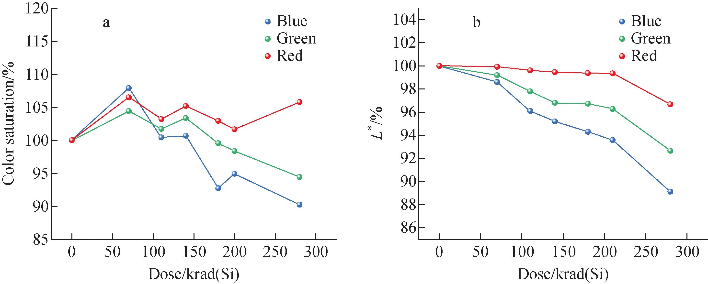
Fig.7 Color saturation (a) and L* value (b) of color blocks varying with cumulative dose under γ-ray irradiation
It can be seen from Fig.7a that the shift of color saturation of the blue block caused by γ-radiation is greater than that of the green block, and the shift of the color saturation of the red block is smallest. Besides, when the cumulative dose is low, there is little difference in the degradation of color saturation of different color blocks. But after the cumulative dose reaches 140 krad(Si), there is significant difference in the variation of different color blocks with the cumulative dose, which is due to the inconsistency of net increase rate of interfacial trap charge density formed on SiO2layer surface in CIS PPD region at different cumulative doses. The analysis of color saturation’s variation is as follows. As shown in Fig.3[23],a*andb*in the LAB color model are two color channels, where the color corresponding toa*is from dark green (low brightness) to gray (medium brightness) to bright pink (high brightness), andb*is from bright blue (low brightness) to gray (medium brightness) to yellow (high brightness). For the blue block, the initial value ofb*is negative, and since the QE at short wavelength of the CIS degraded distinctly during irradiation, which inducesb*drifts significantly toward to positive value. Similarly, for green block, the initial value ofa*is negative, and since the QE at wavelength of green light of the CIS has a certain degradation after irradiation. The degree of this degradation is smaller than that of blue, so it drifts toward positive with smaller amplitude than the blue block. While the two color-channel values ofa*andb*of the red block both drift positive, but just the value ofa*of the green block slowly drifts positive during irradiation, thus, the amplitude of variation of the red block after irradiation is slightly greater than that of the green block at lower dose. As the cumulative dose increases, the degradation amplitude of different color block is mainly affected by the QE response at different wavelengths. To sum up, there presented different amplitudes and directions of degradation of color blocks after irradiation, they are mainly related to the distribution in the LAB color model, the degradation of QE(λ), and the increase rate of interface trap charge density at different doses.
In the LAB color model, the brightness channelL*only contains brightness information, and the value ofL*ranges from 0 to 100, reflecting the brightness degree of the image. The main reason for the decrease of correspondingL*values of different color blocks in Fig.7b after irradiation is that the FWC of CIS degrades to different degrees after irradiation.
3.5 Effect of optical lens radiation damage on camera color reducibility
It can be seen from the variation in the spectral transmittance of the lens after irradiation in section 3.3 that the degradation degree of the lens transmittance is inconsistent under different wavelengths of incident light. The results of color block sampling and analysis after the camera replaced with lenses with different cumulative doses are shown in Fig.8 and Fig.9.
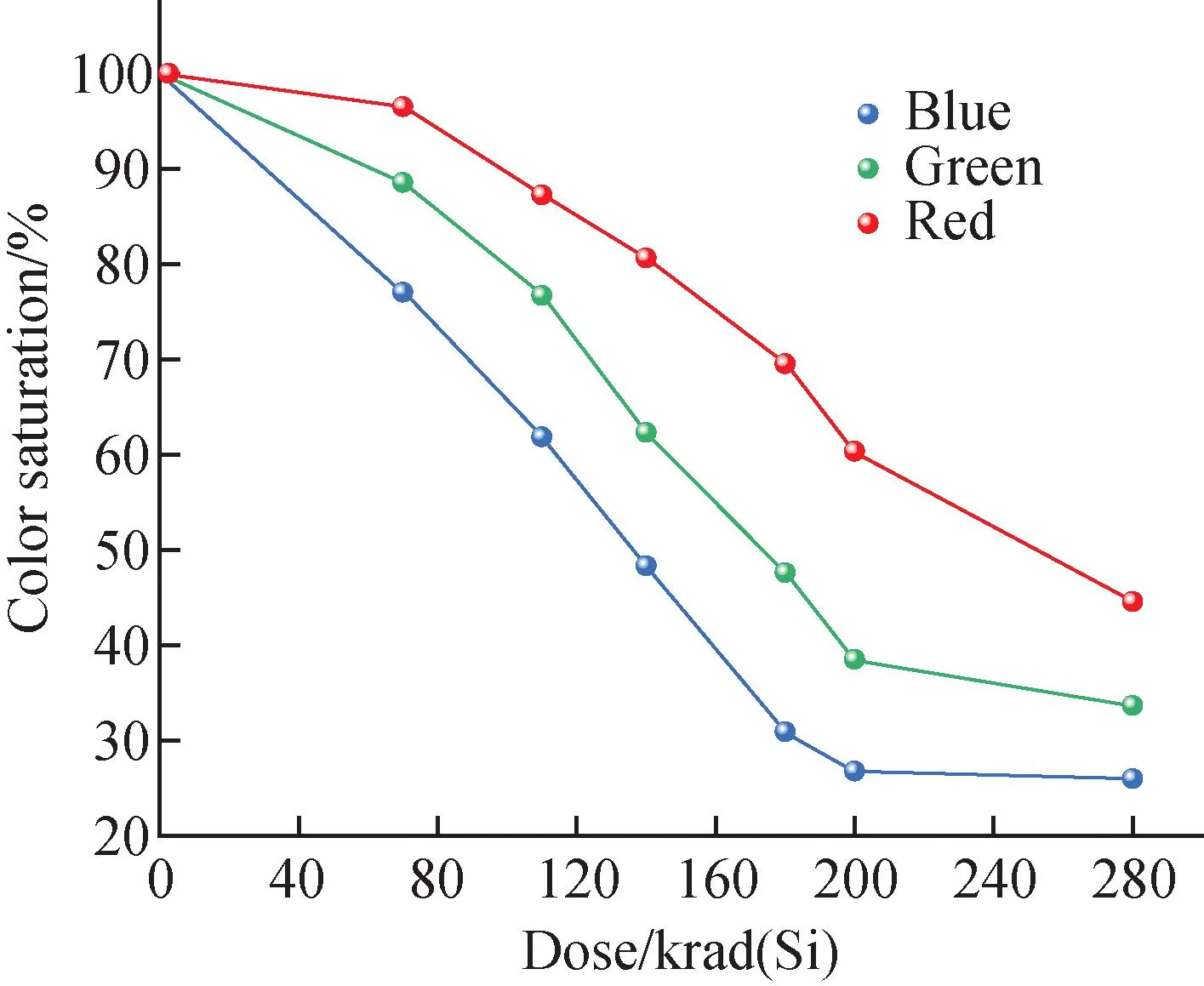
Fig.8 Effect of dose on color saturation under γ-ray irradiation
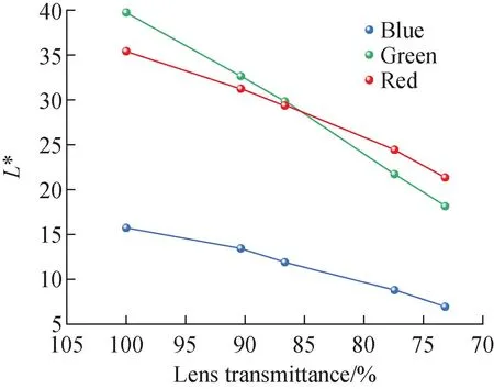
Fig.9 Relationship between lens transmittance and color block L* value under γ-ray irradiation
It can be seen from Fig.8 that the degradation of color saturation of different color blocks is different, in which the degradation degree is blue block, green block and red block in order from large too small. This difference is mainly caused by the obvious degradation of the spectral transmittance of the irradiated lens, but the degradation in different bands is inconsistent. For the red block, the main change is that the overall spectral transmittance of the lens decreases seriously after irradiation, and the degree of red degradation in the red block is greater than that of green reduction and red enhancement, resulting in the decrease ofa*value in LAB coordinates.
TheL*value of the color block is used to describe the depth of the color, which is affected by the lighting source, camera parameters, and the light reflectivity of the corresponding area of the test card. As can be seen from Fig.9, under the condition that the external test environment is consistent, the degradation ratio of light transmittance of the lens after irradiation has a linear relationship with theL*value of different color blocks, and the slope of the linear line corresponding to different color blocks is similar. Among them, since CIS uses a combination of RGGB and Baer array filter, the degradation of lens transmittance will cause the change of pixel unit gray value, resulting in the degradation ofL*value of green block is greater than that of blue block and red block.
4 Conclusions
In this paper, the γ-ray irradiation experiments of CMOS cameras were carried out to analyse the degradation law of the camera’s color reducibility and reveal the degradation mechanism of the color information with cumulative γ-ray irradiation. The following conclusions can be drawn from the above analysis.
1) The spectral response of CIS and the spectral transmittance of lens after irradiation affect thea*andb*values in the LAB color model, while the FWC of CIS and transmittance of lens affect theL*value in the LAB color model, thus increase color difference and reduce brightness.
2) The combined effect of color difference and brightness degradation will degenerate the color reducibility of CMOS cameras.
3) The degradation of camera color information can be attributed to the defects in CIS and color centers in the lens which both induced by irradiation.