游仆虫(原生动物,纤毛门)皮层银线系的形态发生模式❋
2016-12-24 01:58谢冬梅范鑫鹏顾福康
中国海洋大学学报(自然科学版) 2016年12期
谢冬梅, 范鑫鹏, 倪 兵, 顾福康
(华东师范大学生命科学学院,上海 200241)
游仆虫(原生动物,纤毛门)皮层银线系的形态发生模式❋
谢冬梅, 范鑫鹏❋❋, 倪 兵, 顾福康
(华东师范大学生命科学学院,上海 200241)
有关研究证实,游仆类的银线系在超微结构水平上对应了由微管束构成的网络骨架结构,关于其在纤毛虫细胞分裂过程中的发生模式鲜有报道。本工作应用银浸染色法和扫描电镜术,以扇形游仆虫(Euplotesvannus)、镰游仆虫(Euplotesharpa)和包囊游仆虫(Euplotesencysticus)为代表,研究了游仆类纤毛虫3种类型皮层银线系的形态发生过程。研究表明,游仆虫3种类型银线系的形态发生过程呈现基本相同的模式:该3种纤毛虫新银线网在棘毛原基基体周围发生,据此推测口围带小膜、棘毛基体复合单元和背触毛基体单元不仅对相应纤毛器结构行使微管组织中心作用,也可能对相应皮层区银线网的发生具有定位和组织中心的作用;围绕在基体左侧的银线网先于右侧开始发育,据此推测银线系的分化可能也具有方向性;腹面银线系发生中离新结构发生区近的老结构先瓦解,由此使新结构不断延伸发展,据此推测老银线网对新结构的形成可能起到诱导及定向作用;背面银线系发生中在每一背触毛列的中部范围相邻2列新背触毛间的老银线网消失,背触毛左、右两侧的新银线网会合形成新银线网,并且前仔虫前半部分和后仔虫后半部分的老银线网保留下来共同组成新细胞的背面银线网,由此表明,其老银线网参与了新结构的组成并对新结构的形成具有物质贡献。
游仆虫;银线系;形态发生;银浸染色法;扫描电镜术
应用银浸染色法(Silver impregnation technique),能使膜口类、前口类、肾形类和游仆类纤毛虫的细胞皮层显示一种网状或条纹状结构,这种结构被称为银线系(Silver-line system)[1]。其中,游仆虫属于纤毛虫中最高等的类群,研究其皮层银线系的形态特征及其分化对揭示纤毛虫皮层结构模式具有重要意义。自60多年前Tuffrau发现游仆虫细胞表面具有银线系以来,对游仆虫银线系在细胞非分裂时期形态的观察中已经积累了较丰富的资料[2-8]。Curds[9]根据游仆虫属不同种类细胞背皮层银线系网格的组成特征,将游仆虫属的背银线系分为5种基本类型,即:相邻2列背触毛间含有1列银线网的单扇形型银线系(Single-vannussilver-line system);相邻2列背触毛间含有2列大小近似相等的银线网的双阔口型银线系(Double-eurystomussilver-line system);相邻2列背触毛间含有2列大小不等的银线网的双盘状型银线系(Double-patellatype);相邻2列背触毛间含有多列排列规则的银线网的多线型银线系(Multiple silver-line system);相邻2列背触毛间含有多列排列不规则的银线网的复杂型银线系(Complex silver-line system)。根据相邻2列背触毛之间银线网的列数,这5种银线系可概括为3种主要类型:单线型、双线型和多线型[10]。近年来,所在实验室通过用生化抽提结合扫描电镜术,以及应用荧光紫杉醇标记法显示,游仆虫背、腹面网状纤维骨架与银浸法获得的银线系是同一种结构,并且也是一类重要的皮层微管骨架[11-13]。但是,目前对游仆虫这类细胞表面骨架仅限于基本形态的描述,尚未深入到其形态发生及其结构形成机理的研究。因此,作者应用银浸染色法及扫描电镜技术,以代表单线型银线系的扇形游仆虫(Euplotesvannus)、代表双线型银线系的镰游仆虫(E.harpa)和代表多线型银线系的包囊游仆虫(E.encysticus)3种游仆虫为材料,对游仆虫皮层银线系的形态及形态发生进行了比较观察,以期为揭示该类纤毛虫生命活动中细胞结构的分化及其调控机理提供新的资料。
1 材料和方法
1.1 材料
镰游仆虫采自上海市崇明岛北湖,扇形游仆虫和包囊游仆虫来自中国海洋大学原生动物学研究室。将3种游仆虫在室温下用表面皿建立纯系培养,扇形游仆虫以煮沸后盐度为30的海水为培养液,其余2种以煮沸过滤后的湖水(盐度0)为培养液,每日喂食3次淡水草履唇滴虫(Chilomonasparamecium),不同盐度的游仆虫均能够以淡水草履唇滴虫为食。一般在每次喂食后数小时内能获得大量的分裂细胞。
1.2 方法
显示细胞表面银线系及纤毛器采用Chatton-Lwoff银浸染色技术[14]并稍作变更:固定液用3%重铬酸钾、1%三氧化铬、2%锇酸按7∶7∶5(体积比)相混合,现配现用。固定10 min后转入Da Fano液中再固定。
扫描电镜样品制备主要步骤如下:将收集的虫体在6份饱和升汞溶液和1份1%的锇酸相混合的固定液中固定10 min;用超纯水清洗,除去固定液;梯度酒精脱水;CO2临界点干燥;将干燥后的样品转移至样品铜台上;喷金;用扫描电子显微镜(Hitachi S-4800)观察和拍照[15]。
2 结果
2.1 细胞分裂间期3种游仆虫皮层银线系的形态
2.1.1 细胞分裂间期扇形游仆虫皮层银线系的形态 扇形游仆虫的细胞腹皮层纤毛器包括位于左前侧的口围带、波动膜,以及按10-5-2-2或10-5-3-2模式分布的额腹棘毛(FC)、横棘毛(TC)、尾棘毛(CC)和左缘棘毛(LMC)。在腹皮层表面布满四边形的或近似四边形的银线网。其中,在额腹棘毛与横棘毛之间的区域含7~8列银线网,其网格排列较整齐,从前到后每列银线网约由9格紧密排列的四边形网格构成;在口围带基部与波动膜基部交叉处的银线网以及位于最右侧横棘毛右后方的伸缩泡孔周围的银线网较小,且网格呈紧密排列(见图1A)。在细胞背面,背皮层纤毛器包括9~10列背触毛(DK),整个背皮层表面布满四边形银线网。在相邻2列背触毛间的银线网均由1列组成,其中在中部范围于每2列背触毛间的银线网约为14格,在其两侧每2列背触毛间的银线网渐少(见图1B)。
2.1.2 细胞分裂间期镰游仆虫皮层银线系的形态 镰游仆虫的细胞腹皮层纤毛器包括位于左前侧的口围带、波动膜,以及按10-5-2-2模式分布的额腹棘毛、横棘毛、尾棘毛和左缘棘毛。在腹皮层表面布满多边形银线网。其中,位于口腔前方,口围带与波动膜之间的皮层表面布满多边形至六边形的银线网;在口围带基部与波动膜基部交叉处的银线网以及位于最右侧横棘毛右后方的伸缩泡孔周围的银线网较小,且网格呈紧密排列(见图1C)。在细胞背面,背皮层纤毛器包括13列背触毛,整个背皮层表面布满四边形银线网。在相邻两列背触毛间的银线网均由2列组成,其中在中部范围于每2列背触毛间的银线网约为48格,在其两侧每2列背触毛间的银线网渐少(见图1D)。
2.1.3 细胞分裂间期包囊游仆虫皮层银线系的形态 包囊游仆虫的细胞腹皮层纤毛器包括位于左前侧的口围带、波动膜,以及按9-5-2-2模式分布的额腹棘毛、横棘毛、尾棘毛和左缘棘毛。在腹皮层表面布满多边形银线网。其中,位于口腔前方,口围带与波动膜之间的皮层表面布满多边形至六边形的银线网;在口围带基部与波动膜基部交叉处的银线网以及位于最右侧横棘毛右后方的伸缩泡孔周围的银线网较小,且网格呈紧密排列(见图1E、G)。在细胞背面,背皮层纤毛器包括7列背触毛,整个背皮层表面布满多边形银线网。在相邻2列背触毛间的银线网排列不规则,其中在中部范围于每2列背触毛间的银线网约为71格,在其两侧每2列背触毛间的银线网渐少(见图1F、H)。
2.2 分裂期3种游仆虫皮层银线系的形态发生
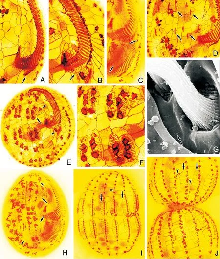
2.2.1 分裂期扇形游仆虫皮层银线系的形态发生 在扇形游仆虫中,首先于口围带基部后端左侧皮层产生成斜向“一”字形排列小群毛基体(见图2A),毛基体快速增殖,形成后仔虫口围带原基区。此时,在口原基区右前方的龛腔壁上出现紧密排列的新银线网(见图2B)。当口原基区自前端开始逐步组装成半弧形排列的小膜时,新银线网在其右侧不断扩展。伴随着龛腔变大,口围带小膜充分发育变宽,其一侧的新银线网也充分扩展,成为处于后仔虫口腔前方,在口围带和波动膜之间皮层表面的新银线网(见图2C)。
新口原基形成时,在额腹横棘毛皮层区形成2组各含5列原基的前、后额腹横棘毛原基区(见图2D),其每列原基不断变粗,并按由后至前、由左至右顺序分化成3、3、3、3、2段棘毛原基(见图2E、F)。与此同时,在每段棘毛原基的左侧和右侧按自后至前的顺序发生细小的银线网,其银线网不断发育伸展,于每列原基的左右两侧各形成1列新银线网(见图2G、H),最终在新的额腹横棘毛原基区形成7~8列四边形或近似四边形的银线网。此外,伴同老口侧膜前方唇和新口围带原基右侧皮层区的前、后2个新棘毛原基的分化,其周围也发生细小的银线网并不断发育伸展,形成四边形或近似四边形的银线网(见图2E)。随着额腹横棘毛原基进一步发育形成新棘毛、虫体演化成前、后仔虫时,腹面残留的老棘毛及老银线网消失,新棘毛及新银线网相继定位,成为前、后仔虫的额腹横棘毛和腹面银线网。
(A~F:银浸染色照片;G,H:扫描电镜照片;标尺:20 μm。A.扇形游仆虫腹面含有排列较整齐的四边形或近四边形银线网;无尾箭头示伸缩泡孔;B.扇形游仆虫背面单扇形型银线系;C.镰游仆虫腹面含有排列不规则的多边形至六边形银线网;无尾箭头示伸缩泡孔;D.镰游仆虫背面双阔口型银线系;E、G.包囊游仆虫腹面含有大小不一且排列不规则的多边形至六边形银线网;有尾箭头示波动膜;无尾箭头示伸缩泡孔;F、H.包囊游仆虫背面复杂型银线系。CC:尾棘毛;DK:背触毛;FC:额腹棘毛;LMC:左缘棘毛;MC:口棘毛;TC:横棘毛。A~F: Silver nitrate impregnation micrographs; G,H: Scanning electron micrographs; Scale bars: 20 μm. A. The ventral side ofE.vannuscontains neatly arranged quadrilateral silver-line system;arrowhead depicts the contractile vacuole pore; B. Dorsal view ofE.vannus, showing the single-vannussilver-line system; C. The ventral silver-line system ofE.harpacontains irregular polygons to hexagons;arrowhead refers to the contractile vacuole pore; D. Dorsal view ofE.harpa, showing the double-eurystomussilver-line system; E, G. The ventral silver-line system ofE.encysticuscontains irregular meshwork of different sized polygons to hexagons;arrow depicts the paroral membrane; arrowhead marks the contractile vacuole pore; F, H. Dorsal view ofE.encysticus, showing the complex silver-line system. CC: Caudal cirri; DK: Dorsal kinety; FC: Frontoventral cirri; LMC: Left marginal cirri; MC: Migratory cirrus; TC: Transverse cirri.)
图1 3种游仆虫细胞分裂间期皮层银线系形态
Fig.1 The silver-line system of threeEuplotesspecies in non-dividing stage
在细胞背面,新背触毛及其周围银线系的发生与腹皮层纤毛器和银线系的分化相伴进行。首先在每个老背触毛列的中部范围各形成1列新背触毛原基,于每列原基中基体间分开成一定距离时,在各个毛基体的周围产生细小的银线网,其中在基体左侧的新银线网先于右侧开始发育,接着在每个背触毛基体两侧各形成1列新银线网(见图2I)。细胞形成分裂沟时,每列新背触毛在其中部分开成前、后两部分,相邻两列新背触毛间的老银线网消失,背触毛左、右两侧的新银线网会合成1列。随着细胞的演化,新形成的背触毛和新银线网与同一列中的其他老背触毛及老银线网,最终成为新仔虫的背触毛列及其背皮层银线网(见图2J)。
2.2.2 分裂期镰游仆虫银线系的形态发生 镰游仆虫腹面纤毛器和银线系的发生与扇形游仆虫相似,但额腹横棘毛原基及其周围银线系的发生却略有不同。在新口原基形成时(见图3A),于额腹横棘毛皮层区形成2组各含5列原基的前、后额腹横棘毛原基区,其每列原基不断变粗,并按由后至前、由左至右顺序分化成3、3、3、3、2段棘毛原基(见图3B、C)。与此同时,在每段棘毛原基的左侧和右侧按自后至前的顺序发生细小的新银线网,其银线网不断发育伸展,于每列原基的周围各形成许多排列不规则的新银线网,最终在新的额腹横棘毛原基区形成多边形银线网(见图3D~F、H)。此外,伴同老口侧膜前方唇和新口围带原基右侧皮层区的前、后2个新棘毛原基的分化,其周围也发生细小的银线网并不断发育伸展,形成多边形银线网(见图3B、D)。随着额腹横棘毛原基进一步发育形成新棘毛、虫体演化成前、后仔虫时,腹面残留的老棘毛及老银线网消失,新棘毛及新银线网相继定位,成为前、后仔虫的额腹横棘毛和腹面银线网(见图3G)。
(A~F和H~J:银浸染色照片;G:扫描电镜照片。A.口原基分化出小群毛基体(箭头所示);B.口原基周围银线网(箭头所示);C.后仔虫口腔前方银线网(箭头所示);D.额腹横棘毛原基出现(箭头所示);E.示分成段的额腹横棘毛原基和其周围银线网(有尾箭头),以及前、后仔虫口棘毛原基(无尾箭头);F、G.分成段的额腹横原基及其周围银线网的细节图;H.腹面新银线网(有尾箭头)和老银线网(无尾箭头);I、J.背触毛原基发生及其周围新银线网。有尾箭头示背面新银线网;无尾箭头示老银线网。A~F&H~J: Silver nitrate impregnation micrographs; G:Scanning electron micrograph. A.Asmall number of basal bodies are differentiating from the oral primordium (arrow);B. Silver-line system around the oral primordium (arrow);C. Silver-line system in front of the buccal cavity of proter (arrows);D. The appearance of frontoventral-transverse anlagen (arrows);E.Fragmented frontoventral-transverse anlagen and the silver-line system around them (arrow); arrowheads mark themigratory cirrus anlagen of proter and opisthe;F, G. Detail of the fragmented frontoventral-transverse anlagen and the silver-line system around them; H.Ventral view of the new (arrows) and the old (arrowheads)silver-line system; I, J. Dorsal bristle basal bodies appear and the new silver-line system around them. Showing the dorsal view of the new (arrows) and the old (arrowheads) silver-line system.)
图2 扇形游仆虫皮层银线系的形态发生
Fig.2 Morphogenesis ofE.vannus
在细胞背面,新背触毛及其周围银线系的发生与扇形游仆虫的基本相似,只是在细胞形成分裂沟时,每列新背触毛在其中部分开成前、后两部分,相邻2列新背触毛间的老银线网消失,背触毛左、右两侧的新银线网汇合成2列(见图3I~L)。随着细胞的演化,新形成的背触毛和新银线网与同一列中的其他老背触毛及老银线网,最终成为新仔虫的背触毛列及其背皮层新银线网。
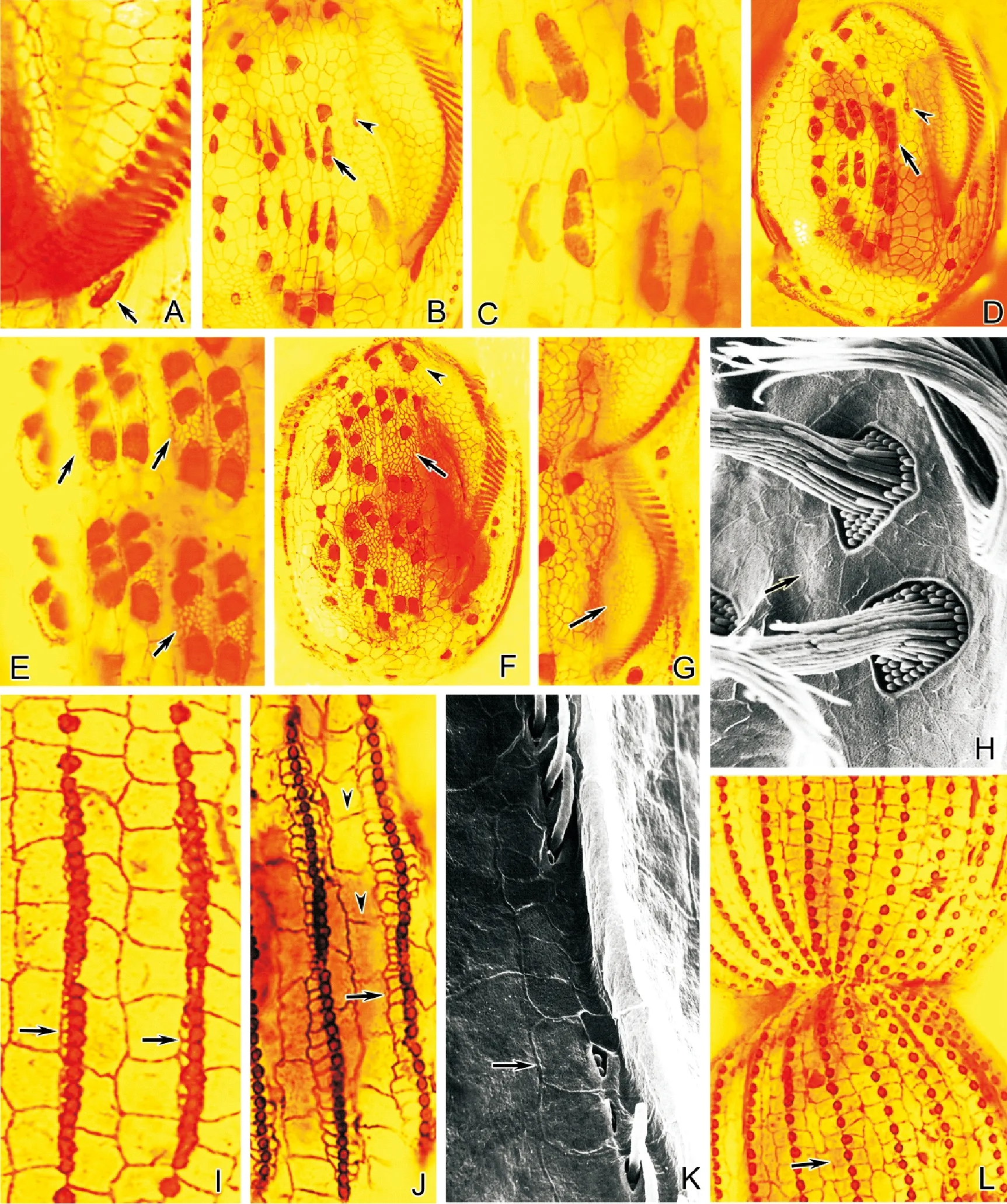
(A~G、I、J、L:银浸染色照片;H,K:扫描电镜照片。A.口原基分化出小群毛基体(箭头所示);B.示开始分段的额腹横棘毛原基(有尾箭头)和前仔虫口棘毛原基(无尾箭头);C.开始分段的额腹横棘毛原基及其左侧新银线网的细节图;D.示分成段的额腹横棘毛原基和其周围新银线网(箭头),以及前仔虫口棘毛原基(无尾箭头);E.示分成段的额腹横棘毛原基及其周围银线网的细节图(箭头);F、H.示腹面新银线网(有尾箭头)和老银线网(无尾箭头);G.后仔虫口腔前方银线网(箭头所示);I. 背触毛原基发生及其周围新银线网;J、K. 示背触毛原基周围新银线网发展扩大(有尾箭头),及老银线网(无尾箭头);L.背面新银线网(箭头所示)。A~G, I, J, L:Silver nitrate impregnation micrographs; H, K: Scanning electron micrographs. A. A small number of basal bodies are differentiating from the oral primordium (arrow);B. The frontoventral-transverse anlagen (arrow)start to fragment;migratory cirrus anlagen of proter (arrowhead); C. Detail of the frontoventral-transverse anlagen start to fragment and the new silver-line system on the left; D. Fragmented frontoventral-transverse anlagen and the new silver-line system around them(arrow); migratory cirrus anlagen of proter (arrowhead); E. Detail of the fragmented frontoventral-transverse anlagen and the silver-line system around them (arrows); F, H.Ventral view of the new (arrows)and the old (arrowhead)silver-line system; G. Silver-line system in front of the buccal cavity of proter (arrow); I. Dorsal bristle basal bodies appear and the new silver-line system around them;J, K. New silver-line system around the dorsal bristle basal bodiesis extending further (arrows);arrowheads depict the old silver-line system;L.Dorsal view of the new (arrow) silver-line system.)
图3 镰游仆虫皮层银线系的形态发生
Fig.3 Morphogenesis ofE.harpa
2.2.3 分裂期包囊游仆虫银线系的形态发生 包囊游仆虫腹面纤毛器和银线系的发生与扇形游仆虫相似, 但额腹横棘毛原基及其周围银线系的发生却略有不同。在新口原基形成时(见图4A),于额腹横棘毛皮层区形成2组各含5列原基的前、后额腹横棘毛原基区,其每列原基不断变粗,并按由后至前、由左至右顺序分化成3、3、3、2、2段棘毛原基(见图4B、C)。与此同时,在每段棘毛原基的左侧和右侧按自后至前的顺序发生细小的新银线网,其银线网不断发育伸展,于每列原基的周围各形成许多排列不规则的新银线网,最终在新的额腹横棘毛原基区形成多边形银线网(见图4E、F)。随着额腹横棘毛原基进一步发育形成新棘毛、虫体演化成前、后仔虫时,腹面残留的老棘毛及老银线网消失,新棘毛及新银线网相继定位,成为前、后仔虫的额腹横棘毛和腹面银线网。

(A、B、D~J:银浸染色照片;C:扫描电镜照片。A.口原基分化出小群毛基体(箭头所示);B.额腹横棘毛原基(箭头所示);C、E.分段的额腹横棘毛原基及其周围银线网(箭头所示);D.后仔虫口围带原基小膜(箭头所示);F.腹面新银线网(箭头所示);G.新背触毛基体发生(箭头所示);H. 背触毛原基及其周围新银线网(有尾箭头);老银线网(无尾箭头);I.背触毛原基周围新银线网(有尾箭头)发展扩大;老银线网(无尾箭头);J.背面新银线网(有尾箭头)和老银线网(无尾箭头)。A, B, D~J: Silver nitrate impregnation micrographs; C:Scanning electron micrographs.A. A small number of basal bodies are differentiating from the oral primordium;B. Frontoventral-transverse anlagen (arrow);C, E.Fragmented frontoventral-transverse anlagen and the silver-line system around them (arrows); D. The new formed membranelles of opisthe (arrow);F. Ventral view of the new silver-line system (arrow); G.New dorsal bristle basal bodies appear (arrow); H.Dorsal bristle basal bodies and the new silver-line system around them (arrow); the old silver-line system (arrowhead);I.New silver-line system around the dorsal bristle basal bodies is extending further (arrow);the old silver-line system (arrowhead); J. Dorsal view of the new (arrow)and the old (arrowhead) silver-line system.)
图4 包囊游仆虫皮层银线系的形态发生
Fig.4 Morphogenesis ofE.encysticus
在细胞背面,新背触毛及其周围银线系的发生与腹皮层纤毛器和银线系的分化相伴进行。首先在每个老背触毛列的中部范围各形成1列新背触毛原基(见图4G),于每列原基中基体间分开成一定距离时,在各个毛基体的周围产生细小的银线网,其中在基体左侧的新银线网先于右侧开始发育,接着在每个背触毛基体两侧各形成多列新银线网。细胞形成分裂沟时,每列新背触毛在其中部分开成前、后两部分,相邻2列新背触毛间的老银线网消失,背触毛左、右两侧的新银线网会合成多列。随着细胞的演化,新形成的背触毛和新银线网与同一列中的其他老背触毛及老银线网,最终成为新仔虫的背触毛列及其背皮层银线网(见图4H~J)。
3 讨论
3.1 游仆虫腹面银线系形态
游仆虫背面银线系因具有种类的特异性,一直以来被应用于游仆虫类的物种鉴定与分类,而关于腹面银线系的特点未见进一步探讨[2,9]。本实验结果显示扇形游仆虫腹面银线网排列整齐且稀疏,而镰游仆虫和包囊游仆虫腹面银线网形状不规则且排列紧密。这种不同的排列特征分别与其背面银线网的排列规律相类似,尤其体现在银线网的紧密程度上;而背腹面银线网的紧密程度又与背触毛列中的基体数目密切相关。这种对应关系亦出现在其他已有银线系报道的游仆虫种类中[16-23],即在所有银线系类型中,背触毛基体数目越多,背面银线网越密集,腹面网格也相应密集。据此推测,游仆虫腹面银线系虽不能如背面银线系以具体类型划分,但其形态特征与背面银线系具有一定程度的一致性,这显示细胞在背腹面银线系结构形成过程中的整体性调控。
3.2 游仆虫银线系的形态发生
游仆虫皮层纤毛器、纤毛器附属微管及皮层银线网等是一类重要的的皮层细胞骨架[12],目前对该类纤毛虫皮层纤毛器及其纤毛器附属微管的形态发生及其细胞调控的研究已经取得较丰富的资料[13,24-35],但对其银线系的形态发生尚未见报道。本文应用银浸染色法及扫描电镜术显示,扇形游仆虫、镰游仆虫和包囊游仆虫3种纤毛虫皮层银线网的形态发生与细胞皮层纤毛器的发生是相伴进行的,其腹皮层银线网在口围带原基区分化形成的小膜原基和额腹横棘毛原基区原基列分段形成的棘毛原基周围发生,背皮层银线网在每列背触毛原基的各个背触毛基体周围发生,由于此时的口围带小膜原基、棘毛原基和背触毛基体已经形成纤毛基体复合单元和基体单元[24,36-40],也即其银线网的形态发生是在纤毛器原基形成口围带小膜原基、棘毛原基基体复合单元和背纤毛基体单元后启动的。根据Shao et al.划分的游仆虫属额腹横棘毛原基的分段模式,扇形游仆虫和镰游仆虫属于charon-type,II至V额腹横棘毛原基分段为3∶3∶3∶3∶2,包囊游仆虫则属于affinis-type,分段为3∶3∶3∶2∶2[41],即前2种游仆虫与包囊游仆虫相比,其第5列额腹横棘毛原基多断裂生成一个棘毛3/V的基体。本文观察到在扇形游仆虫和镰游仆虫中,新银线网于棘毛3/V基体周围产生并随该基体的迁移而持续分化;而对于该部位无此棘毛基体的包囊游仆虫,新银线网产生仅限于2/V和1/V 2个棘毛原基附近。这进一步验证了在非纤毛区无新银线网独立发生,新银线网的发生与分化与纤毛基体密切相关。据以上资料推测,所述的口围带小膜、棘毛基体复合单元和背触毛基体单元不仅对相应纤毛器结构行使微管组织中心作用,也可能对相应皮层区银线网的发生具有定位作用和组织中心的作用。结果还显示,3种游仆虫围绕在基体左侧的银线网先于右侧开始发育,与游仆虫新棘毛原基发生和分化过程相一致[25],据此推测银线系的分化可能也具有一致的方向性。因此,以上所得结果可为揭示纤毛虫生命活动中细胞皮层结构的分化及其调控机理提供新的资料。
3.3 老结构在游仆虫新银线系形成过程中的作用
关于游仆虫皮层纤毛器形态发生过程中新、老结构的演化及其作用关系已有较多研究[25,42],但对银线系发生中新、老结构的更替及其作用机理等方面尚未取得相关资料。本文观察到,3种游仆虫腹面银线系发生中老纤毛结构及其周围银线网按序先后瓦解,其中离新结构发生区近的老结构先瓦解,由此使新结构不断延伸发展,据此推测老银线网对新结构的形成可能起到了诱导定向作用;细胞出现分裂沟时,在每一背触毛列的中部范围相邻2列新背触毛间的老银线网消失,背触毛左、右两侧的新银线网会合形成新银线网,并且前仔虫前半部分和后仔虫后半部分的老银线网保留下来共同组成新细胞的背银线网,结果表明,游仆虫的老银线网不仅参与了新结构的组成,并对新结构的形成具有物质贡献。
4 结语
本文揭示了游仆虫银线系形态发生过程中的结构特征,为研究这类微管类细胞骨架提供了新的资料。但本研究并未揭示新银线网在发生时,皮层纤毛器及其附属微管和银线系在皮层下的变化情况,因此,在本研究的基础上,可以通过透射电镜等方法进一步研究探讨银线系、皮层纤毛器及其附属微管之间的关系,构建其在形态发生时的三维模型,为游仆虫皮层模式形成提供更多资料。
[1] Tuffrau M. Les caractères specifiques dans le genreEuplotes(Note Préliminaire) [J]. Bulletin de la Societe Zoologique de France, 1954, 79: 463-465.
[2] Tuffrau M. Revision of genusEuploteson the bases of structural comparison [J]. Hydrobiologia, 1960, 15(1): 1-77.
[3] Tuffrau M, Fryd-Versavel G, Tuffrau H, et al. Description ofEuplotesversatilisn. sp., a marine tropical ciliate exhibiting an unusually extensive phenotypic plasticity [J]. European Journal of Protistology, 2000, 36(4): 355-366.
[4] Chen X, Zhao Y, Al-Farraj S A,et al. Taxonomic descriptions of two marine ciliates,Euplotesdammamensisn. sp. andEuplotesbalteatus(Dujardin, 1841) Kahl, 1932 (Ciliophora, Spirotrichea, Euplotida), collected from the Arabian Gulf, Saudi Arabia [J]. Acta Protozoologica, 2013, 52(2): 73-89.
[5] Dai R, Xu K, He Y. Morphological, physiological, and molecular evidences suggest thatEuplotesparawoodruffiis a junior synonym ofEuploteswoodruffi(Ciliophora, Euplotida) [J]. The Journal of Eukaryotic Microbiology, 2013, 60(1): 70-78.
[6] Chen X, Ma H, Al-Rasheid K A S. Taxonomic description of a new marine ciliate,Euplotesqingdaoensisn. sp. (Ciliophora: Euplotida) [J]. Chinese Journal of Oceanology and Limnology, 2014, 32(2): 426-432.
[7] Giuseppe G D, Erra F, Frontini F P, et al. Improved description of the bipolar ciliate,Euplotespetzi, and definition of its basal position in theEuplotesphylogenetic tree [J]. European Journal of Protistology, 2014, 50(4): 402-411.
[8] Giuseppe G D, Dini F, Vallesi A, et al. Genetic relationships in bipolar species of the protist ciliate,Euplotes[J]. Hydrobiologia, 2015, 761(1): 71-83.
[9] Curds C R. A guide to the species of the genusEuplotes(Hypotrichida, Ciliatea) [J]. Bulletin of the British Museum (Natural History) Zoology, 1975, 28: 5-9.
[10] Gates M A, Curds C R. The argyrome of the genusEuplotes[J]. Bulletin of the British Museum (Natural History) Zoology, 1979, 35: 127-200.
[11] 朱慧, 邹士法, 李艺松, 等. 用非离子去垢剂抽提获得的小游仆虫皮层细胞骨架的构形[J]. 动物学研究, 2004, 25(5): 422-428. Zhu H, Zou S, Li Y, et al. Cortical cytoskeleton ofEuplotesgracilis(Protozoa, Ciliophora) by non-ionic detergent extraction [J]. Zoological Research, 2004, 25(5): 422-428.
[12] 李艺松, 柳伟君, 顾福康. 伍氏游仆虫皮层微管类细胞骨架的荧光标记[J]. 复旦学报 (自然科学版), 2008, 47(3): 364-369. Li Y, Liu W, Gu F. Cortical microtubular cytoskeleton inEuploteswoodruffirevealed by fluorescent labeling [J]. Journal of Fudan University (Natural Science), 2008, 47(3): 364-369.
[13] 林钦, 范鑫鹏, 孙磊, 等. 小腔游仆虫 (Euplotesaediculatus) 皮层微管胞器的荧光标记[J]. 华东师范大学学报 (自然科学版), 2013(5): 53-60. Lin Q, Fan X, Sun L, et al. Microtubular organelles inEuplotesaediculatus (Ciliophora: Hypotrichida) revealed by fluorescent labeling [J]. Journal of East China Normal University (Natural Science), 2013(5): 53-60.
[14] 史新柏. 银浸法在纤毛虫研究中的应用[J]. 哈尔滨师范学院学报 (自然科学版), 1963, 1: 79-83. Shi X. Application of silver-impregnation methods in the study of ciliates [J]. Journal of Harbin Normal University (Natural Science), 1963, 1: 79-83.
[15] 顾福康, 倪兵. 原生动物扫描电镜样品制备方法的探讨[J]. 电子显微学报, 1993(6): 525-529. Gu F, Ni B. The exploration of preparing protozoan specimens for scanning electron microscopy [J]. Journal of Chinese Electron Microscopy Society, 1993(6): 525-529.
[16] Borror A C.EuplotesminutaYocom (Ciliophora, Hypotrichida) [J]. Journal of Protozoology, 1962, 9(3): 271-273.
[17] Pan Y, Li L, Shao C, et al. Morphology and ontogenesis of a marine ciliate,Euplotesbalteatus(Dujardin, 1841) Kahl, 1932 (Ciliophora, Euplotida) and definition ofEuploteswilbertinov. spec [J].Acta Protozoologica, 2012, 51(1): 29-38.
[18] Jong O, Mann K. Redescription of newly recorded ciliate,Euplotesmuscorum(Ciliophora: Polyhymenophora: Hypotrichida) and comparison with related species from Korea [J]. Korean Journal of Systematic Zoology, 2003, 19(2): 227-235.
[19] Song W, Warren A, Hill B F. Description of a new freshwater ciliate,Euplotesshanghaiensisnov. spec. from China (Ciliophora, Euplotidae) [J]. European Journal of Protistology, 1998, 34 (2): 104-110.
[20] Liu M, Fan Y, Miao M, et al. Morphological and morphogenetic redescriptions and SSU rRNA gene-based phylogeny of the poorly-known speciesEuplotesamietiDragesco, 1970 (Ciliophora, Euplotida) [J]. Acta Protozoologica, 2015, 54: 171-182.
[21] Fan X, Huang J, Lin X, et al. Morphological and molecular characterization ofEuplotesencysticus(Protozoa: Ciliphora: Euplotida) [J]. Journal of the Marine Biological Association of the United Kingdom, 2010, 90(7): 1411-1416.
[22] Jiang J, Zhang Q, Warren A, et al. Morphology and SSU rRNA gene-based phylogeny of two marineEuplotesspecies,E.orientalisspec. nov. andE.raikoviAgamaliev, 1966 (Ciliophora, Euplotida) [J]. European Journal of Protistology, 2010, 46(2): 121-132.
[23] Jiang J, Zhang Q, Hu X, et al. Two new marine ciliates,Euplotessinicussp. Nov. andEuplotesparabalteatussp. nov., and a new small subunit rRNA gene sequence ofEuplotesrariseta(Ciliophora, Spirotrichea, Euplotida) [J]. International Journal of Systematic and Evolutionary Microbiology, 2010, 60: 1241-1251.
[24] 顾福康, 庞延斌, 张作人. 一种游仆虫无性生殖的研究II.无性分裂过程中皮层结构的形态发生[J]. 动物学报, 1987, 33(4): 362-369. Gu F, Pang Y, Zhang Z. On the asexual reproduction of the genusEuplotesII. Morphogenesis of cortical structures during the asexual division [J]. Acta Zoologica Sinica, 1987, 33(4): 362-369.
[25] 顾福康, 张作人. 一种游仆虫棘毛基部纤维的形态及其在形态发生过程中的演化[J]. 动物学研究, 1989, 10(2): 89-96. Gu F, Zhang Z. The morphology of cirral-base-associated fibers inEuplotessp. and their morphogenesis [J]. Zoological Research, 1989, 10(2): 89-96.
[26] 牛延宁, 吴月华, 倪兵, 等. 包囊游仆虫纤毛器微管在不同生理状态下的分化[J]. 华东师范大学学报 (自然科学版), 2007(6): 106-111. Niu Y, Wu Y, Ni B, et al. Ciliature microtubule differentiation ofEuplotesencysticusin different physiological conditions [J]. Journal of East China Normal University (Natural Science), 2007(6): 106-111.
[27] 余齐耀, 张萌, 陈季武, 等. 显示纤毛虫细胞微管骨架的两种荧光标记方法[J]. 生物学杂志, 2012, 29(1): 92-94. Yu Q, Zhang M, Chen J, et al. Two fluorescent labeling methods for revealing microtubular cytoskeleton in ciliates [J]. Journal of Biology, 2012, 29(1): 92-94.
[28] 吴娜, 周慧琳, 范鑫鹏, 等. 澳洲管膜虫纤毛器微管的荧光标记及其激光扫描共聚焦显微观察[J]. 生物学杂志, 2015, 32(1): 30-33. Wu N, Zhou H, Fan X, et al. Observation on ciliature microtubules ofCyrtohymenaaustralisusing fluorescent labeling and laser scanning confocal microscopy [J]. Journal of Biology, 2015, 32(1): 30-33.
[29] Jiang J, Shao C, Xu H, et al. Morphogenetic observations on the marine ciliateEuplotesvannusduring cell division (Protozoa: Ciliophora) [J]. Journal of the Marine Biological Association of the United Kingdom, 2010, 90(4): 683-689.
[30] Rashmi F, Thorsten S, Sabine F, et al. Description of the HalophileEuplotesqatarensisnov. spec. (Ciliophora, Spirotrichea, Euplotida) isolated from the Hypersaline Khor Al-Adaid Lagoon in Qatar [J]. Journal of Eukaryotic Microbiology, 2016. DOI:10.111/jeu.12305.
[31] Fleury A. Dynamics of the cytoskeleton during morphogenesis in the ciliateEuplotesI. Basal bodies related microtubular system [J]. European Journal of Protistology, 1991, 27: 99-114.
[32] Fleury A. Dynamics of the cytoskeleton during morphogenesis in the ciliateEuplotesII. Cortex and continuous microtubular systems [J]. European Journal of Protistology, 1991, 27(3): 220-237.
[33] 毕红卫, 庞延斌. 卡龙游仆虫Euplotescharon的形态和形态发生的研究[J]. 华东师范大学学报 (自然科学版), 2001(2): 71-77. Bi H, Pang Y. The studies on the morphology and morphogenesis ofEuplotescharon[J]. Journal of East China Normal University (Natural Science), 2001(2): 71-77.
[34] 王梅, 宋微波. 卡龙游仆虫无性生殖期间的形态发生学研究[J]. 动物学研究, 1995, 16(3): 233-238. Wang M, Song W. Morphogenetical studies on the marine ciliateEuplotescharon[J]. Zoological Research, 1995, 16(3): 233-238.
[35] 庞延斌, 魏红兵. 小腔游仆虫Euplotesaediculatus形态和形态发生的研究[J]. 华东师范大学学报 (自然科学版), 1999(1): 103-109. Pang Y, Wei H. Studies on the morphology and morphogenesis inEuplotesaediculatus[J]. Journal of East China Normal University (Natural Science), 1999(1): 103-109.
[36] Jerka-Dziadosz M, Frankel J. An analysis of the formation of ciliary primordia in the hypotrich ciliateUrostylaweissei[J].Journal of Protozoology, 1969, 16(4): 512-537.
[37] Jerka-Dziadosz M. Ultrastructural study on development of the hypotrich ciliateParaurostylaweisseiI. Formation and morphogenetic movements of ventral ciliary primordial [J]. Protistologica, 1980, 16: 571-589.
[38] Jerka-Dziadosz M. Ultrastructural study on development of hypotrich ciliateParaurostylaweisseiII. Formation of the adoral zone of membranelles and its bearing of problems of ciliate morphogenesis [J]. Protistologica, 1981, 17: 67-81.
[39] Jerka-Dziadosz M. Ultrastructural study on development of hypotrich ciliateParaurostylaweisseiIII. Formation of paroral membranelles and essary on comparative morphogenesis [J]. Protistologica, 1981, 17: 83-97.
[40] Jerka-Dziadosz M. Ultrastructural study on development of the hypotrich ciliateParaurostylaweisseiIV. Morphogenesis of dorsal bristles and caudal cirri [J]. Protistologica, 1982, 18: 237-251.
[41] Shao C, Ma H, Gao S, et al. Reevaluation of cortical developmental patterns inEuplotes(s.1.), including a morphogenetic redescription ofE.charon(Protozoa, Ciliophora, Euplotida) [J]. Chinese Journal of Oceanology and Limnology, 2010, 28(3): 593-602.
[42] 顾福康, 张作人. 腹毛目纤毛虫无性生殖周期中形态发生的研究[J]. 华东师范大学学报 (自然科学版), 1990, 2: 85-93. Gu F, Zhang Z. Study on morphogenesis of hypotrich ciliates during asexual reproduction [J]. Journal of East China Normal University (Natural Science), 1990, 2: 85-93.
责任编辑 朱宝象
更正声明
《中国海洋大学学报(自然科学版)》第11期中“DNA条形码技术在鲻科鱼类鉴定中的应用”论文刊登的作者信息、单位信息的中英文有误,在此进行更正。正确的作者信息、单位信息的中英文如下:
刘 璐1, 孙典荣2, 李纯厚2, 韩志强3, 高天翔3, 宋 娜1❋❋
(1.中国海洋大学水产学院,山东 青岛 266003;2.中国水产科学研究院南海水产研究所,广东 广州 510300;3.浙江海洋大学水产学院,浙江 舟山 316022)
LIU Lu1, SUN Dian-Rong2, LI Chun-Hou2, HAN Zhi-Qiang3, GAO Tian-Xiang3, SONG Na1
(1.College of Fisheries, Ocean University of China, Qingdao 266003, China;2.South China Sea Fisheries Research Institute, Chinese Academy of Fishery Sciences, Guangzhou 510300, China;3.Fishery College, Zhejiang Ocean University, Zhoushan 316022, China)
特此声明!
中国海洋大学学报(自然科学版)编辑部
Morphogenesis of the Cortical Silver-Line Systems in the Ciliate Genus Euplotes (Protozoa, Ciliophora)
XIE Dong-Mei, FAN Xin-Peng, NI Bing, GU Fu-Kang
(School of Life Sciences, East China Normal University, Shanghai 200241, China)
The silver-line system ofEuplotescorresponds to the cytoskeleton network, which is composed of microtubules at the ultrastructure level, and its morphogenesis pattern during cell division is rarely reported. Based on the application of silver nitrate impregnation and scanning electron microscopy, and usingE.vannus,E.harpaandE.encysticusas the representatives, the morphogenesis of three types of cortical silver-line systems in genusEuploteswas examined in this study.The result showed that the morphogenesis of three types of cortical silver-line systems in the genusEuplotesshared the following features: the ventral silver-line system occurred around the new membranelles and the fragmented frontoventral-transverse anlagen; and the dorsal silver-line system occurred around the basal bodies of the dorsal bristle primordia. Thus, it was speculated that the composite element of basal bodies was not only microtubule organizing center, but also the center of the silver-line system. The new meshwork on the left of the basal bodies developed ahead of the meshwork on the right, thus, it was speculated that the silver-line system oriented similarly with ciliary organelles during their morphogenesis. In addition, the old meshwork near new silver-line first disintegrated, which led to the new silver-line system to develop constantly. It was assumed that the old silver-line system played a role of induction and orientation in the formation of new structure. The old silver-line system locating in the first half of the proter and in the latter part of the opisthe was reserved to form the dorsal silver-line system in new cells. It was supposed that the old silver-line system participated in the formation of new structure and also had a material contribution to it.
Euplotes; silver-line system; morphogenesis; silver nitrate impregnation; scanning electron microscopy
国家自然科学基金项目(31172042;31572223)资助 Supported by National Natural Science Foundation of China (31172042;31572223)
2015-11-06;
2016-03-16
谢冬梅(1989-),女,硕士生。E-mail: zynx9194@sina.com
❋❋ 通讯作者:E-mail: xpfan@bio.ecnu.edu.cn
Q952;Q954
A
1672-5174(2016)12-041-10
10.16441/j.cnki.hdxb.20150385
谢冬梅, 范鑫鹏, 倪兵, 等. 游仆虫(原生动物,纤毛门)皮层银线系的形态发生模式[J]. 中国海洋大学学报(自然科学版), 2016, 46(12): 41-50.
XIE Dong-Mei, FAN Xin-Peng, NI Bing, et al. Morphogenesis of the cortical silver-line systems in the ciliate genusEuplotes(Protozoa, Ciliophora)[J]. Periodical of Ocean University of China, 2016, 46(12): 41-50.
猜你喜欢
自然杂志(2022年3期)2022-08-18
医学研究生学报(2021年4期)2021-12-02
临床与实验病理学杂志(2021年3期)2021-04-25
植物研究(2020年6期)2020-03-05
中华皮肤科杂志(2019年5期)2019-06-24
小学生必读(高年级版)(2019年10期)2019-03-28
都市(2018年11期)2018-11-22
计算机应用(2018年10期)2018-11-22
中成药(2017年12期)2018-01-19
浙江大学学报(农业与生命科学版)(2017年2期)2017-05-19