血肿形态预测急性高血压脑出血血肿再扩大的研究
2019-04-21 13:43:50王琼
中外医学研究
2019年28期
关键词:高血压脑出血
王琼
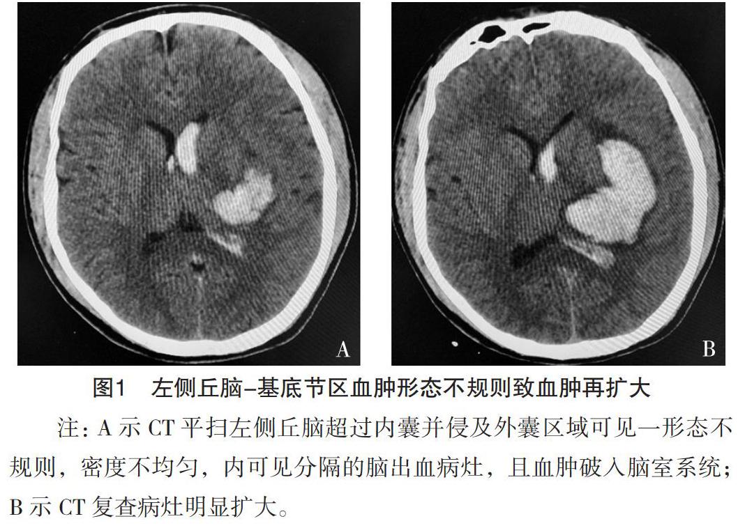
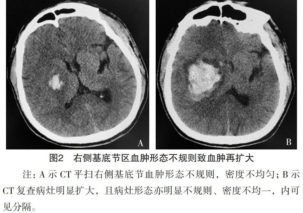
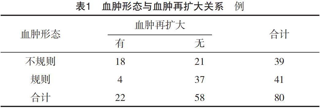
【摘要】 目的:分析血肿形态与急性高血压脑出血血肿再扩大的关系。方法:所有急性高血压脑出血患者均接受基线及(24±6)h内CT平扫复查。一位影像科医生采用半自动血肿体积测量软件计算基线及复查CT平扫的大小及体积,另一名影像科医生独立评估CT平扫血肿形态信息。比较血肿形态不同者血肿再扩大情况的差异。结果:经统计,血肿再扩大者22例,血肿形态分为规则41例、不规则39例,血肿形态不规则致血肿再扩大率明显高于血肿形态规则者,差异有统计学意义(字2=13.281,P=0.000 1)。血肿形态预测血肿再扩大的敏感度和特异度分别为81.8%(18/22)和63.8%(37/58)。结论:血肿形态不规则可作为血肿再扩大的影像指征,血肿的形态预测血肿再扩大的灵敏度较高。
【关键词】 血肿形态 高血压脑出血 血肿再扩大 预测指标
doi:10.14033/j.cnki.cfmr.2019.28.027 文献标识码 B 文章编号 1674-6805(2019)28-00-03
Hematoma Morphology Predicting for Hematoma Re-enlargement of Acute Hypertensive Intracerebral Hemorrhage/WANG Qiong. //Chinese and Foreign Medical Research, 2019, 17(28): -68
[Abstract] Objective: To investigate the relationship in hematoma morphology predicting hematoma re-enlargement of acute hypertensive intracerebral hemorrhage. Method: All acute hypertensive intracerebral hemorrhage patients underwent the admission of baseline CT examination and follow-up (24±6) h CT examination using the semi-automatic software. One radiologic doctor calculates the volume of the hematoma in the baseline and follow-up CT images. Another radiologic doctor independently evaluates the morphology of the hematoma. Compared the difference of hematoma re-enlargement in different hematoma forms. Result: After calculating the volume, 22 cases had significant hematoma expansion. Hematoma morphology can divided into the regular and irregular, revealing 41 cases and 39 cases respectively. The re-enlargement rate of hematoma caused by irregular shape of hematoma was significantly higher than that caused by regular shape of hematoma (字2=13.281, P=0.000 1). The sensitivity and specificity of hematoma morphology in predicting hematoma re-enlargement were 81.8% (18/22) and 63.8% (37/58), respectively. Conclusion: It is revealed that hematoma morphology can be considered as imaging indictor for hematoma re-enlargement. Hematoma morphology has high sensitivity in predicting hematoma expansion.
[Key words] Hematoma morphology Acute hypertensive intracerebral hemorrhage Hematoma re-enlargement Predictor
First-authors address: Capital Medical University Affiliated Shijingshan Teaching Hospital, Beijing 100043, China
高血壓脑出血性卒中较缺血性卒中后果严重,预后差,死亡率较高。血肿大小在一定程度上是预测高血压脑出血死亡率的重要指标[1]。而高血压脑出血血肿再扩大是造成病情恶化的主要原因之一,国外很多研究证实了血肿再扩大对于高血压脑出血病情恶化是有预测性的,并且是病死率的独立预测因素[2-3]。因此,如果有影像信息能够预测血肿再扩大,这将对患者的个体化治疗提供依据。本研究中主要目的为CT平扫血肿形态与急性高血压脑出血血肿再扩大的关系,现报告如下。
1 资料与方法
1.1 一般资料
回顾性分析2018年1月-2019年5月收集的急性高血压脑出血患者资料,所有入组患者都经过严格的临床及影像选择。……
登录APP查看全文
猜你喜欢
中国实用医药(2016年29期)2016-12-26 10:15:32
中国实用医药(2016年23期)2016-12-26 08:18:30
中国实用医药(2016年28期)2016-12-07 08:01:29
中国实用医药(2016年27期)2016-11-30 11:57:32
中国实用医药(2016年27期)2016-11-30 11:19:39
中外医学研究(2016年26期)2016-11-30 02:48:03
中外医学研究(2016年28期)2016-11-28 07:20:12
中外医学研究(2016年28期)2016-11-28 06:54:19
今日健康(2016年12期)2016-11-17 12:11:26
上海医药(2016年13期)2016-07-26 23:19:07